Chapter 29
Conjunctiva
J. ERIC PEPPERL, TOM GHUMAN, KULJIT S. GILL, JAMES D. ZIESKE and STEFAN D. TROCME
Main Menu   Table Of Contents

Search

ANATOMY
CONJUNCTIVAL GLANDS
CONJUNCTIVAL EPITHELIUM
EPITHELIAL CYSTS
GOBLET CELLS
SUBSTANTIA PROPRIA
CONJUNCTIVAL INFLAMMATION
CONJUNCTIVAL WOUND HEALING
CONJUNCTIVAL HEALING IN GLAUCOMA FILTERING SURGERY
ARTERIES
VEINS
LYMPHATICS
NERVE SUPPLY
THE CARUNCLE
PLICA SEMILUNARIS
REFERENCES

The conjunctiva is a vascularized mucous membrane that covers the anterior surface of the globe (bulbar and forniceal conjunctiva) and the posterior surface of the upper and lower eyelids (palpebral conjunctiva). Its superficial layer, the conjunctival epithelium, is continuous with the epidermis of the lids and the outermost layer of the cornea, the corneal epithelium. The conjunctiva is responsible for the production of mucous, which is essential for tear film stability and corneal transparency.1 The conjunctiva also has enormous potential for combating infection for four reasons: (1) it is highly vascular; (2) the different cell types contained in it can initiate and participate in defensive inflammatory reaction; (3) it has many immunocompetent cells that contribute a rich supply of immunoglobulins; and (4) the surface anatomy (microvilli) and biochemistry (enzymatic activity) of the conjunctival cells enable that tissue to engulf and neutralize foreign particles, such as viruses.2,3

Clinically, the conjunctiva is an extremely valuable ally of the ophthalmic surgeon and diagnostician. The bulbar surface, because of its loosely adherent properties, is used to great advantage in glaucoma surgery. Its capacity to heal rapidly ensures the success of many surgical procedures. A conjunctival flap placed over a nonhealing or infected cornea promotes healing and preserves corneal integrity.4 Autologous conjunctival transplantation has been carried out from one eye to the other to repair leaky filtration blebs and to hasten corneal epithelial covering after chemical burns have destroyed the corneal surface.5 Characteristic conjunctival changes occur in many systemic diseases. For example, pathognomonic vascular signs are present in the conjunctival vessels in sickle cell anemia, scleral icterus is an early sign of jaundice, distinctive pigmentary changes are present in ochronosis, Bitot's spot appears in the conjunctiva in vitamin A deficiency, and typical crystalline deposits are present in the conjunctiva in cystinosis.6,7 Another exceptional advantage of the conjunctiva is the ease with which conjunctival biopsy material can be obtained, causing little discomfort to the patient and producing virtually no loss of tissue integrity.

Under normal circumstances, the mucocutaneous junction is a well-defined border. As the epidermis approaches the conjunctiva, the cornified acellular layer becomes thinner, and at the junction the superficial cells of the epidermis are no longer present in large numbers in the border region. Only a few keratohyalin granules remain.8 The superficial cells in the border region retain only a few well-defined nucleated cells. In the conjunctiva proper, however, all cells are nucleated and contain many cytoplasmic organelles. The superficial cells begin to show microvilli, contain numerous mucous granules, and have wider intercellular spaces. Goblet cells are also apparent. The conjunctiva becomes keratinized only in certain diseases, such as the Stevens-Johnson syndrome, cicatricial pemphigoid, and vitamin A deficiency.9

Back to Top
ANATOMY
The conjunctiva lines the posterior surface of the upper and lower lids and the anterior surface of the globe. From the inner surface of the lid it is reflected forward onto the globe above and below, forming two recesses: the superior and inferior fornix. The superior fornix is located at the level of the orbital margin 8 to 10 mm from the limbus (Fig. 1A, B, and C). The inferior fornix is approximately 8 mm from the limbus (Fig. 2A, B, and C). On the medial side, the forniceal structures are replaced by the caruncle and the plica semilunaris (Fig. 3). The absence of the fornix on the medial side is necessary in order to allow the inferior punctum to dip and drain from the superficial tear fluid layer.10 Laterally, the fornix extends just behind the equator of the globe (Fig. 4). It is quite deep and approximately 14 mm from the limbus.

Fig. 1. Low-power view of the globe. A. Arrow pointing to the region of the superior fornix. B. Superior fornix (F) showing epithelium and substantia propria. Conjunctival sac (CS). C. Higher-power view of epithelium showing goblet cells on the surface (arrows). (B, × 50; C, × 170)

Fig. 2. A. Region of the inferior fornix (arrow). B. Inferior fornix showing epithelium, goblet cells, and a follicle (F). C. Inferior fornix showing Müller's muscle (MM) in the substantia propria. (B, × 60; C, × 80)

Fig. 3. Medical region of the eye showing the caruncle (C) and plica semilunaris (P).

Fig. 4. Region of the lateral fornix (arrow).

At the posterior end of the eyelid margin at the mucocutaneous junction, the skin epidermis of the eyelid abruptly transforms into the palpebral conjunctiva and continues on the posterior aspect of the eyelid.11 The palpebral conjunctiva is markedly adherent to the tarsal plate of the lids. The palpebral conjunctiva is an area where reactive pathology of the conjunctiva may be seen clinically. There are two types of changes that can occur in this region: follicle formation and papilla formation. Follicles are thought to be identical to lymphoid follicles found elsewhere in the body (Fig. 5).7 Follicle formation is characteristic of viral and chlamydial infections as well as toxic conjunctivitis due to application of certain topical medications.12 Papillae are composed of chronic inflammatory cells such as lymphocytes and plasma cells and are distinguished from follicles by the presence of blood vessels at their center.8 Giant papillae are found in certain allergic diseases (e.g., vernal catarrh) and after long-term use of contact lenses, keratoprostheses, ocular postenucleation prostheses, and cosmetic shells (Fig. 6).7,13

Fig. 5. Follicles on the upper tarsal surface.

Fig. 6. Giant papillae present on the upper tarsal surface of a patient who wore a cosmetic shell.

The bulbar conjunctiva extends from the limbus to the forniceal area. It is so thin and translucent that the underlying sclera can be seen through it. The bulbar conjunctiva is loosely adherent to the sclera to allow the eye free movement in all directions. It is attached to the tendons of the rectus muscles, which in turn are covered by Tenon's capsule. Approximately 3 mm from the limbus, the bulbar conjunctiva, Tenon's capsule, and sclera become firmly attached, and the conjunctiva cannot be easily picked up.14 This attachment is routinely encountered during the dissection of a limbal-based conjunctival flap in ocular surgery.

Back to Top
CONJUNCTIVAL GLANDS
Krause's glands are accessory lacrimal glands found in the deep subconjunctival connective tissue of the upper fornix. There are 42 in the upper fornix and approximately 6 to 8 in the lower fornix. Because accessory lacrimal glands present in this region may be inadvertently excised, causing a dry eye problem, the integrity of the superior border of the upper tarsus is extremely important to preserve during operations (e.g., Fasanella-Servat procedure for upper lid ptosis).

The glands of Wolfring are also accessory lacrimal glands. There are two to five on the upper lid along the upper border of the tarsus.10 Two glands are present along the inferior edge of the lower tarsus. The excretory duct is lined with basal cuboidal epithelial cells, similar to the conjunctival epithelium onto which it opens. The fine structure of Krause's gland is essentially the same as that of the lacrimal gland in the orbit (Fig. 7A and B). Manz's glands, which produce mucinous secretions and are present at the superior limbus, have been identified in the pig, calf, and ox, but they are thought to be absent in humans.10,15

Fig. 7. A. Inferior fornix showing papillary projection (P). B. Inferior fornix demonstrating Krause's glands (KG). (A, × 40; B, × 80)

Back to Top
CONJUNCTIVAL EPITHELIUM
The conjunctiva, like other mucous membranes, is composed of two layers: the stratified epithelial layer and the substantia propria layer, which is composed of an adenoid fibrous layer (Fig. 8). The lid margins are covered anteriorly by dry, keratinized epithelium, which merges into the moist, nonkeratinized epithelium posteriorly covering the tarsus. In many ocular surface disorders, the normal epithelium is modified and becomes nonsecretory and keratinized. This pathologic transition is called squamous metaplasia. The severity of ocular surface alteration is parallel to the degree of metaplasia.16 The stratified epithelium varies in thickness from 2 to 4 layers in the upper tarsal portion, to 6 to 8 layers at the corneoscleral junction, to 8 to 10 layers at the conjunctival margins.17 Epithelial cells are columnar in the fornix and tend to be cuboidal on the bulbar and tarsal conjunctiva.18,19 Because of the increased mechanical pressure on the limbal and marginal epithelial zones, these superficial cells at the limbus have adapted by producing a flat surface cell layer.19

Fig. 8. Bulbar conjunctival epithelium, composed of irregularly piled polygonal epithelial cells. The surface is uneven and beset with microvilli. The basal line also is undulated. GC, goblet cells; ST, stroma. (× 4300)

At the corneal periphery, the conjunctival epithelium and stroma form rete pegs and papillae. The limbal stroma, with its overlying epithelium, is arranged in radial fibrovascular elevations termed the palisades of Vogt. These palisades alternate with epithelial rete ridges. Palisades are present all around the cornea, but are most defined superiorly and inferiorly. Small nerves, vessels, and lymphatics run the length of the papillae. The nerves are unmyelinated and branch considerably on entering the conjunctival stroma and basal epithelial layer. The epithelial layer becomes increasingly similar to corneal epithelium. Furthermore, limbal epithelial cells contain the stem cell population for corneal epithelial cellular proliferation and differentiation,20–23 which is evidenced by the following: (1) Limbal cells migrate centripetally during healing of large corneal epithelial defects; (2) human corneal epithelium has a lower proliferative capacity in cell culture than limbal epithelium; (3) limbal basal cells do not express the differentiation marker keratin 3; (4) only limbal basal cells retain [3H] thymidine for extended periods; and (5) the limbal basal cells express unique proteins including α-enolase (Fig. 9).23–28

Fig. 9. Immunolocalization of α-enolase in the basal cell layer of human limbal epithelium. (Courtesy of James D. Zieske)

The structure of the conjunctival epithelium can be related to its functions. Smaller interepithelial openings measure 1 to 3 μm, which can be appreciated with a scanning electron microscope (Fig. 10), and these are openings of interepithelial goblet cells. Larger openings (10 to 60 μm) are the openings of the epithelial rugae, which are produced by numerous interepithelial glands. At the surface, the intercellular spaces are completely closed by tight junctions. Beneath the surface, extensions of the cytoplasm (microvilli) protrude into the intercellular spaces, which are occasionally connected by desmosomes.20 These microvilli can also be seen on the epithelial cell surface. The basal epithelial cells are attached to the quite thick basement membrane by hemidesmosomes.20

Fig. 10. Middle layer of bulbar conjunctival epithelium showing widened intercellular spaces, into which small cytoplasmic processes are protruding. Relatively few desmosomes are shown. Tonofilaments (t) tend to form bundles. IS, intercellular space; d, desmosomes; p, cytoplasmic processes. (× 20,800)

One function of the conjunctival epithelium is resorption. One study showed that in patients with occluded efferent tear ducts, 30% of the technetium placed in the conjunctival sac was absorbed within 15 minutes.29 Superficial application of medication is also absorbed by the conjunctiva.30 These properties are attributed to phagocytic capabilities of the conjunctival epithelium and the leakiness of the tight junctions. Another important function of the epithelium is its contribution to the tear film. Histochemical and immunochemical studies have shown that the conjunctival epithelium is capable of producing protein and cytokines.31,32 In addition, the conjunctival epithelium synthesizes MUC1 mucin, a membrane spanning mucin that may anchor the tear film and MUC4 mucin, a secreted mucin that may form a portion of the mucous layer of the tear film.33,34 The contribution of the conjunctiva to the tear film is also indicated by the findings that many substances, including proteolytic and glycolytic enzymes, various antibiotic glycoproteins, and glucose, are less concentrated in reflex tear secretions than in basic tear secretions.31,32 Reflex tear secretion affects only the volume secreted by the lacrimal and accessory lacrimal glands, so that the above substances are probably of conjunctival origin.

Ultrastructures at the surface of the epithelium can be imaged by scanning electron microscope. The surface cells are hexagonal and completely covered with microvilli (Fig 11). The diameter and height of the microvilli are 0.5 μm and 1 μm, respectively. These structures are important not only for enlarging the resorption surface of the epithelium, but also for stabilizing and anchoring the tear film.17,22,28 The anchoring of the tear film may also be aided by a mucin-like protein, which has been shown to localize in these microvilli.35 Many believe that the conjunctival microvilli play an important role in absorbing viral particles during infection.19 These microvilli have a high alkaline phosphatase activity. Branched microvilli with or without giant papillary conjunctivitis can also be appreciated. Scanning electron microscope further differentiates surface epithelium into light-, medium-, and dark-colored cells. These qualities are also found in the corneal epithelial surface. The light-colored cells are most numerous. The medium- and dark-colored cells are less frequent, and they have more compact microvilli than the light-colored cells.

Fig. 11. Transmission electron micrographs of the microvilli (mv) in the bulbar conjunctival epithelial surface (A) and in the forniceal conjunctival epithelial surface (B), showing the length difference. They are short in the former and long and slender in the latter. mg, mucous granules—those in B exhibit fibrillogranular contents. (A, × 26,000; B, × 26,000)

Surface epithelial cells can be further divided into five different cell types in the conjunctiva. These differentiations are based on the number and kind of organelles found in the cells and on the arrangement of these organelles in the cytoplasm.

Type I cells are the goblet cells. These cells produce the mucinous layer of the tear film. Thus, these cells' cytoplasm is filled with large, electron-dense granules. In most cases, a well-differentiated Golgi system is found in the perinuclear spaces of cells of this type.18 Goblet cells are found throughout the conjunctiva except at the limbus. They are most frequently found in the fornix. Goblet cells appear to be derived from epithelial stem cells.36 Although the site of conjunctival stem cells is not as well defined as the site of the corneal epithelial stem cells, the available data indicate that they are concentrated in the fornix.37

Type II cells are defined by the numerous 60- to 300-nm electron-dense granules. These large granules are usually present in the apical cytoplasm of the cells. Rough endoplasmic reticulum (rER) and Golgi material also define these cells.18 In the apical area of the cells, the vesicles partly coalesce with the cell membrane and release their contents onto the surface of the epithelium. Type II cells are the most common cells in the human conjunctiva, and they are distributed throughout the conjunctiva. The highest amount is found in the tarsal and forniceal conjunctiva.

Type III cells are recognizable by their welldeveloped Golgi complex.18 Numerous vesicles are often collected on the concave or convex side of the Golgi complexes. It is well known that polysaccharides and proteins combine to form glycoproteins within the Golgi system.38 On the concave side of a Golgi complex, the finished product is then presented in the form of vacuoles that can reach the epithelial surface. Here the contents of the vesicles are released outward through fusion of the vesicle membrane with the plasma membrane. For this reason, it is thought that type III cells also belong to the functional complex that contributes to the mucinous secretions of the tear film. Type III cells are equally distributed throughout the conjunctival epithelium.

Large quantities of rER characterize type IV cells.18 These cells are most frequent in the nasal part of the tarsal conjunctiva; they constitute 35% to 40% of epithelial surface cells, slightly more in subjects who are more than 60 years old. A small amount of mitochondria and Golgi apparatus is present in these cells, but they do not determine the cellular structure. From previous studies, it is known that protein content of up to 20 g/L is present in the aqueous layer of tear film.18 Human tear fluid contains a specific tear albumin, immunoglobulins, plasminogen activators, proteases, lysozymes, complement factors, and lactoferrin.39 At this point, further studies are needed to identify the proteins secreted by type IV cells.

Type V cells are identified by the high content of mitochondria, which are typically located in the apical part of the cell. These cells are most frequent among epithelial surface cells in the bulbar and upper limbal region. They constitute more than 50% of the epithelial cells in the upper limbus. Resorption of substances requires active transport processes, which in turn require energy. Because type V cells contain numerous mitochondria, it can be deduced that these cells provide a morphologic basis for such processes. This theory is supported by the fact that animal species with high numbers of mitochondria-rich epithelial cells have accelerated rates of absorption of horseradish peroxidase.18 The cytoplasm of these cells are often more electron dense than that of their neighboring epithelial cells. Other cells, including lymphocytes, Langerhans' cells, and melanocytes can also be found in the conjunctival epithelium.

Conjunctiva-associated lymphoid tissue is similar to mucosa-associated lymphoid tissue of the gut and respiratory tract. Mucosa-specific lymphocytes (CD8 T-cells) are found maximally in epithelium of epibulbar conjunctiva and in the lacrimal glands.40 Such mucosa-specific lymphocytes express the human mucosal lymphocyte (HML-1) antigen. HML-1 antigen is a membrane antigen expressed on more than 95% of intraepithelial CD8 lymphocytes. Lymphoid follicles containing CD4 and CD8 T-cells are found within the lamina propria. Modified epithelium (M-cells) overlie these follicles and are specialized to capture and present antigens to underlying immune cells.41

In chronic inflammation or normal aging, conjunctival concretions that appear as yellow spots are found in the upper tarsal conjunctiva.42 These concretions are composed of finely granular material and membranous debris. Histochemically they stain strongly for phospholipid and elastin. Plasmin and phosphate are absent, suggesting that the concretions are products of cellular degeneration without any calcific deposits. Other changes seen in the epithelium with increasing age include flatter epithelium, intracellular hyaline substance deposit, and decreased number of microvilli.43

Langerhans' cells are found in the basal and -suprabasal portion of the limbal epithelium. These cells have dendritic processes and characteristic granules. They have no desmosomal connections with the epithelial cells. Melanocytes are scattered in the basal layer of both limbal and bulbar epithelium (Fig. 12).44 A conjunctival melanoma is uncommon but potentially lethal. The ascent of atypical melanocytes to the surface of the conjunctival epithelium is indicative of malignancy.45

Fig. 12. Basal portion of the epithelium of bulbar conjunctiva, showing bundles of tonofilaments (t) in the epithelial cells and a melanocyte (M) containing melanin granules (arrow). Note also a long process (P) extending from the melanocyte between epithelial cells. Although epithelial cells are joined by desmosomes (d), the melanocyte has no desmosomes around the cell. hd = hemidesmosome; bm = basement membrane (× 12,700)

Back to Top
EPITHELIAL CYSTS
Epithelial cysts found in the conjunctiva under normal circumstances are classified according to their location. Intraepithelial cysts, such as cystic goblet cells, occur exclusively in the upper quadrant of the bulbar conjunctiva. Cystic subepithelial cysts occur on the semiluminar fold. Solitary subepithelial cysts occur in the lower and upper fornix.46 Polycystic mucous cysts are found mainly in the upper fornix. Various theories have been proposed to account for their presence. These cysts are generally thought to arise from (1) dilatation of extra ducts of accessory lacrimal glands; (2) lumen formation in epithelium that has grown into the substantia propria after an inflammatory process; (3) agglutination of mucosal infoldings in inflammatory diseases; or (4) a traumatic injury leading to the formation of implantation cysts. Clinically, these cysts vary in size and may produce a mucoid substance.47
Back to Top
GOBLET CELLS
Goblet cells are present in the middle and superficial layers of the epithelium and constitute 15% of human epithelial surface cells (Fig. 13).18 Intraepithelial collections of goblet cells, known as Manz's glands, are located 3 to 7 mm nasally from the corneal limbus on the bulbar conjunctiva.48 The presence of Manz's glands in humans is controversial. Another structure formed by goblet cells is Henle's crypts, which are 0.5-mm appendix-shaped invaginations.48 These large structures contain goblet cells and are most prominently developed in the nasal half of the tarsal area. In 1867, Steida49 described conjunctival crypts with elaborate morphology. He described them as net-shaped, saccular, branched crypts lying in the tarsal regions, especially temporally in the upper tarsal areas, and lined with goblet cells. Kessing described mucus crypts that were strictly intraepithelial.48 These intraepithelial crypts were more numerous on the nasal side, particularly in the bulbar and inferior forniceal region. In the elderly, these crypts often show mucin stagnation, which forms small cystic structures of various shapes.

Fig. 13. Conjunctival epithelium of fornix, showing many goblet cells (GC); one goblet cell protrudes above the epithelial surface (arrow). n, nucleus of a goblet cell; mv, microvilli. (× 4000)

Goblet cells are relatively large cells and can measure up to 25 by 25 μm. The entire cell is composed of membrane-bound mucus packets that may or may not be filled with mucin. A welldeveloped Golgi system can be found in the perinuclear space of these cells. Here, the Golgi apparatus assembles mucus packets. These mucin packets fill the cell and give the cell its goblet-shaped appearance. The organelles and nucleus of a fully developed goblet cell are pushed into the marginal—especially basal—region by the numerous mucus packets. Lysosomes, microsomes, and mitochondria are also found in the cytoplasm.

Ultrastructural studies of goblet cells suggest an apocrine secretory mechanism that releases mucus in the form of packets; however, this has not been demonstrated conclusively (Fig. 14A and B).50 This secreted mucus forms the posterior layer of the tear film. Other layers of the tear film include an aqueous layer containing soluble proteins and mucins and a thin anterior layer consisting of meibomian gland oil.51 Mucus is released rapidly in response to surface irritants, trauma, or toxins. This reflexive response is necessary to replenish the mucous layer and to protect the ocular surface. Recent evidence indicates that parasympathetic and sympathetic nerves are located adjacent to the goblet cells. It is not clear whether the cells are directly innervated (Figs. 15 and 16).52,53 However, corneal debridement causes goblet cell secretion,54 suggesting that ocular damage stimulates the reflex sensory nerves of the cornea to activate a local reflex arc. In turn, the efferent neurons in the conjunctiva activate and release neurotransmitters, which stimulate the goblet cells. This is supported by the fact that topical application of vasoactive intestinal peptide (VIP), serotonin, epinephrine, dopamine, or phenylephrine stimulates conjunctival goblet cell mucus secretions.52,54

Fig. 14. A. Low-power SEM of the epithelial surface of bulbar conjunctiva, showing relatively distinct cell borders. Large white spots (arrows) are the mucous substance secreted by goblet cells representing the sites of the openings of these cells. The tissue was treated with 20% acetylcysteine for 10 minutes, but mucoid substance still remained in places on the other epithelial surface as well. B. Higher-power SEM of the same epithelial surface as shown in A. The entire surface is covered with microvilli, but the cell borders are clearly distinguished from the adjacent areas because of different distribution densities of microvilli. Arrows indicate the surface of goblet cells; an abundant mucous substance still remains here after treatment with 20% acetylcysteine for 10 minutes. (A, × 2300; B, × 6500)

Fig. 15. Immunolocalization of vasoactive intestinal peptide in conjunctival flat mount. Fluorescence micrographs are a montage of sections imaged parallel to the conjunctival surface at 1-μm intervals with a confocal microscope. Vasoactive intestinal peptide-containing nerves appear as green lines.

Fig. 16. Fluorescence micrograph of section from inferior conjunctiva showing tyrosine hydroxylase (TH)-containing nerve fibers. Presence of TH indicates that sympathetic nerve fibers surround individual goblet cells. (Original magnification; × 600. Dartt DA, McCarthy DM, Mercer HJ et al: Localization of nerves adjacent to goblet cells in rat conjunctiva. Curr Eye Res 14:993, 1995)

Goblet cells normally appear to be present in the middle and superficial layer of the epithelium. Most if not all goblet cells, however, are attached to the basement membrane by a thin cytoplasmic stalk.37 Goblet cells are attached to neighboring epithelial cells by desmosomes. The question of whether goblet cells and the stratified conjunctival epithelial cells share a common stem cell precursor or derive from two separate stem cell pools is unclear. Morphologic data suggest, however, that the basal epithelial cells of the conjunctiva may differentiate into mucin-producing goblet cells, since immature goblet cells with few mucus packets in the cytoplasm can be observed in the basal cell layer among other squamous epithelial cells. As the goblet cell fills with mucous, the apical portion of the cell moves upward to the epithelial surface, where it secretes mucus. Although it takes approximately 3 to 6 days for basal epithelial cells to reach the surface of the conjunctival epithelium, it appears that goblet cells have a much longer life span. This may allow these cells to secrete their mucus and then refill with newly synthesized mucin several times during their life span.37

Goblet cells are most numerous in the lower nasal fornix, lower middle fornix, and lower palpebral site; goblet cells are scarce in the bulbar conjunctiva temporal to the cornea and usually absent adjacent to the cornea.18,48,55 The density of the goblet cells is variable in different age groups. In adults older than 37 years, the number of goblet cells remains fairly constant, but it can be modified by external factors that may cause either an increase or a decrease in cell count at any age. After an initially rapid period of development during the first year of life, the density of goblet cells slowly decreases through childhood and then reaches a fairly constant level (30 to 70 goblet cells per 0.1 mm2 mucosal surface). Qi56 demonstrated that in the nasal inferior fornix conjunctiva, the mean number of goblet cells per 100 epithelial cells was 10.17 ± 2.81 in the younger group (average age 25 years) and 5.27 ± 3.38 in the older group (average age 62 years). Although little is known about the factors that influence normal conjunctival goblet cell density and distribution, the degree of conjunctival hydration has been proposed as a significant exogenous factor. Some believe that gravitation of aqueous tears into the lower conjunctival sac, formation of a lacrimal lake, and accumulation of tears at the median canthus results in maximal hydration of the lower nasal fornix and lower nasal palpebral conjunctiva, therefore resulting in maximal density of goblet cells.55 This is also supported by the fact that the goblet cell count in patients with keratitis sicca has been found to be significantly lower than similar counts in normal patients.9,57

It has been known for more than 140 years that goblet cells produce mucus secretions.48 We now know that goblet cells may produce up to 2.2 μL of mucus daily.58 Mucus is crucial for ocular surface integrity because it lubricates and protects the epithelial cells. Mucin acts to reduce the surface tension of the tear film to ensure its stability. The normal preocular tear film comprises a complex mixture of lipids, polysaccharides, and proteins that continually bathe the ocular surface. The lipid component also reduces the surface tension of the tear film for stability, and it also prevents desiccation of the underlying epithelium.18,59,60

Goblet cell mucus has many other functions in addition to preserving the stability of the tear film. It contributes to local immunity by providing a medium for adherence of immunoglobulins (IgA) and microbicidal lysozyme.55 Mucus also aids in the cleansing mechanism of the eye. The mucus network arrangement traps cell debris, foreign bodies, and bacteria. Upon blinking, this network apparently collapses into mucous strands that are then moved to the medial canthus, where it dries out on the skin. Mucus also plays a role in the inflammatory response. The mucus thread that lies in the inferior fornix in normal persons contains a superoxide-producing system. Peroxidase activity has also been reported in rat conjunctival goblet cells.61

Biochemical and histochemical analysis have shown that the secreted material of goblet cells consists of high-molecular-weight sulfated and nonsulfated glycoproteins. These glycoproteins include sialomucins and sulphomucins.62,63 Recently one of these mucins has been identified as MUC5 mucin. In situ hybridization data suggest that this mucin is made only in goblet cells.33 Neuraminidase, chondro-6-sulfatase, and hyaluronidase can digest most mucin. Mucin can also be differentiated into acidic and neutral mucin. In the newborn, the majority of mucin is acidic; the amount of neutral mucin increases with postnatal development.55 This is important to know because the different biochemical compositions of mucin may have different functional implications. It is also important to note that sequential staining reactions with aldehyde fuchsin and Alcian blue also demonstrate that goblet cells from different organ systems of the same organism may not be chemically similar to the goblet cell of the conjunctiva.

In many ocular surface disorders, the normal epithelium—secretory or nonsecretory—is modified and becomes nonsecretory keratinized epithelium. This pathologic transition is called squamous metaplasia.64 The decrease in goblet cell density is associated with a decrease in tear mucin; the tear film becomes unstable and causes keratoconjunctivitis sicca (dry eye syndrome).65–67 “Snake-like” nuclear chromatin and other nuclear changes are also seen in conjunctival nonsecretory cells from patients with dry eyes.68 These nuclear changes are also seen in normal subjects who wear contact lenses. A deficiency of either the aqueous or mucin components of tears causes a drying of the conjunctiva and cornea. A deficiency of the aqueous component is seen in keratoconjunctivitis sicca, whereas the mucin component is deficient in conditions causing loss of goblet cells (e.g., chemical burns, Stevens-Johnson syndrome, hypovitaminosis A, ocular pemphigoid, Sjögren's syndrome, acute alkali burns).9,18,59

A recent study has shown that asymptomatic wearers of soft contact lenses who had been using their lenses for several years have a decreased density of goblet cells.57 Medications such as topical beta-blockers also cause a pronounced reduction of goblet cells. For this reason, patients who must be treated with this drug should undergo regular checks of tear secretions and stability of their tear film. Although a decrease of mucin content has been used to indicate the severity of various mucin deficiency disorders, goblet cell density may be a better indicator of ocular surface integrity,55,69,70 especially in determining the severity of keratoconjunctivitis sicca. In this condition, patients may have an excess of mucus as well as a relative decrease in goblet cell density. Other conditions associated with increased mucous production are lagophthalmos and blepharitis. Decreased mucus production is associated with pemphigoid and infectious conjunctivitis.

Non-goblet cell epithelium is another important source of mucus production by the conjunctiva.62,63,71 Electron microscopy of human and rat conjunctiva reveals numerous small granules that stain specifically for mucopolysaccharides, but not for lysosomes (Figs. 17 and 18). The non-goblet cells' secretory vesicles contain sulfomucin, sialomucin, and neutral mucins,63 a mucin profile similar to that of goblet cells. Although goblet cells are the main source of the conjunctival mucin, non-goblet cell epithelium is also an important source of mucin. This epithelial source may be responsible for the tenacious or sticky mucus found in patients who have giant papillary conjunctivitis or ocular allergies, or who wear contact lenses, ocular prostheses, or cosmetic shells.13 This tenacious mucus is also seen in diseases such as keratoconjunctivitis sicca, Stevens-Johnson syndrome, and ocular pemphigoid, where goblet cell density is below normal.72,73

Fig. 17. Superficial layer of epithelium of bulbar conjunctiva showing numerous mucous granules (arrows) of the epithelial cells and granules (g) of goblet cells. mv, microvilli. (× 24,700)

Fig. 18. Forniceal epithelium showing a goblet cell (GC) protruding into the epithelial surface (conjunctival sac). Numerous mucous granules (mg) are found near the epithelial cell surface. Finely fibrillar substance within the goblet cell granules (g) is often partially condensed in bundles and exhibits a faint fingerprint-like pattern. mv, long microvilli cut in cross-sections. (× 9000)

Back to Top
SUBSTANTIA PROPRIA
The conjunctival epithelium rests on a connective tissue layer called the substantia propria. This tissue has enormous anti-infectious potential. Numerous mast cells (6000/mm3), lymphocytes, plasma cells, and neutrophils are normally present in this layer.74–76 Extracellular IgG, IgA, and IgM were found in the substantia propria of conjunctivas of 16 persons without ocular disease.3

The substantia propria is divided into two layers: a superficial lymphoid layer and a deeper fibrous layer. The lymphoid layer is not present at birth, but is formed a few months afterward. The lymphocytes in this layer are aggregated into nodules, but they are not true lymphoid follicles. The deeper fibrous layer consists of thick, collagenous, elastic tissue and contains the vessels and nerves of the conjunctiva in addition to Krause's glands.

In many persons, localized, elevated, yellowish-white excrescences are observed medially and laterally, close to the limbal margin. These elevations are called pingueculae, and histologically they are very similar to pterygia, which also exist in the same perilimbal area but involve the cornea as well. Both pingueculae and pterygia show senile elastotic degeneration—a process involving a breakdown of the collagen. The breakdown products stain with elastin but are not sensitive to elastase.77

Biomicroscopic examination of the peripheral bulbar conjunctiva in many persons has revealed lipid nodules that vary in diameter from 30 to 80 nm.78 These lipid deposits are conjunctival and episcleral, and the number of deposits increases with age. They are present nasally and temporally; they usually are found adjacent to blood vessels, but occasionally they occur in more isolated foci.

Back to Top
CONJUNCTIVAL INFLAMMATION
Inflammation of the conjunctiva or conjunctivitis may be induced by a large variety of exogenous and endogenous infectious and toxic agents. The clinical and histopathologic findings in conjunctivitis are highly variable and depend on the severity, duration, and inciting agent.79 Nonetheless, hyperemia, edema (chemosis), and formation of papillae are almost always features of conjunctival inflammation. Clinically, hyperemia presents as increased conjunctival redness or “injection.” Hyperemia occurs when neurogenic mechanisms or vasoactive substances produce blood vessel dilatation. Inflammatory edema results from direct endothelial cell injury or the release of vasoactive substances. Vasoactive mediators, such as histamine, serotonin, and bradykinin, increase vascular permeability by causing endothelial cells to contract.80 This contraction probably involves only those epithelial cells that line postcapillary venules.79 Clinically, conjunctival edema is characterized by swelling of the bulbar conjunctiva, rugae (folds) in the fornices, and formation of papillae.81

Papillae are small (less than 1 mm), fairly regular, hyperemic projections that develop in areas where the conjunctiva is firmly attached to the underlying tissue by connective tissue septa. These fibrous attachments are present on the tarsus and semilunar fold and at the limbus. Papillae contain a central fibrovascular core of vessels that branch in a spoke-like pattern upon reaching the surface. The valleys between the projections are pale and relatively avascular. Papillae confer a slightly irregular or velvety appearance to the tarsal conjunctiva.81 Histologically, papillae are covered with hyperplastic epithelium. The stromal tissue surrounding the vascular core is edematous and infiltrated with chronic inflammatory cells.82 Papillae are a nonspecific sign of inflammation and may result from virtually any etiologic agent.

Large or giant papillae have a more specific clinical significance. Moderately severe conjunctival inflammation may disrupt the connective tissue anchoring septa in the tarsal and limbal areas. This allows small papillae to coalesce and form larger projections (greater than 1 mm).79 The appearance of giant papillae varies according to their cause. In atopic and palpebral vernal keratoconjunctivitis, giant papillae are frequently large, polygonal, and flat-topped.13,83 They bestow a cobblestone appearance to the tarsal conjunctiva. In limbal vernal conjunctivitis, giant papillae assume a smooth, round gelatinous appearance and are frequently associated with Trantas dots (clumps of eosinophils or degenerated epithelial cells).84 The giant papillae on the upper tarsal conjunctiva associated with contact lens use (giant papillary conjunctivitis) range from slightly raised, symmetric, pale lesions to large, polygonal, flat-topped lesions.81,85

Another common feature of conjunctival inflammation is follicle formation. Follicles are frequently seen in the fornix and have little clinical significance in this area. Follicular hypertrophy is significant when it involves the bulbar or palpebral conjunctiva. Most follicles are small (0.5 to 1.5 mm), pale, round-oval, elevated structures.79,81 Unlike papillae, which have a central vascular tuft, follicles are avascular lesions; however, they are often bypassed or encircled by small conjunctival vessels. Histologically, follicles consist of aggregated lymphocytes in the superficial substantia propria. Some are organized into germinal centers and contain histiocytic cells with phagocytized nuclear debris. The stromal tissues surrounding the follicles are frequently infiltrated with lymphocytes and plasma cells.79 Follicle formation is most commonly associated with viral and chlamydial infections.

Another sign of conjunctival inflammation that may suggest a specific cause is membrane formation. True membranes consist of fibrin, fibrinous byproducts, leukocytes, and necrotic debris, which become firmly interlaced around superficial epithelial cells. Clinically, they have a translucent and porcelain-like appearance. When true membranes are removed, strands of fibrin tear away the epithelium, resulting in bleeding.81 True membrane formation may be seen in diphtheria conjunctivitis, Neisseria gonorrhoeae conjunctivitis, β-hemolytic streptococcal conjunctivitis, and Stevens-Johnson syndrome.82,85 Pseudomembranes are similar in composition and appearance to true membranes, but they are much less adherent to the underlying epithelium and bleeding usually does not occur when they are removed. Important causes of pseudomembranous conjunctivitis include viral conjunctivitis, bacterial conjunctivitis, and alkali burns. Ligneous conjunctivitis is an unusual childhood form of membranous conjunctivitis. It typically presents as a thick, whitish, wood-like induration on the upper tarsal conjunctiva. Histologic examination reveals a thickened and sometimes dyskeratotic epithelium. The subepithelial tissue contains fibrin, acute and chronic inflammatory cells, and amorphous eosinophilic material.82

Conjunctivitis may present with various types of cellular exudate. Examination of Giemsa-stained conjunctival scrapings may help suggest or confirm a specific diagnosis. A polymorphonuclear leukocytic response is seen in bacterial or fungal conjunctivitis, neonatal inclusion conjunctivitis, acute toxic drug reactions, and any conjunctivitis with inflammatory membranes or necrosis. Viral infections such as adenoviral or herpes simplex conjunctivitis, as well as molluscum contagiosum and chronic toxic drug reactions, usually provoke a mononuclear response. A mixed response consisting of both polymorphonuclear and mononuclear cells is characteristic of conjunctivitis caused by chlamydial or trachomatous infection or chemical burns. The presence of eosinophils can be demonstrated on cytologic examination in conjunctival allergic responses to allergens such as dust and pollen. Multinucleated giant cells are elicited by herpes, trachoma, chlamydia, and neoplasia.85

Persistent or recurrent inflammation of the conjunctiva causes a series of reactive and degenerative changes. Initially, the epithelium and goblet cells undergo hyperplasia. Focal crypt-like infoldings of the proliferated epithelium and goblet cells (pseudoglands of Henle) may develop.79 The surface openings of these pseudoglands may become clogged with cellular debris, chronic inflammatory cells, and mucin, forming clear or yellow cysts called pseudoretention cysts.82 Eventually, in-flam-mation will cause atrophy and epidermalization of the conjunctival epithelium. Epidermalization consists of goblet cell loss, keratinization, and the formation of rete ridges. It imparts a white, plaque-like appearance (leukoplakia) to the conjunctival epithelium. Ectropion of the lower lid is commonly associated with epidermalization of the palpebral conjunctiva. Vitamin A deficiency, dry eye syndrome, and various conjunctival neoplasms can provoke keratinization of the bulbar conjunctiva.79

Back to Top
CONJUNCTIVAL WOUND HEALING
Surgical incisions and traumatic lacerations of the conjunctiva provoke a rapid healing response. The conjunctival epithelium heals by migration of cells and mitotic proliferation. The bare sclera, tarsus, or residual subepithelial tissue provides the scaffold for epithelial wound healing. Initially, epithelial cells from the suprabasal layers migrate and slide inward to cover the defect. Subsequently, the basal cells lose their desmosomal attachments and slide inward. Proliferation of the basal layer reestablishes the normal thickness of the epithelium. In this way, conjunctival wounds as large as 1 cm2 can be re-epithelialized within 48 to 72 hours.79,82

The wound healing response in the conjunctival stroma is similar to vascularized tissue in other body sites. Stromal wound healing can be divided into four phases: (1) clot phase; (2) proliferation phase; (3) granulation phase; and (4) collagen phase.86 The clot phase occurs almost immediately after surgical or traumatic injury to the conjunctiva. It is characterized by blood vessel constriction and the leakage of blood cells and plasma proteins (fibrinogen, fibronectin, and plasminogen). A fibrin-fibronectin matrix or clot forms when the extravascular blood or plasma is exposed to certain tissue factors. During the proliferative phase, fibroblasts, new capillaries, and various inflammatory cells such as monocytes and macrophages migrate into the clot and replicate. Inflammatory cells degrade the fibrin-fibronectin clot. Fibroblasts originate from the wound margins, subconjunctival tissue, and episclera.87 Monkey studies suggest that fibroblast proliferation begins at about the 5th day after surgical injury. Fibroblasts synthesize fibronectin, interstitial collagen and glycosamino-glycans to form fibrovascular connective tissue or granulation tissue. The granulation phase occurs by day 10 in the monkey model. Finally, the collagen phase is characterized by the aggregation of tropocollagen molecules to form immature soluble collagen fibrils which then undergo cross-linking to form mature collagen.86 Initially, type III collagen is synthesized; this is replaced by type I collagen as the wound matures.88 With time, capillaries and fibroblasts largely disappear, leaving a dense, collagenous scar.87

The conjunctival response to corneal wounding has been known since Mann first observed that peripheral corneal abrasions heal by the sliding of limbal cells to cover the epithelial defect.89 This response should be split into two phases: (1) the response of the limbal epithelium, which is the source of the corneal epithelial stem cells; and (2) the response of the conjunctival epithelium itself. Under normal circumstances, the limbal epithelium acts as a barrier and is able to exert an inhibitory growth pressure that prevents migration of conjunctival epithelial cells onto the cornea.89 Like the rest of the body surface, the conjunctiva and cornea are in a constant state of turnover. Corneal epithelial cells are continuously shed into the tear pool and simultaneously replenished by cells moving centrally from the limbus and anteriorly from the basal layer of the epithelium. Movement from the basal to superficial layers is relatively rapid, requiring 7 to 10 days; however, movement from the limbus to the center of the cornea is slow and may require months.

This normal physiologic process is exaggerated in the case of a corneal abrasion. During corneal healing of a lesion, corneal epithelial cells flatten, spread, and actually move across the defect until it is completely covered.90 Cell proliferation, which is independent of cell migration, begins to occur approximately 24 hours after the injury has been inflicted.91 Stem cells from the limbus also respond to heal the corneal defect by proliferating to give rise to daughter cells termed transient amplifying cells.92 These cells migrate to heal the wound and also undergo proliferation to replenish the wound area.92 Further evidence of this was provided by observation of migrating limbal pigment onto the clear cornea.90 The concept that the limbal cells form a barrier to conjunctival cells was further supported by the observation that rabbit eyes treated for 120 seconds with n-heptanol, which removed both the corneal and conjunctival epithelium but left the limbal basal cells intact, healed with corneal epithelium and had unvascularized corneas. However, when the entire limbal zone was surgically removed along with n-heptanol treatment, corneal vascularization and conjunctivalization was observed.93 Demonstration of the centripetal migration of limbal cells (marked by India ink) provided more direct evidence of this concept.94,95 The rate of migration has been established to be 17 μm/day in the mouse94 and 64 μm/h in the rabbit model.96 These cells migrate in masses as a continuous, coherent sheet with most cells retaining their positions relative to each other, much like the movement of a “herd of cattle.”95

Rearrangement of intracellular actin filaments plays a role in movement. Cell migration can be inhibited by blocking polymerization of actin, indicating that actin filaments actively participate in the mechanism of cell motion.97 Some authors believe that conjunctival as well as limbal epithelial cells may contribute to the regeneration of corneal epithelium. Marked proliferative responses in the conjunctiva after a central corneal epithelium abrasion have been described.98,99 Why the conjunctival epithelium should proliferate in response to a central corneal wound is unknown. One possibility is that the proliferation acts to replenish the goblet cell number, which decreases by up to 50% after corneal wounding.52 However, proliferation occurs at high levels in the bulbar conjunctiva, which contains few if any goblet cells. Also, the apparent decrease in cell number is more likely the result of mucin secretion, rather than actual loss of goblet cells. Alternately, conjunctival cells may migrate into the limbus or cornea to help replenish the wound area. No firm data exist, however, that conjunctival epithelium migrates onto the corneal surface in the presence of intact limbal epithelium. Finally, the corneal epithelial wound healing is not complete until the newly regenerated epithelium has anchored itself firmly to the underlying connective tissue. Permanent anchoring units are not formed until the wound defect is completely covered. Epithelial cells migrate rapidly and develop strong, permanent adhesions within 1 week when the basement membrane is intact. When this membrane is disrupted, the cells must secrete new membrane, and then normal adhesions are established. Although transient attachments are regularly formed and released during the cell migration process, according to Dua and associates90 it takes 6 weeks for the formation of normal adhesions. Their study suggests that tiny buds of corneal epithelium are present all along the contact line between the normal corneal epithelium and the migrating conjunctival epithelium. They observed these buds arising from the corneal epithelium and reported that normal corneal epithelium appears to replace the conjunctival epithelium by gradually pushing it toward the limbus.90 The magnitude and extent of both the conjunctival and corneal regenerative responses to a corneal abrasion correlate with the size of the wound. Larger erosions were reported to induce a more pronounced response in the epithelial cell migration and mitotic rate at the limbus.29 Insults caused by chemical injuries, Stevens-Johnson syndrome, contact lens-induced keratopathy, and aniridia result in limbal damage. These insults cause delayed healing of the cornea, recurrent epithelial erosions, corneal vascularizations, and conjunctival epithelial ingrowth.93

The stability of the wound healing response differs when the replacement cells are conjunctival versus corneal in origin. Experiments indicate that healing accomplished by conjunctival cells results in erosion or regression of healing in many eyes, whereas corneal cells proceed smoothly to cover the corneal defect.100 Epithelial growth factor receptors are present on the conjunctival and corneal epithelial cells and are an indispensable agent in corneal wound healing.101,102 Epithelial growth factor is found in the tear film. Further investigation is needed to fully understand the regulatory mechanism by which stem cells differentiate and proliferate.

Back to Top
CONJUNCTIVAL HEALING IN GLAUCOMA FILTERING SURGERY
Glaucoma filtering surgery is the primary surgical procedure for the treatment of glaucoma. Unlike cataract surgery, the success of glaucoma filtering surgery depends partly on the inhibition of conjunctival wound healing. The basic mechanism of filtering surgery is the creation of a fistula between the anterior chamber and the subconjunctival space, thereby avoiding the pathologic obstruction to aqueous outflow.86 Aqueous that enters the subconjunctival space has two possible routes of egress: (1) reabsorption by blood vessels or conjunctival lymphatics, or both; and/or (2) movement through the conjunctival epithelium into the tears.87

A subconjunctival accumulation of aqueous called a filtering bleb is commonly seen after successful glaucoma filtering surgery. The diameter, elevation, and vascularity of filtering blebs are highly variable. Functioning blebs may be thin and polycystic, or they may have a flatter, thicker, and more diffuse appearance. Nearly all functioning blebs are relatively avascular and contain small cystic spaces (microcysts).86 Microcysts are best seen with indirect illumination and probably represent channels for the passage of aqueous humor. Histologically, functioning blebs show normal conjunctival epithelium with no junctions between the cells that would limit fluid movement. The subepithelial connective tissue is loosely organized and contains many histologically clear spaces. The clear spaces probably correspond to the microcysts seen clinically.103

In failed blebs, the conjunctiva is scarred to the underlying episcleral tissue.87 Failed blebs are typically low to flat and heavily vascularized with no microcysts. Both light and electron microscopy of failed blebs reveals normal epithelium, but abnormally dense and thickened subepithelial connective tissue due to large amounts of collagen. Also, fibroblasts and blood vessels are present in the bleb wall.103 Failed blebs must be differentiated from encapsulated blebs (Tenon's capsule cysts). Encapsulated blebs are smooth, dome-shaped, conjunctival elevations with large vessels separated by avascular spaces. Microcysts are not usually present. Encapsulated blebs trap aqueous over the filtering site, thereby raising intraocular pressure. Unlike failed blebs, however, most of these blebs will recover function within a few months.86 Histologically, encapsulated blebs consist of thin, almost avascular sheets of fibrous connective tissue with areas of proliferating fibroblasts. The inner surfaces of the bleb walls are lined with acellular material (probably fibrin).87

Pharmacologic agents that inhibit fibroblast proliferation have been shown to decrease the risk of bleb failure in high-risk patients.104 5-Fluorouracil (5-FU) and mitomycin-C (MMC) have been studied most extensively. 5-FU is a pyrimidine analogue that blocks DNA synthesis by inhibiting thymidylate synthesis.86 5-FU has been shown to inhibit fibroblast proliferation in tissue culture and animal models. Additional studies have demonstrated that subconjunctival injections of 5-FU improve the success rate of filtering surgery in high-risk eyes.87 A major disadvantage of 5-FU is the need for frequent postoperative injections. Complications associated with 5-FU administration include corneal epithelial defects, conjunctival wound leaks, suprachoroidal hemorrhages, subepithelial scarring, and endophthalmitis.104 MMC is an antibiotic derived from the fermentation of Streptomyces caespitosus. MMC appears to have greater inhibitory effects on fibroblasts than 5-FU.88 A single intraoperative application of MMC is effective in increasing the success rate of filtering surgery in high-risk eyes. Disadvantages associated with its use include conjunctival wound leaks, choroidal detachments, and prolonged hypotony.104 A recent randomized clinical trial compared the efficacy and complication rate of 5-FU versus MMC in high-risk eyes. Patients were followed for nearly 3 years after glaucoma filtering surgery. Eyes treated with MMC had lower intraocular pressure and required fewer medications than eyes treated with 5-FU. Apart from an increased incidence of Tenon's cyst formation in the MMC treated eyes, late postoperative complications were similar in the two groups.104

Back to Top
ARTERIES
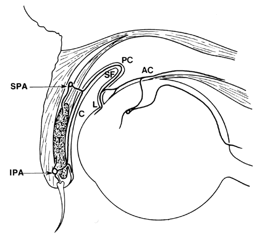
The conjunctival arteries are derived from two sources: (1) the palpebral branches of the nasal and lacrimal arteries of the lid; and (2) the anterior ciliary artery. Both vessels are derived from the ophthalmic artery, which is derived from the internal carotid artery (Fig. 19).14 The post-tarsal plexus of the lid, which is supplied by the marginal and peripheral artery of the upper lids, supplies the palpebral conjunctiva. The perforating arteries from the marginal palpebral arcade pass through the tarsus, reaching the subconjunctival space in the region of the subtarsal sulcus to form the marginal and tarsal vessels. The perforating vessels from the peripheral palpebral arcade perforate Müller's muscle and supply most of the forniceal conjunctiva. This arcade sends descending branches to supply the tarsal conjunctiva and also anastomoses with vessels from the marginal arcade and ascending branches that pass into the superior or inferior fornix to continue around the fornices to the bulbar conjunctiva as the posterior conjunctival arteries.

Fig. 19. Arterial supply of the conjunctiva. AC, anterior ciliary artery; C, palpebral conjunctiva; IPA, inferior (marginal) palpebral arcade; L, limbus; PC, posterior conjunctival arteries; SF, superior fornix; SPA, superior palpebral arcade. (Modified from Duke-Elder S: System of Ophthalmology, Vol II, p 547. St Louis, CV Mosby, 1976)

The second major source of supply, the anterior ciliary arteries, travel along the tendon of the rectus muscles and give off anterior conjunctival arteries just before piercing the globe. These arteries send branches to the pericorneal plexus and to the neighboring regions of the bulbar conjunctiva in the limbal area. In this region, there is free anastomosis in the subconjunctival and episcleral tissue between the anterior conjunctival vessels and terminal branches of the posterior conjunctival vessels, resulting in the zone of palisades of Busacca. Thus, the superficial and deep systems in the limbal area are closely connected. Clinically, this is an area of diagnostic importance. With inflammation and infections of the conjunctiva, the superficial posterior vessels are engorged; in deep keratitis or iritis, the deeper ciliary vessels are hyperemic.

Back to Top
VEINS
On close surface examination of the conjunctiva, it may be possible to see aqueous veins originally described by Ascher.105,106 These veins vary in diameter from 0.01 to 0.1 mm and are easily identifiable. They are usually found near the limbus, most often nasally, and appear hook shaped when they first come off the sclera. They contain a clear fluid and run a short course for approximately 1 cm. They are joined by an episcleral vein, the blood of which may become diluted; alternatively, the clear fluid and the blood may run side by side unmixed, forming a laminated vein. These aqueous veins are the exit channels of the aqueous.

The conjunctival veins are more numerous than the arteries.10 For the most part, the major portion of the drainage from the tarsal conjunctiva and the bulbar conjunctiva is directed to the palpebral veins. Some of the tarsal veins empty independently into the superior and inferior ophthalmic veins. Outflow is from the circum-corneal region to the veins that serve the extraocular muscles.

Small blood vessels of the bulbar conjunctiva have arteriovenous communications.14 The communicating vessels may be tortuous and uneven in caliber, but they usually are larger in diameter than capillaries. Each gives off capillary branches proximally and receives capillaries distally. They are not true arteriovenous anastomoses because they do not possess muscular walls that would render them capable of responding to chemical agents. Occasionally, the conjunctival blood vessels may become damaged, causing a spontaneous subconjunctival hemorrhage. These hemorrhages have very few associations with systemic diseases and usually absorb in a matter of a week or two. Electron microscopy has revealed that a majority of the capillaries often have a thick, continuous wall but few fenestrations (Fig. 20). Larger vessels with smooth muscles are also present.

Fig. 20. A blood capillary in the stroma of bulbar conjunctiva. This is a nonfenestrated type, but there also is a fenestrated type in the conjunctival stroma. Both endothelium (E) and pericytes (P) are surrounded by a basement membrane (bm), and the lumen contains erythrocytes (RC). (× 6300)

Back to Top
LYMPHATICS
The lymphatic channels in the conjunctiva14 are arranged in two plexuses: (1) a superficial plexus consisting of small vessels placed below the capillaries; and (2) a deeper plexus consisting of larger vessels in the fibrous portion of the substantia propria (Fig. 21A and B). These vessels are important in the mediation of immunologic reactions that occur in certain ocular diseases and surgical conditions.

Fig. 21. A. Photomicrograph of bulbar conjunctival epithelium. Note goblet cells (arrow). The substantia propria is composed of loose connective tissue and diverse cellular elements. B. A lymph channel (L) in the substantia propria. The channel is lined with endothelial cells (arrows). (A and B, × 240)

The superficial plexus receives lymphatic drainage from the limbal area. It has larger collector channels that run circumferentially 7 to 8 mm behind the limbus, forming an incomplete pericorneal lymphatic ring. Its other lymphatic drainage channels include a recurrent nasal group that drains the upper nasal quadrant and a descending temporal group. Both groups drain by way of the medial canthus. A large collecting vessel from the inferior fornix empties by way of the lateral canthus. Laterally placed lymph vessels flow to the preauricular lymph nodes; medially placed vessels flow to the submaxillary lymph nodes. Clinically, these vessels may be visible with a biomicroscope in certain conditions such as scleredema adultorum, and some of these lymph vessels may fill with blood and look like dilated veins.107,108 Electron microscopy reveals that these lymphatics differ from blood capillaries in some respects. For example, the endothelium is very thin but has no fenestrae, the intercellular junctions are less well formed than in capillaries, the basement membrane is interrupted or absent, and pericytes are usually absent (Fig. 22).

Fig. 22. A lymphatic capillary in the substantia propria of bulbar conjunctiva. Endothelium (E) is thin, its basement membrane is often not well developed (as in this picture), and the lumen (L) usually contains no erythrocytes. (× 13,700)

Back to Top
NERVE SUPPLY
The nerve supply to the conjunctiva is derived entirely from the first division of the trigeminal nerve.10 The nerves to the lid supply most of the conjunctiva. These nerves comprise the infratrochlear branch of the nasociliary nerve, the lacrimal nerve, the supratrochlear and supraorbital branches of the frontal nerve, and the infraorbital nerve from the maxillary division of the trigeminal nerve. The limbal area is supplied by branches from the ciliary nerves. All nerves form a network in the conjunctiva and terminate either peripherally in various forms of specialized endings or on blood vessels and epithelial cells. The majority of nerve endings in the conjunctiva are free, unmyelinated nerve endings (Figs. 23 and 24). They form a sub-epithelial plexus in the superficial part of the substantia propria. Many of these fibers end on blood vessels, and others form an intraepithelial plexus around the base of epithelial cells and send free nerve endings between cells.10

Fig. 23. A nerve fiber bundle in conjunctival stroma (substantia propria) composed of several unmyelinated nerve fibers (arrows) surrounded by a layer of perineurium (P); there are also intervening collagen fibrils (c). Each unmyelinated nerve fiber is composed of axons (A) wrapped with Schwann's cells (SC). (× 13,700)

Fig. 24. Substantia propria of bulbar conjunctiva, showing myelinated (MN) and unmyelinated (UN) nerve fibers. In both fibers, axons (A) are wrapped with Schwann's cells (SC) that have a basement membrane (bm). However, the axons are single and have a myelin sheath (ms) in the former; they are multiple and have no myelin sheath in the latter. nf, neurofilament; nt, neurotubule; m, mitochondria. (× 20,800). Inset. Higher power of a portion (arrow) of the myelin sheath, showing the lamellar structure. (× 40,000)

Also present in the conjunctiva are the end bulbs of Krause, which are specialized, compact nerve endings 0.2 to 0.1 mm in length and surrounded by a connective tissue capsule. The bulb itself may be single but of complex structure, or it may be compound. These end bulbs are relatively rare and vary in distribution. Their exact function remains unknown. One theory is that they are really a stage in the growth cycle of specialized nerve end organs.10

Back to Top
THE CARUNCLE
The caruncle is a small, flesh-like body that lies in the lacus lacrimalis and to the medial side of the plica semilunaris. It is part of the margin of the lower lid that becomes cut off by the development of inferior canaliculus. The caruncle is covered by a stratified squamous epithelium similar to skin, but it does not undergo keratinization. Like skin, it contains hair as well as sebaceous and sweat glands, but unlike skin, it contains accessory lacrimal glands similar to Krause's glands. The deep connective tissue in the caruncle is made up of parts of the septum orbitale and the medial check ligament. Numerous goblet cells can be found singly or in groups (Fig. 25). The blood supply of the caruncle comes only from the superior palpebral arteries. Because the supply is so abundant to this tissue, bleeding may be profuse if the caruncle is inadvertently damaged. Nerve supply is provided by the infratrochlear nerve. The lymphatics from here drain along with the rest of the medial part of the conjunctiva into the submaxillary lymph nodes.

Fig. 25. Epithelium of caruncle. It has many goblet cells (GC), some protruding toward the conjunctival sac. mv, microvilli of goblet cells. (× 12,200)

Back to Top
PLICA SEMILUNARIS
The plica semilunaris is a fold of conjunctiva lying lateral to the caruncle. It is more or less vertical, with its concavity facing laterally. Because the lateral border is free, a cul-de-sac of approximately 2 mm in depth is formed when the globe is adducted. It is practically nonexistent when the globe is abducted. The plica corresponds to the nictitating membrane in lower vertebrates. In humans, it is a vestigial structure consisting of a mere fold of conjunctiva. There are 8 to 10 layers of epithelial cells containing many goblet cells. Langerhans' cells may also be present in the epithelium. The substantia propria may have some nonstriated muscle supplied by sympathetic nerves and may contain fatty tissue. The connective tissue stroma of the plica is loose and highly vascular (Fig. 26).

Fig. 26. Plica semilunaris showing goblet cells (arrows), epithelium and fibrovascular connective tissue (× 50). (Fine BS, Yanoff M: Ocular Histology, 2nd ed, p 315. Hagerstown, Harper & Row, 1979)


Portions of the original chapter, including many of the illustrations, have been retained in this revision.
The authors, editors, and publisher wish to recognize the work of B. D. Srinivasan, Frederick A. Jakobiec, and Takeo Iwamoto for their contributions from the original chapter.
Back to Top
REFERENCES

1. Holly RJ: Formation and stability of the tear film. Int Ophthalmol 13:73, 1973

2. Pfister RR: The normal surface of conjunctiva epithelium: A scanning electron microscopic study. Invest Ophthalmol 14:267, 1975

3. Allansmith MR, O'Connor GR: Immunoglobulins: Structure, function and relation to the eye. Surv Ophthalmol 14:367, 1970

4. Gundersen T, Pearlson HR: Conjunctival flaps for corneal disease: Their usefulness and complications. Trans Am Ophthalmol Soc 67:78, 1969

5. Thoft RA: Conjunctival transplantation. Arch Ophthalmol 95:1425, 1977

6. Nagpal KC, Asdourian GK, Goldbaum MH: The conjunctival sickling sign, hemoglobin S, and irreversibly sickled erythrocytes. Arch Ophthalmol 95:808, 1977

7. Duke-Elder S: Diseases of the Outer Eye, Vol 8, p 1061. St Louis, CV Mosby, 1965

8. Parakkal PF, Alexander NJ: Keratinization, pp 44–45. New York, Academic Press, 1972

9. Ralph RA: Conjunctival goblet cell density in normal subjects and in dry eye syndromes. Invest Ophthalmol 14:299, 1975

10. Warwick R: Eugene Wolff's Anatomy of the Eye and Orbit, Seventh Edition. Philadelphia, WB Saunders, 1976

11. Kurpakus MA, Maniaci MT, Esco M: Expression of keratins K12, K4 and K14 during development of ocular surface epithelium. Curr Eye Res 13:805, 1994

12. Spinak M: Cytological changes of the conjunctiva in immunoglobulin-producing dyscrasias. Ophthalmology 88:1207, 1981

13. Trocme SD, Raizman MB, Bartley GB: Medical therapy for ocular allergy. Mayo Clin Proc 67:557, 1992

14. Duke-Elder S: System of Ophthalmology, Vol 2, p 543. St Louis, CV Mosby, 1961

15. Jordan DR, Anderson RB, Mamalis N: Accessory lacrimal glands. Ophthalmic Surg 21:146, 1990

16. Rivas L, Oroza MA, Perez-Esteban A, Murube-delCastillo J: Topographical distribution of ocular surface cells by the use of impression cytology. Acta Ophthalmol 69:371, 1991

17. Oduntan AO: The inferior conjunctiva of the monkey. Acta Anat 143:178, 1992

18. Steuhl KP: Ultrastructure of the conjunctival epithelium. Dev Ophthalmol 19:1, 1989

19. Weingeist TA: Fine structure and function of ocular tissues: The conjunctiva. Int Ophthalmol Clin 13(3):85, 1973

20. Nicolaissen B Jr, Eidal K, Haaskjold E et al: Outgrowth of cells from human conjunctival explants onto cornea in vitro. Acta Ophthalmol 69:723, 1991

21. Sandvig KU, Haaskjold E, Bjerknes R et al: Cell kinetics of conjunctival and corneal epithelium during regeneration of different-sized corneal epithelial defects. Acta Ophthalmol 72:43, 1994

22. Tseng SC, Tsai RJ: Limbal transplantation for ocular surface reconstruction—a review. Fortschr Ophthalmol 88:236, 1991

23. Zieske JD: Perpetuation of stem cells in the eye. Eye 8:163, 1994

24. Zieske JD, Bukusoglu G, Yankauckas MA: Characterization of a potential marker of corneal epithelial stem cells. Invest Ophthalmol Vis Sci 33:143, 1992

25. Schermer A, Galvin S, Sun T-T: Differentiation-related expression of a major 64K corneal keratin in vivo and in culture suggests limbal location of corneal epithelial stem cells. J Cell Biol 103:49, 1986

26. Cotsarelis G, Cheng S-Z, Dong G et al: Evidence of slow-cycling limbal epithelial basal cells that can be preferentially stimulated to proliferate: Implications on epithelial stem cells. Cell 57:201, 1989

27. Lindberg K, Brown ME, Chaves HV et al: In vitro propagation of human ocular surface epithelial cells for transplantation. Invest Ophthalmol Vis Sci 34:2672, 1993

28. Sandvig KU, Haaskjold E: The proliferative response during regeneration of a ringshaped defect in the corneal epithelium. Acta Ophthalmol 71:39, 1993

29. Sorensen T, Jensen FT: Conjunctival transport of technetium-99m pertechnetate. Acta Ophthalmol 57:691, 1979

30. Ahmed I, Patton TF: Importance of the noncorneal absorption route in topical ophthalmic drug delivery. Invest Ophthalmol Vis Sci 26:584, 1985

31. Lantz E, Andersson A: Release of fibrinolytic activators from the cornea and conjunctiva. Graefes Arch Clin Exp Ophthalmol 219:263, 1982

32. Liu SH, Tagawa Y, Prendergast RA et al: Secretory component of IgA: A marker for differentiation of ocular epithelium. Invest Ophthalmol Vis Sci 20:100, 1981

33. Inatomi T, Spurr-Michaud S, Tisdale AS et al: Mucin genes MUC4 and MUC5 are expressed by human conjunctival epithelia. Invest Ophthalmol Vis Sci (in press)

34. Inatomi T, Spurr-Michaud S, Tisdale AS, Gipson IK: Human corneal and conjunctival epithelia express MUCI mucin. Invest Ophthalmol Vis Sci 36:1818, 1995

35. Watanabe H, Fabricant M, Tisdale AS et al: Human corneal and conjunctival epithelia produce a mucin-like glycoprotein for the apical surface. Invest Ophthalmol Vis Sci 36:337, 1995

36. Tsai RJ, Ho YS, Chen JK: The effects of fibroblasts on the growth and differentiation of human bulbar conjunctival epithelial cells in an in vitro conjunctival equivalent. Invest Ophthalmol Vis Sci 35:2865, 1994

37. Wei Z-G, Cotsarelis G, Sun T-T, Lavker RM: Labelretaining cells are preferentially located in forniceal epithelium: Implications on conjunctival epithelial homeostasis. Invest Ophthalmol Vis Sci 36:236, 1995

38. Farquhar MG, Palade GE: The Golgi apparatus (complex) (1954-1981): From artifact to center stage. J Cell Biol 91(suppl 3, Pt 2):77S, 1981

39. Janssen PT, van Bijsterveld OP: Origin and biosynthesis of human tear fluid proteins. Invest Ophthalmol Vis Sci 24:623, 1983

40. Dua HS, Gomes JA, Jindal VK et al: Mucosa specific lymphocytes in the human conjunctiva, corneoscleral limbus and lacrimal gland. Curr Eye Res 13:87, 1994

41. Latkovic S: Ultrastructure of M cells in the conjunctival epithelium of the guinea pig. Curr Eye Res 8:751, 1989

42. Chin GN, Chi EY, Bunt AH: Ultrastructural and histochemical studies of conjunctival concretions. Arch Ophthalmol 98:720, 1980

43. Abdel-Khalek LM, Williamson J, Lee WR: Morphological changes in the human conjunctival epithelium: I. In the normal elderly population. Br J Ophthalmol 62:792, 1978

44. Wanko T, Lloyd BJJ, Matthews J: The fine structure of human conjunctiva in the perilimbal zone. Invest Ophthalmol 3:285, 1964

45. Paridaens AD, McCartney AC, Curling OM et al: Impression cytology of conjunctival melanosis and melanoma. Br J Ophthalmol 76:198, 1992

46. Kessing SV: Epithelial cysts in the conjunctiva. Acta Ophthalmol 47:642, 1969

47. Srinivasan BD, Jakobiec FA, Iwamoto T, Devoe AG: Epibulbar mucogenic subconjunctival cysts. Arch Ophthalmol 96:857, 1978

48. Kessing SV: Mucous gland system of the conjunctiva: A quantitative normal anatomical study. Acta Ophthalmol (Copenh) (suppl 95), 1968

49. Steida L: Ueber den Bauder Augenlidbindehaut des Menschen. Arch Mikr Anat 3:357, 1867

50. Apple DJ, Rabb MF: Ocular Pathology: Clinical Applications and Self-Assessment, Fourth Edition, pp 454–473. St Louis, CV Mosby, 1991

51. Dilly PN: Structure and function of the tear film. Adv Exp Med Biol 350:239, 1994

52. Kessler TL, Mercer HJ, Zieske JD et al: Stimulation of goblet cell mucous secretion by activation of nerves in rat conjunctiva. Curr Eye Res 14:985, 1995

53. Dartt DA, McCarthy DM, Mercer HJ et al: Localization of nerves adjacent to goblet cells in rat conjunctiva. Curr Eye Res 14:993, 1995

54. Kessler TL, Dartt DA: Neural stimulation of conjunctival goblet cell mucous secretion in rats. Adv Exp Med Biol 350:393, 1994

55. Huang AJ, Tseng SC, Kenyon KR: Morphogenesis of rat conjunctival goblet cells. Invest Ophthalmol Vis Sci 29:969, 1988

56. Qi LP: Determination of the conjunctival goblet cell density in 43 normal subjects. Chung Hua Yen Ko Tsa Chih 25(3):161, 1989

57. Knop E, Brewitt H: Conjunctival cytology in asymptomatic wearers of soft contact lenses. Graefes Arch Clin Exp Ophthalmol 230:340, 1992

58. Ehlers N, Kessing SV, Norn MS: Quantitative amounts of conjunctival mucous secretion and tears. Acta Ophthalmol 50:210, 1972

59. Jones DT, Monroy D, Ji Z et al: Sjogren's syndrome: Cytokine and Epstein-Barr viral gene expression within the conjunctival epithelium. Invest Ophthalmol Vis Sci 35:3493, 1994

60. Carney LG, Hill RM: Human tear pH: Diurnal variations. Arch Ophthalmol 94:821, 1976

61. Iwata T, Ohkawa K, Uyama M: The fine structural localization of peroxidase activity in goblet cells of the conjunctival epithelium of rats. Invest Ophthalmol 15:40, 1976

62. Srinivasan BD, Worgul BV, Iwamoto T, Merriam GRJ: The conjunctival epithelium: Part 2. Histochemical and ultrastructural studies on human and rat conjunctiva. Ophthalmic Res 9:65, 1977

63. Greiner JV, Weidman TA, Korb DR, Allansmith MR: Histochemical analysis of secretory vesicles in nongoblet conjunctival epithelial cells. Acta Ophthalmol 63:89, 1985

64. Rivas L, Oroza MA, Perez-Esteban A, Murube-delCastillo J: Morphological changes in ocular surface in dry eyes and other disorders by impression cytology. Graefes Arch Clin Exp Ophthalmol 230:329, 1992

65. Driot JY, Bonne C: Beneficial effects of a retinoic acid analog, CBS-211 A, on an experimental model of keratoconjunctivitis sicca. Invest Ophthalmol Vis Sci 33:190, 1992

66. Tseng SC: Topical tretinoin treatment for dry-eye disorders. Int Ophthalmol Clin 27:47, 1987

67. Nelson JD, Wright JC: Conjunctival goblet cell densities in ocular surface disease. Arch Ophthalmol 102:1049, 1984

68. Marner K: ‘Snake-like’ appearance of nuclear chromatin in conjunctival epithelial cells from patients with keratoconjunctivitis sicca. Acta Ophthalmol 58:849, 1980

69. Kinoshita S, Kiorpes TC, Friend J, Thoft RA: Goblet cell density in ocular surface disease: A better indicator than tear mucin. Arch Ophthalmol 101:1284, 1983

70. Friend J, Kiorpes T, Thoft RA: Conjunctival goblet cell frequency after alkali injury is not accurately reflected by aqueous tear mucin content. Invest Ophthalmol Vis Sci 24:612, 1983

71. Versura P, Maltarello MC, Bonvicini F et al: Detection of mucus glycoconjugates in human conjunctiva by using the lectin colloidal gold technique in TEM: I. A quantitative study in normal subjects. Acta Ophthalmol 64:445, 1986

72. Srinivasan BD, Jakobiec FA, Iwamoto T, DeVoe AG: Giant papillary conjunctivitis with ocular prostheses. Arch Ophthalmol 97:892, 1979

73. Greiner JV, Kenyon KR, Henriquez AS et al: Mucus secretory vesicles in conjunctival epithelial cells of wearers of contact lenses. Arch Ophthalmol 98:1843, 1980

74. Allansmith MR, Kajiyama G, Abelson MB et al: Plasma cell contents of main and accessory lacrimal glands and conjunctiva. Am J Ophthalmol 82:819, 1976

75. Allansmith MR, Greiner JV, Baird RS: Number of inflammatory cells in the normal conjunctiva. Am J Ophthalmol 86:250, 1978

76. Baum JL: Ocular infections. N Engl J Med 299:28, 1978

77. Yanoff M, Fine BS: Ocular Pathology. New York, Harper & Row, 1975

78. Fraunfelder FT, Garner A, Barras TC: Subconjunctival and episcleral lipid deposits. Br J Ophthalmol 60:532, 1976

79. Spencer WH: Ophthalmic Pathology: An Atlas and Textbook, Third Edition. Philadelphia, WB Saunders, 1985

80. Robbins SL, Cotran RS, Kumar V: Pathologic Basis of Disease, Third Edition. Philadelphia, WB Saunders, 1984

81. Ostler HB: Diseases of the External Eye and Adnexa: A Text and Atlas. Baltimore, Williams & Wilkins, 1993

82. Yanoff M, Fine BS: Ocular Pathology: A Text and Atlas, Second Edition. Philadelphia, JB Lippincott, 1982

83. Trocmé SD, Kephart GM, Allansmith MR et al: Conjunctival deposition of eosinophil granule major basic protein in vernal keratoconjunctivitis and contact lens-associated giant papillary conjunctivitis. Am J Ophthalmol 108:57, 1989

84. Trocmé SD, Aldave AJ: The eye and the eosinophil. Surv Ophthalmol 39:241, 1994

85. Liesgang T: Disorders of the cornea, conjunctiva and lens. In Bartley GB, Liesgang TJ (eds): Essentials of Ophthalmology. Philadelphia, JB Lippincott, 1992

86. Shields MB: Textbook of Glaucoma, Third Edition. Baltimore, Williams & Wilkins, 1992

87. Skuta GL, Parrish RK II: Wound healing in glaucoma filtering surgery. Surv Ophthalmol 32:149, 1987

88. Kook MS, Lee DA: Improving the success of glaucoma surgery by controlling wound healing. Ophthalmol Clin North Am 8:393, 1995

89. Mann I: Study of epithelial regeneration in living eye. Br J Ophthalmol 28:26, 1944

90. Dua HS, Gomes JA, Singh A: Corneal epithelial wound healing. Br J Ophthalmol 78:401, 1994

91. Haaskjold E, Sandvig KU, Bjerknes R, Kravik K: The early cell kinetic response during healing of corneal epithelial wounds. Ophthalmic Surg 23:680, 1992

92. Chung EH, DeGregorio PG, Wasson M, Zieske JD: Epithelial regeneration after limbus-to-limbus debridement: Expression of alpha-enolase in stem and transient amplifying cells. Invest Ophthalmol Vis Sci 36:1336, 1995

93. Kruse FE, Chen JJ, Tsai RJ, Tseng SC: Conjunctival transdifferentiation is due to the incomplete removal of limbal basal epithelium. Invest Ophthalmol Vis Sci 31:1903, 1990

94. Buck RC: Measurement of centripetal migration of normal corneal epithelial cells in the mouse. Invest Ophthalmol Vis Sci 26:1296, 1985

95. Buck RC: Cell migration in repair of mouse corneal epithelium. Invest Ophthalmol Vis Sci 18:767, 1979

96. Crosson CE, Klyce SD, Beuerman RW: Epithelial wound closure in the rabbit cornea: A biphasic process. Invest Ophthalmol Vis Sci 27:464, 1986

97. Soong HK, Cintron C: Disparate effects of calmodulin inhibitors on corneal epithelial migration in rabbit and rat. Ophthalmic Res 17:27, 1985

98. Chen JJ, Tseng SC: Abnormal corneal epithelial wound healing in partial-thickness removal of limbal epithelium. Invest Ophthalmol Vis Sci 32:2219, 1991

99. Danjo S, Friend J, Thoft RA: Conjunctival epithelium in healing of corneal epithelial wounds. Invest Ophthalmol Vis Sci 28:1445, 1987

100. Srinivasan BD, Worgul BV, Iwamoto T, Eakins KE: The reepithelialization of rabbit cornea following partial and complete corneal denudation. Exp Eye Res 25:343, 1977

101. Murata T, Ishibashi T, Inomata H: Localizations of epidermal growth factor receptor and proliferating cell nuclear antigen during corneal wound healing. Graefes Arch Clin Exp Ophthalmol 231:104, 1993

102. Zieske JD, Wasson M: Regional variation in distribution of EGF receptor in developing and adult corneal epithelium. J Cell Sci 106:145, 1993

103. Addicks EM, Quigley HA, Green WR, Robin AL: Histologic characteristics of filtering blebs in glaucomatous eyes. Arch Ophthalmol 101:795, 1983

104. Katz GJ, Higginbothan EJ, Lichter PR et al: Mitomycin C versus 5-fluorouracil in high-risk glaucoma filtering surgery. Ophthalmology 102:1263, 1995

105. Ascher KW: Aqueous veins and their significance for pathogenesis of glaucoma. Arch Ophthalmol 42:66, 1949

106. Ascher KW: Aqueous veins: Preliminary note. Am J Ophthalmol 25:31, 1942

107. Jampol LM, Nagpal KC: Hemorrhagic lymphangiectasia of the conjunctiva. Am J Ophthalmol 85:419, 1978

108. Breinin GM: Scleredema adultorum: Ocular manifestations. Arch Ophthalmol 50:155, 1953

Back to Top