|
|
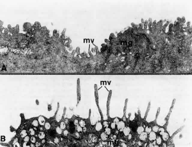
| Fig. 11. Transmission electron micrographs of the microvilli (mv) in the bulbar conjunctival epithelial surface (A) and in the forniceal conjunctival epithelial surface (B), showing the length difference. They are short in the former and long and slender in the latter. mg, mucous granules—those in B exhibit fibrillogranular contents. (A, × 26,000; B, × 26,000) |