|
|
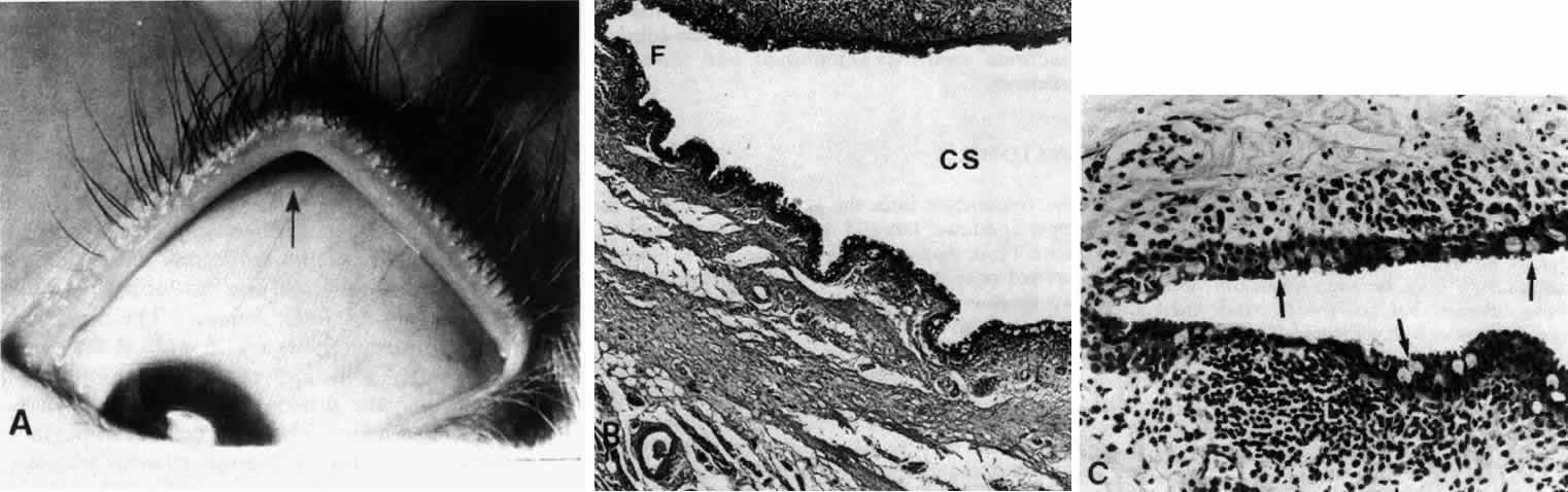
| Fig. 1. Low-power view of the globe. A. Arrow pointing to the region of the superior fornix. B. Superior fornix (F) showing epithelium and substantia propria. Conjunctival sac (CS). C. Higher-power view of epithelium showing goblet cells on the surface (arrows). (B, × 50; C, × 170) |