|
|
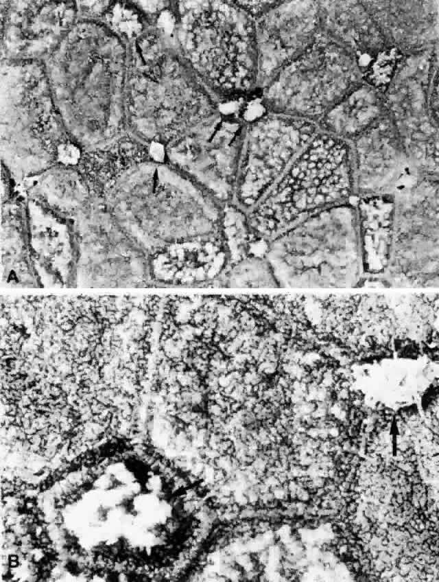
| Fig. 14. A. Low-power SEM of the epithelial surface of bulbar conjunctiva, showing relatively distinct cell borders. Large white spots (arrows) are the mucous substance secreted by goblet cells representing the sites of the openings of these cells. The tissue was treated with 20% acetylcysteine for 10 minutes, but mucoid substance still remained in places on the other epithelial surface as well. B. Higher-power SEM of the same epithelial surface as shown in A. The entire surface is covered with microvilli, but the cell borders are clearly distinguished from the adjacent areas because of different distribution densities of microvilli. Arrows indicate the surface of goblet cells; an abundant mucous substance still remains here after treatment with 20% acetylcysteine for 10 minutes. (A, × 2300; B, × 6500) |