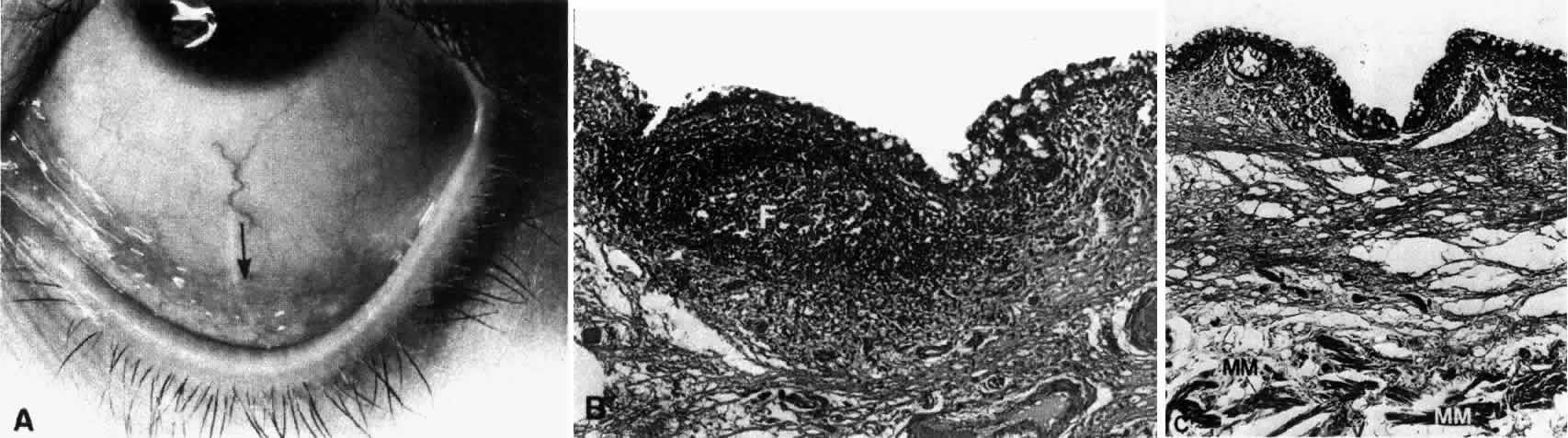
Fig. 2.
A.
Region of the inferior fornix (
arrow
).
B.
Inferior fornix showing epithelium, goblet cells, and a follicle (
F
).
C.
Inferior fornix showing Müller's muscle (
MM
) in the substantia propria. (
B,
× 60;
C,
× 80)