|
|
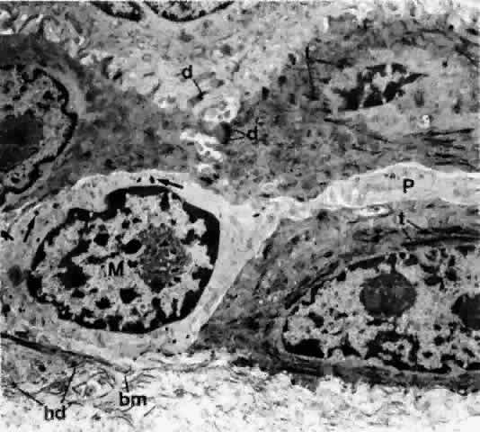
| Fig. 12. Basal portion of the epithelium of bulbar conjunctiva, showing bundles of tonofilaments (t) in the epithelial cells and a melanocyte (M) containing melanin granules (arrow). Note also a long process (P) extending from the melanocyte between epithelial cells. Although epithelial cells are joined by desmosomes (d), the melanocyte has no desmosomes around the cell. hd = hemidesmosome; bm = basement membrane (× 12,700) |