Chapter 49
Inflammatory Choroiditis
JOSE S. PULIDO and C. RICHARD BLAKE
Main Menu   Table Of Contents

Search

WHITE DOT SYNDROMES
MULTIPLE EVANESCENT WHITE DOT SYNDROME
ACUTE IDIOPATHIC BLIND SPOT ENLARGEMENT SYNDROME
ACUTE ZONAL OCCULT OUTER RETINOPATHY
ACUTE ANNULAR OUTER RETINOPATHY
ACUTE POSTERIOR MULTIFOCAL PLACOID PIGMENT EPITHELIOPATHY
PRESUMED OCULAR HISTOPLASMOSIS
MULTIFOCAL CHOROIDITIS
DIFFUSE SUBRETINAL FIBROSIS
DIFFERENTIAL DIAGNOSIS OF INFLAMMATORY CHOROIDITIS
PUNCTATE INNER CHOROIDOPATHY (PIC)
OTHER CHOROIDITIDES
BIRDSHOT RETINOCHOROIDITIS
VOGT-KOYANAGI-HARADA DISEASE
RETINAL PIGMENT EPITHELIITIS
ACUTE MACULAR NEURORETINOPATHY
UNIFOCAL HELIOID CHOROIDITIS
ACKNOWLEDGMENTS
REFERENCES

There are a group of ocular diseases that appear to be inflammatory reactions in the choroid. The cause of the inflammatory reaction is unknown in all of these entities. Some of these entities are considered infectious, but there has been no histologic evaluation to show the presence of infectious agents that would be required to confirm that is the case. Another possibility is that they are immunologic in origin but there also is no definite confirmatory determination of this.

The reason the diseases are considered infectious is because they appear to cause acute visual loss and some are associated with white blood cells in the vitreous or a retinal vasculitis. In addition, the lesions may crop up and have an acute, chronic, or semiacute course similar to systemic infectious diseases. Many of these entities affect persons in the third to fifth decade of life, which is when many of the immunologic reactions occur elsewhere. It may be that there is an initial infectious agent that in a host with certain predisposing characteristics triggers an immunologic reaction, or it could be that the agent stays indefinitely in the host causing a host-induced reaction.

On physical examination, the patients may have iritis, vitreitis, or retinitis, although in many of the choroiditic entities they may not manifest these findings. Because the choroid or the retinal pigment epithelium is affected, they may show a serous retinal detachment that presents as a blister of fluid that is loculated or as an exudative retinal detachment with shifting subretinal fluid. Acutely there may be a gray-yellow plaque of varying size. It may then resolve with only mild almost imperceptible pigmentary changes or there can be dense white chorioretinal scarring. Because the photoreceptor/retinal pigment epithelial complex is affected, many note photopsias or colored swirling lights and also they may have visual field defects.

It is important to determine carefully that the disease is truly an inflammatory disease and not a known infectious or neoplastic disease. Infectious choroiditides have to be considered in all circumstances of inflammatory choroiditis. A specific test for Treponema pallidum, for instance, the fluorescent treponema antibody-absorbed (FTA-ABS) test or the microhemaglutination-treponema pallidum test (MHA-TP) should be performed to determine if the choroiditis is caused by syphilis. Other diseases, including tuberculosis, cat-scratch disease, coccidioidomycosis, West Nile encephalitis, and nocardiosis among others need to be considered. In addition, certain noninflammatory diseases, for instance, central serous chorioretinopathy may have an acute time course similar to that of an inflammatory condition and eclampsia, thrombotic thrombocytopenia purpura, and disseminated intravascular coagulation may also mimic an acute inflammatory choroiditis.

Back to Top
WHITE DOT SYNDROMES
One group of these inflammatory choroiditides are subclassified under the auspices of white dot syndromes. These tend to have multiple lesions. The patients tend to be young women and in addition to visual loss they may have photopsias. The photopsias are caused by photoreceptor damage during acute episodes of choroiditis. This group of diseases consists of multiple evanescent white dot syndrome (MEWDS), acute zonal occult outer retinopathy (AZOOR), acute annular outer retinopathy (AAOR), punctate inner choroiditis (PIC), multifocal choroiditis, acute posterior multifocal placoid pigment epitheliopathy (APMPPE), birdshot chorioretinopathy, and diffuse subretinal fibrosis. Presumed ocular histoplasmosis syndrome is sometimes included in this group although in direct contradistinction to the other diseases, the patients are usually asymptomatic until they develop a choroidal neovascular membrane.
Back to Top
MULTIPLE EVANESCENT WHITE DOT SYNDROME

HISTORY AND CLASSIC FINDINGS

MEWDS was first described by Jampol and associates1 and in the same year by Takeda and coinvestigators.2 The first cases involved acute unilateral visual loss and scotomas in 12 young women and 3 men. All had multiple white lesions at the level of the retinal pigment epithelium and approximately half had a previous flu-like syndrome. Macular granularity was seen commonly in these cases.

EPIDEMIOLOGY

Most affected persons tend to be young women in their 20s and thirties with an average age of 26 years although there have been reports of a female who was 14 years old and several in their sixties.2-4 Males are less often affected in a ratio of 3:1.

PATHOPHYSIOLOGY

The etiology of this form of white dot syndrome is unknown. There is a history of a precedent upper respiratory tract infection in 25% to 50% of cases.5 It could be that in certain predisposed individuals, exposure to a certain infectious organism can trigger an immune reaction to the choriocapillaris or the overlying retinal pigment epithelium. There is one case of murine typhus associated with MEWDS-like fundus findings.6 There is also a case with MEWDS-like findings that occurred after hepatitis B vaccination.7

INITIAL CLINICAL FINDINGS

Presenting Symptoms

The most common presenting symptoms are unilateral loss of vision and photopsias. These photopsias are described as flashing colored lights. Occasionally they may complain of a unilateral blind spot. Rarely do they have bilateral symptoms.

There may be a history of a preceding upper respiratory tract infection. Usually this is not volunteered by the patient because if it had been present, it was mild and so the patients do not consider it related to the present problem.

Signs

VISION.

The vision is usually only mildly diminished although the vision may range from 20/20 to 20/400. Color vision may be slightly or moderately diminished as well.

PUPILS AND FIELDS.

The visual fields are variable but most often they show an enlarged blind spot. There may be an afferent papillary defect as well.

BIOMICROSCOPY.

Iritis is rare but has occasionally been described. Vitreitis is more common although if it is present, it tends to be very mild.

FUNDUS FINDINGS.

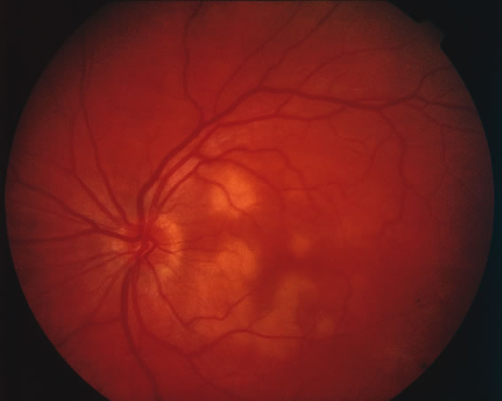
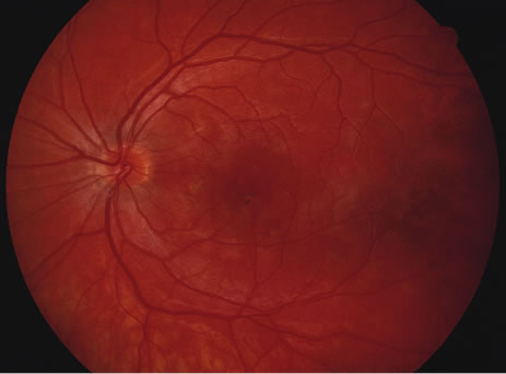
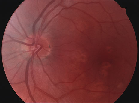
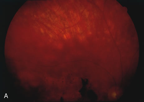
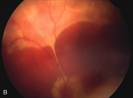
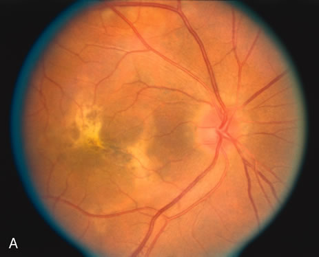
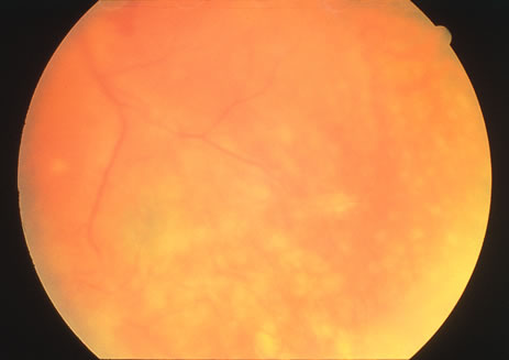
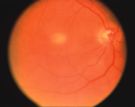
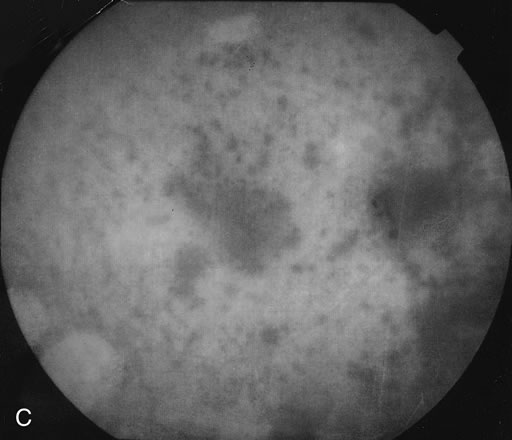
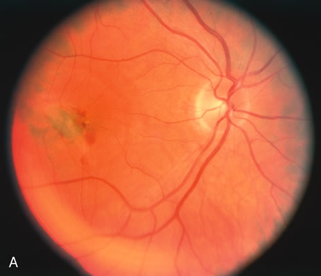
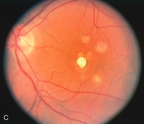
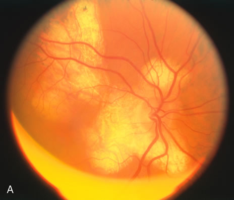
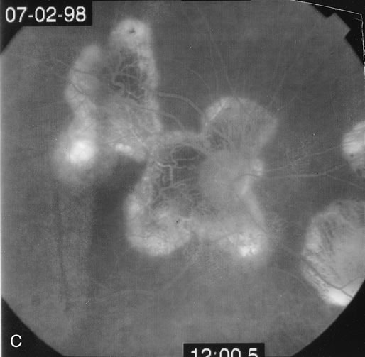
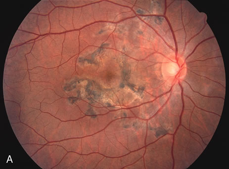
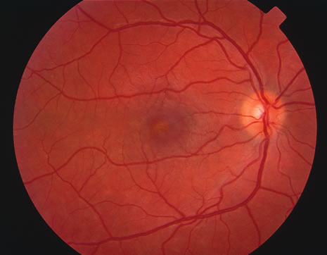
The classic fundus findings are small (500 μm), ill-defined, and sometimes difficult to see grayish-white dots that are dispersed throughout the posterior pole. These spots rarely may become confluent and if this occurs to spots that are around the disc, it may present as a case of giant blind spot syndrome or of an entity that probably is a variant of MEWDS called AZOOR.8 In addition to the dispersed small spots, there is a fine granularity of the fovea. This finding is pathognomonic for this disease, and it can exist in the absence of the white dots (Figs. 1 and 2).

Fig. 1. Color fundus photograph of a patient with multiple evanescent white dot syndrome. Notice that the foveal spot and irregularity is much easier to detect than the subtle white dots.

Fig. 2. Red free black and white photograph of a different patient with MEWDS. This technique accentuates the white dots but does not show the foveal changes as well.

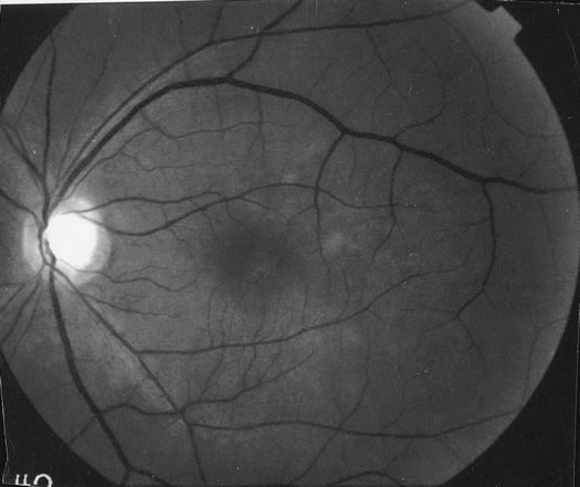
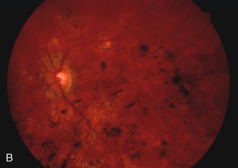
Usually there is resolution of the fundus findings, although careful fundus evaluation sometimes shows some residual fine retinal pigment epithelial mottling. In rare cases, there may be long-standing pigment clumping with surrounding pigment atrophy similar to the lesions seen in multifocal choroiditis or punctate inner choroidopathy although that has been contended. The foveal granularity may take longer to resolve than the white spots.

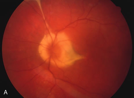
There may be mild optic disc swelling. This may lead someone unfamiliar with this disease to consider that the cause of visual loss is optic neuritis. There also may be mild retinal phlebitis that may be more easily seen by fluorescein angiography than by ophthalmoscopy. Choroidal neovascularization rarely develops.9

ANCILLARY TESTING

Fluorescein

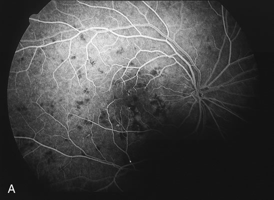
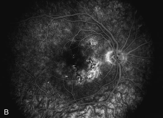
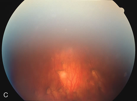
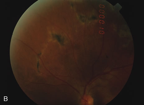
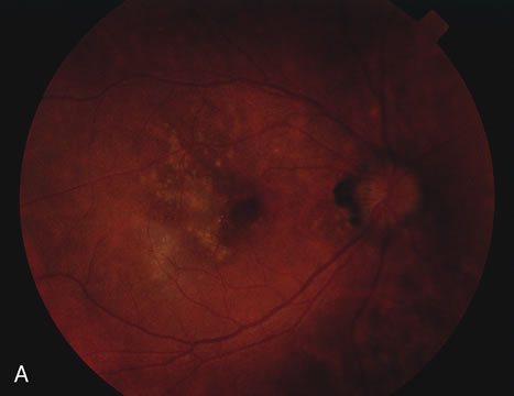
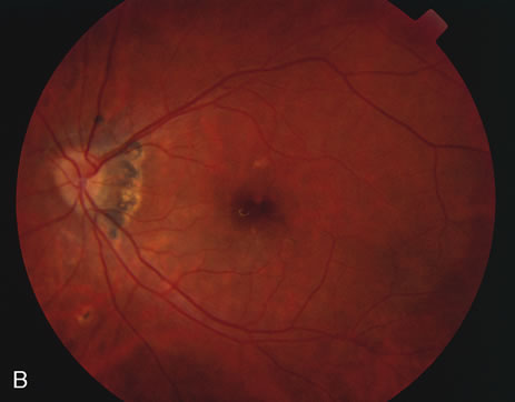
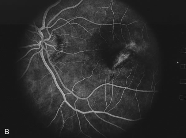
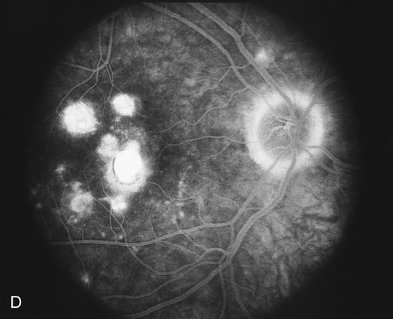
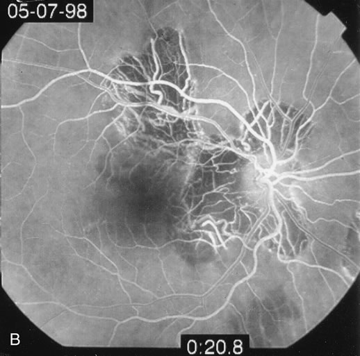
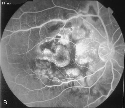
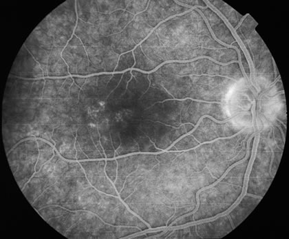
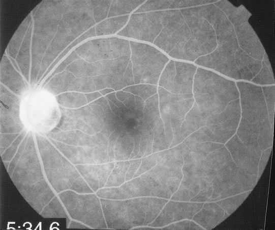
The fluorescein angiograms of the white dots show a fine halo appearance in a wreath-like swirl configuration to some of the white dots, and this halo diffuses in the later stages of the angiogram, first developing a stippled pattern of fluorescence and then becoming a diffuse white spot that leaks only mildly. There may be leakage of fluorescein from the optic nerve and occasionally, there may be late staining of retinal vessels because of a mild vasculitis (Figs. 3 and 4).

Fig. 3. Venous phase fluorescein angiogram of fundus shown in Figure 1. It demonstrates the hyperfluorescent dots coalesced near the fovea and in a subtle wreath-like fashion elsewhere.

Fig. 4. Recirculatory state fluorescein angiogram of fundus shown in Figure 2. It demonstrates the leakage from the disc and the fluorescence of the white dots is more evident.

Indocyanine Green

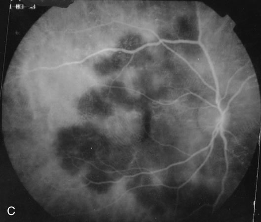
In the early stages of the angiogram, the indocyanine green (ICG) angiogram may not show any distinct abnormalities. In the late phases, ICG angiography can show more lesions than can be seen by ophthalmoscopy or by fluorescein angiography. These lesions tend to block or show absence of fluorescence.10,11 In addition, there can be many hypofluorescent lesions around the disc. Sometimes the spots are so extensive that they become confluent. It is thought that it is a confluence of these small lesions that cause the blind spot enlargement seen on visual field testing. At the peripheral edge of the confluent spots however, it is still possible to see single spots that are characteristic with the stippled halo-like fluorescence.12 The hypofluorescent spots noted by ICG can last longer than the symptoms, or ophthalmoscopic or fluorescein angiographic findings.13

Electroretinography

Electroretinography (ERG) shows a diffuse loss of the early receptor potential and a decrease of the a wave, indicating that the disease appears to affect the retinal pigment epithelium-photoreceptor complex.14 Multifocal ERG shows reduced a and b waves in the areas that correspond to the visual field scotomas as well as showing areas of dysfunction where the visual field does not seem affected.15 In addition, the reduced amplitudes may be noted months after the visual fields and visual acuity have returned to normal. In patients with acute idiopathic blind spot enlargement (AIBSE) syndrome, a disease that may be related to MEWDS, the multifocal ERG is similarly affected in the peripapillary area.16 These patients may actually have had MEWDS in later stages of resolution because the white lesions may resolve much earlier than the scotomatous changes.17 Electrooculography may also show a diminution of the dark peak to light trough ratio.

Laboratory Tests/Immune Testing

There is a small study that showed a higher incidence of the HLA B51 haplotype in persons with MEWDS compared to the normal population. There is a relative risk of 5.86. This study needs to be confirmed in a larger cohort.18 It is interesting to note that this haplotype is also seen more frequently in patients with Behçet's disease, but in Behçet's disease the relative risk is almost twice as high as in MEWDS.

Testing for immunoglobulin staining in patients with different causes of outer retinopathy have not shown reactivity with the retina. This does not necessarily mean that the disease does not involve an immune response to the retina. It means that the immune reaction does not involve autoantibodies. Cell-mediated immunity may still be present.19

Differential Diagnosis/Mimics

The differential diagnosis tends to involve the other causes of the white dot syndromes. Most commonly the diagnoses that are most often confused with MEWDS are APMPPE, birdshot retinochoroidopathy, multifocal choroiditis, and retinal pigment epitheliopathy. In addition, acute macular neuroretinopathy and diffuse unilateral subacute neuroretinitis are sometimes considered in the differential diagnosis. None of the other white dot syndrome diseases have the peculiar foveal granularity that is seen with MEWDS.

APMPPE is usually a bilateral disease that affects both young men and women. The creamy lesions tend to be larger than those seen in MEWDS, and fluorescein angiography reveals classic early blocked fluorescence and late staining of the lesions.

Birdshot chorioretinopathy usually affects older women between 40 and 60 years of age. It causes deep creamy lesions the borders of which are sometimes difficult to discern. In the late phases of fluorescein angiography some of the lesions show hyperfluorescence while others do not show up on the angiograms. The optic nerve tends to be swollen and shows leakage by fluorescein angiography. Cystoid macular edema may be present.

Many times the lesions in multifocal choroiditis show lesions in different stages of scarring. Choroidal neovascularization, bilaterality, and recurrences are common.

Acute retinal pigment epitheliopathy shows a few small spots that are noted to have pigment clumping in their center with surrounding hypopigmentation. Fluorescein angiography shows window defect with a central spot of blocked fluorescence. The electroretinogram is unaffected.

Acute macular neuroretinopathy tends to be unilateral. There is a mild diminution of vision associated with a reddish discoloration centered under the fovea. This pattern is most easily seen with red free photography.

Diffuse unilateral subacute neuroretintis is caused by a nematode. It can be associated with creamy-white small lesions in the macula. This is a progressive disorder unless the nematode is identified in the subretinal space and treatment is given.

There are three cases of two men and one woman all in their 50s who had whitish spots that were consistent with those seen in MEWDS. The age was atypical and there was progressive vision loss in these cases. These patients had primary intraocular large-cell (non-Hodgkin's) lymphoma.20 In no case were there the pathognomonic foveal changes seen in MEWDS.

Natural History

The symptoms usually resolve within 6 to 15 weeks although some may have the blind spot enlargement and photopsias for up to 1 year after the initial presentation.5,21 Recurrence is rarely noted and choroidal neovascularization is also a rare occurrence. Punched-out lesions, similar to those seen in multifocal choroiditis, can rarely be seen in a few cases.

Treatment and History with Treatment

Treatment is not indicated because the disease spontaneously improves. Laser photocoagulation or possibly photodynamic therapy may be of help in cases that develop choroidal neovascularization.

Back to Top
ACUTE IDIOPATHIC BLIND SPOT ENLARGEMENT SYNDROME
AIBSE is probably a variant of MEWDS. The patients tend to be women between the ages of 20 and 50 years. There may be no to moderate vision loss. An enlarged blind spot is invariably present. The patients have photopsias as well. Although some cases have the white dots similar to MEWDS in the retina, others do not and that is why it is argued that AIBSE is a different disease. In addition, some cases do not have the macular pigment granularity classically seen in MEWDS.22 Some may develop recurrences. It may be that AIBSE are actually cases of MEWDS that have had resolution of the fundus lesions.
Back to Top
ACUTE ZONAL OCCULT OUTER RETINOPATHY
AZOOR was the term used by Gass and colleages23 to describe the ocular findings in a disease noted more commonly in women than men (73% vs. 27%). The patients tend to be young and myopic (66%), and the disease starts unilaterally in 60% of the cases with the fellow eye ultimately becoming involved in 60% of the unilateral cases. There can be a delay of up to 15 years between the first and the second eye becoming involved, although the mean time for the involvement of the fellow eye was 50 months. Bilateral disease is more common in males than in women (100% vs. 68%). Recurrences occur in approximately one third of cases and the mean time to each recurrence was 52 months. An antecedent flu-like illness was noted in 20% of cases and 13% had headaches prior to development of the ocular findings. Approximately 25% of patients had a history of a systemic autoimmune disease.

Most patients develop acute visual loss followed by stabilization after 6 months. Only a rare patient has stepwise progression of visual loss. Patients note the visual loss as scotomas that involve one or more parts of the visual field with the most common finding being an enlarged blind spot. The vast majority of patients (88%) notice photopsias that may be colored or noncolored. Most patients have visual acuity of 20/40 or better on initial evaluation, and the majority retain 20/40 or better at the time of the final evaluation. Vitreous cells are present in approximately one half of the cases and approximately 10% of eyes were noted to have retinal vascular sheathing. The eyes with vitreous cells may develop focal areas of mottling of the retinal pigment epithelium that is similar to the mottling seen in mild cases of retinitis pigmentosa although many eyes appear to have a normal fundus. Rarely, patients may develop chorioretinal scars that appear similar to those seen in multifocal choroiditis. Fluorescein angiography in the acute phase is usually unremarkable. In the chronic phase there may be mottling fluorescence at the level of the retinal pigment epithelium. An abnormal full-field electroretinogram was noted in more than 60% of the patients. The multifocal electroretinogram may also be abnormal.24 The differential diagnosis in this group of patients includes the white dot syndrome diseases, diffuse unilateral subacute neuroretinitis, retinitis pigmentosa, carcinoma associated retinopathy, and syphilis.

Back to Top
ACUTE ANNULAR OUTER RETINOPATHY
AAOR may be a variant of AZOOR.25,26 The first case was reported by Luckie et al.27 The second case was reported by Gass and Stern.28 These cases are characterized by visual loss and visual field loss in young adults associated with a demarcation ring of gray-white outer retinal coloration that separates the area of the retina with visual field loss and normal retina. There are no discernible retinal findings within the annulus of whitening. Usually the diametric center of the annulus was the optic disc. With time the grayish discoloration abates and there is some thinning of the retinal vessels within the area of affected retina. In some cases there was mottling of the retinal pigment epithelium. There were no vitreous cells noted and the fellow eye has not been affected in the few cases that have been reported. Fluorescein angiography may be normal initially and later there may be mottled fluorescence at the level of the retinal pigment epithelium.

There appears to be a group of diseases that involve the retinochoroidal junction and these include multifocal choroiditis, acute macular neuroretinopathy, AIBSE, PIC, AZOOR, AAOR, and MEWDS. Because there have been some patients that have two of these diseases together or in progression, Gass and Stern28 have suggested they are all related. Others have seen cases of overlap between these syndromes as well.29 AZOOR disorders are characterized by rapid visual field loss and electroretinographic findings that show fundus involvement far in excess of that seen by fundoscopy.30 All of these patients tend to have photopsias as well. There is one case of AZOOR associated with a relapsing-remitting acute case of cervical myelitis.9,19,31-32 Many cases have a history of an antecedent viral illness, and it may be that these diseases are caused by a transient viral infection of the photoreceptors and/or the retinal pigment epithelium or there may be an immunologic reaction that affects these layers following the viral infection. Treatment with antiviral agents does not appear to be efficacious and many of these diseases are self-limited.

Back to Top
ACUTE POSTERIOR MULTIFOCAL PLACOID PIGMENT EPITHELIOPATHY

HISTORY AND CLASSIC FINDINGS

APMPPE was first described by Gass.33 He described three patients between the ages of 19 and 22 years in whom both eyes were affected. There were creamy white spots that were associated with mild to marked visual loss. The lesions typically blocked early and stained late by fluorescein angiography, and usually there was resolution with only mild pigment clumping. Recurrences are rare.

EPIDEMIOLOGY

Most cases affect young persons both men and women between the ages of 20 and 40 years. There has been an association with other diseases as noted below.

PATHOPHYSIOLOGY

The fact that there have been cases after known exposure to other diseases or antigens means that this may be a focal response to the systemic inflammatory process. There is an association with cerebral vasculitis, which also supports this possibility. There may be an HLA predisposition that would lend credence to the idea that if a person with a certain immunogenetic haplotype is exposed to certain antigens, he or she may develop APMPPE as a response. Approximately 25% to 40% of patients report a previous prodromal viral illness. It may be that there is a choroidal vasculitis with a secondary reaction in the overlying retinal pigment epithelium.

INITIAL CLINICAL FINDINGS

Presenting Symptoms

SIGNS.

The patient may present with complaints of photopsias, scotomas, and possible vision loss. Both eyes tend to be affected although they can be asymmetrically affected.

VISION.

The vision is variable and depends on whether the lesions are under the fovea. The vision may be normal or reduced to 20/200 or worse.

PUPILS AND FIELDS.

There are visual field defects that correspond to some of the fundus lesions. There can be an afferent papillary defect if both eyes are very asymmetrically affected.

BIOMICROSCOPY.

There may be a mild iritis although most cases do not show it. More commonly there may be a mild vitreitis.

FUNDUS FINDINGS.

Acutely, there may be creamy white lesions in the posterior pole. These lesions tend to be deep and poorly demarcated. Sometimes these lesions may become confluent. The lesions clear by losing their yellow-white opacification and clearance occurs from centrally to the periphery. With time the lesions resolve leaving mottling of the retinal pigment epithelium (Figs. 5, 6, and 7).

Fig. 5. Fundus photograph showing the acute creamy lesions of a recent case of acute multifocal posterior placoid pigment epitheliopathy.

Fig. 6. Fundus photograph of the same case as in Figure 5. The findings at 1 month after the initial presentation now shows mild pigment clumping and mild retinal pigmentary atrophy.

Fig. 7. Fundus photograph of the same case as in Figure 5. The findings at 7 months following initial presentation show more pigment clumping and retinal pigment atrophy in small circular lesions.

SYSTEMIC ASSOCIATIONS OF ACUTE POSTERIOR MULTIFOCAL PLACOID PIGMENT EPITHELIOPATHY

There have been multiple reports of a cerebral vasculitis in association with APMPPE. These patients may have a cerebral spinal fluid pleocytosis. In patients with APMPPE who complain of headaches or have other central nervous system findings, magnetic resonance imaging should be strongly considered and the patient should be placed on high doses of corticosteroids. The interval between the APMPPE and the onset of symptomatic cerebral vasculitis may be up to 3 months.

Mumps have been associated with APMPPE as well as immunization with hepatitis B virus vaccine. In the case of the immunizations, the APMPPE occurred after the booster injection. There has been a case of APMPPE after streptococcal infection.34

ANCILLARY TESTING

Fluorescein

The classic findings in the acute cases are lesions of variable size that are hypofluorescent in the early phase of the fluorescein angiogram. In the late phases of the angiogram there is hyperfluorescence of the lesions. In the late phases, there also may be leakage from the disc as well, and there also may be staining of the retinal vessels because of the vasculitis. With resolution of the disease, there still may be a mottled pattern of fluorescence correlating with the areas of pigment clumping and pigment atrophy (Fig. 8).

Fig. 8. A. Early fundus angiogram showing hypofluorescence of the acute posterior multifocal placoid pigment epitheliopathy (APMPPE) lesions. B. Later stage of the angiogram showing staining of the APMPPE lesions.

Indocyanine Green

ICG angiography shows an absence of fluorescence in the early as well as in the late phases of the angiogram. This is consistent with the possibility that the disease is caused by a choroidal occlusive vasculitis. There may be more lesions than seen ophthalmoscopically or by fluorescein angiography. In addition, there may be the development of new lesions in the first few weeks that can be seen by indocyanine green that cannot be seen by fundoscopy.35 This appears to imply that the disease is first a choroidal vasculitis, and in the more severe cases it can then affect the overlying retinal pigment epithelium and outer retina.

Other Diagnostic Findings

The electroretinogram and electrooculography may both be reduced in APMPPE. There may also be a prolonged dark-adaptation, color match abnormalities, and an abnormal Stiles-Crawford effect. These abnormalities resolve after 1 year. By fundus reflection densitometry, there is decreased cone pigment in the acute phase and pigment regeneration is prolonged; these findings also resolve with time.36,37

Laboratory Tests/Immune Testing

The HLA B7 and the HLA DR2 haplotype were seen more often in patients with APMPPE in one report, but that has not been confirmed.

Differential Diagnosis/Mimics

Although the differential diagnosis includes all other white dot syndromes, the most common diseases that closely resemble APMPPE are serpiginous choroiditis, MEWDS, birdshot choroidopathy, diffuse metastatic cancerous lesions, non-Hodgkin's lymphoma, and Pneumocystis choroiditis. Subacute sclerosing panencephalitis caused by chronic measles infection can have similar fundus findings. If the patient is older than the normal group of patients, if the lesions are not exactly typical, or if there are systemic findings, then a complete systemic work-up should be considered.

Serpiginous choroiditis is associated with recurrences that are rare or probably do not occur with APMPPE. In addition, multiple separate lesions are rare in serpiginous choroiditis but common in APMPPE. By contrast, choroidal neovascularization is rare in APMPPE but common in serpiginous choroiditis. Both tend to be bilateral but there is asymmetry in the time course of the lesions in serpiginous choroiditis whereas in APMPPE the lesions of both eyes tend to follow a similar time course. Finally, patients with serpiginous choroiditis tend to be middle-aged compared to young adults in APMPPE.

Natural History

Usually, the lesions resolve leaving only mild pigment mottling and the vision also improves. Resolution begins after a few weeks and may continue for up to 1 year. Choroidal neovascularization may occur with APMPPE but is a rare finding. Sometimes there may be significant pigment changes and even mild chorioretinal scarring. In cases where the pigmentary changes are under the fovea, visual improvement may be limited. In one series, 11% had visual acuity of 20/200 or worse on follow-up, although another series had 94% of affected eyes having a final visual acuity of 20/30 or better.38 Usually if there is marked chorioretinal scarring, serpiginous choroidopathy should be considered a possible diagnosis.

Treatment and History with Treatment

Treatment is only indicated in cases with concomitant cerebral vasculitis or in cases where the vision is markedly diminished. Corticosteroids are used in those cases. It is not certain that corticosteroids affect the visual outcome but it appears to affect the course of the cerebral vasculitis. In cases where the cerebral vasculitis does not appear to improve or in cases where there is marked bilateral visual loss that is unresponsive to corticosteroids, consideration for using cyclosporine should be given.

Back to Top
PRESUMED OCULAR HISTOPLASMOSIS

HISTORY AND CLASSIC FINDINGS

Presumed ocular histoplasmosis (POH) is probably caused by dissemination of the histoplasma capsulotum after acute exposure to the organism. It is characterized by punched-out chorioretinal lesions, peripapillary scarring, and choroidal neovascular membranes, with no vitreous or anterior chamber inflammation.

This disease is seen most often in people who come from the Ohio-Mississippi River Valley area where the fungus is ubiquitously found. Most often patients present in their fourth and fifth decade of life because of the development of a choroidal neovascular membrane. There may be a genetic predisposition and patients that have the HLA-B7 or the HLA-DR2 haplotype may be more susceptible to developing the ocular form of the disease.

It is hypothesized that after acute exposure to the organisms by inhalation in susceptible persons, there may be a pulmonary infection associated with symptoms of a lower respiratory tract infection including mild fever and a cough. This elicits a granulomatous response and the organisms are probably destroyed by this cell-mediated response.

As opposed to the inflammatory choroiditides, POH tends to be asymptomatic unless there is a choroidal neovascular membrane that is causing visual symptoms. There have been a few cases reported that appear to show that there can be symptomatic exacerbations of the inflammatory component of the choroidal scars. These cases are rare and they appear to respond to corticosteroids. There are also rare cases of intrascar neovascularization that may imitate the exacerbation of inflammation. It is hard to distinguish intrascar neovascularization from a true exacerbation of inflammation because they both show subretinal fluid, the patient is symptomatic in both, and the lesions respond to corticosteroids many times in both cases or at least temporarily in cases of intrascar neovascularization. If there is subretinal hemorrhage, the cause of the symptoms is neovascularization. Once the neovascularization extends to the edge of the scar, then laser photocoagulation or photodynamic therapy should be considered. These intrascar neovascularizations are different from classic cases where the neovascularization begins at the edge of the scar.39

BIOMICROSCOPY

Rarely, there are some cases of documented choroiditis associated with histoplasmosis. There are however rare cases and if there is choroiditis, other diagnoses, especially multifocal choroiditis, should be considered.

Back to Top
MULTIFOCAL CHOROIDITIS

HISTORY AND CLASSIC FINDINGS

Multifocal choroiditis and panuveitis are also considered in the differential diagnosis of inflammatory white dot syndromes. This disease is usually seen in middle-aged women who present with visual loss with the presence or absence of photopsias, vitreitis, and sometimes iritis, multiple chorioretinal scars that are of variable size but usually no larger than 1 mm unless they coalesce. The lesions can develop choroidal neovascularization.

EPIDEMIOLOGY

Women are affected approximately fourfold more often than men.40 The mean age of the patients at the time of initial presentation is approximately 35 years of age.

Signs

Disease tends to be bilateral on presentation in approximately 80% of the cases. There can be significant asymmetry between the two eyes at the initial presentation.

Vision

On presentation, the initial visual acuity of affected eyes tends to be approximately 20/50.

Pupils and Fields

There may be enlarged blind spots secondary to confluence of lesions around the disc. In addition there can be scotomas associated with the choroidal lesions and these can enlarge if there is a choroidal neovascular membrane around the scar. The size of the visual field defects tend to stay constant as opposed to visual field defects associated with PIC or MEWDS that can become smaller.

Biomicroscopy

MULTIFOCAL CHOROIDITIS

Anterior Segment Findings.

If there are corneal infiltrates then there should be consideration that the multifocal choroiditis is caused by Epstein-Barr virus. Syphilis and sarcoidosis should be considered if there is a deep interstitial keratitis in the presence of a choroiditis.

If there is iritis in cases of idiopathic multifocal choroiditis, it is usually mild without large keratic precipitates. Large mutton fat keratic precipitates are most commonly associated with sarcoidosis or syphilis. Iris angiography has shown irregularities including leakage and neovascularization that may be too mild to be seen clinically.41 Iris angiography is rarely if ever necessary in the clinical management of idiopathic multifocal choroiditis.

VITREOUS

There tends to be cells in the vitreous cavity especially in the acute cases, but in inactive cases the vitreitis may have cleared in which case it is difficult to distinguish these cases from presumed ocular histoplasmosis. The vitreous cells tend to be no more than 1 to 2+ in amount and snowballs are extremely rare and in their presence, there should be consideration of other diagnoses including sarcoidosis and bacterial or fungal diseases. Snowbanks are not seen in multifocal choroiditis and pars planitis should be considered in the presence of a snowbank.

RETINAL FINDINGS

The fundus findings tend to depend on the chronicity of the disease. In new-onset cases, there are just a few choroidal lesions most of which are in the posterior pole. The newer lesions tend to be yellowish-white in coloration and with time they become punched-out. The size of the lesions can be variable ranging from one third to 1 disc diameter in size. They can become confluent and so they may appear larger than the typical size. The newer lesions are associated with visual distortions. Rarely subretinal fluid may be present over fresh choroidal lesions. In addition, choroidal neovascularization may be seen in one third of cases. Subretinal hemorrhage and subretinal fluid should be considered as signs of the development of a possible choroidal neovascular membrane. Retinal phlebitis is rarely present but may be seen as well (Fig. 9).

Fig. 9. A and B. Fundus photographs of the right and left eye showing the classic circular choroidal lesions with marked pigment clumping in a case of multifocal choroiditis.

Fluorescein angiography may show hyperfluorescent spots that increase slightly in the later phases in active lesions. In inactive lesions, these spots become window defects. If there is a choroidal neovascular membrane it is usually seen at the edge of the scar and tends to have classic features. ICG angiography shows hypofluorescent lesions some of which cannot be seen by fundoscopy or by fluorescein angiography. In addition, there may be hypofluorescence surrounding the optic disc in cases of enlarged blind spots in conjunction with multifocal choroiditis.42

ASSOCIATIONS

Tubulointersitial nephritis and uveitis are more common among young women. Uveitis usually occurs a few months after the development of the systemic findings that include proteinuria, fever, fatigue, weight loss, and abdominal pain. Although the uveitis is often an anterior uveitis, there has been a case of multifocal choroiditis associated with tublointerstitial nephritis.43 There have been a few cases of multifocal choroiditis associated with West Nile disease (William F. Mieler, personal communication). In addition, the Epstein-Barr virus has been associated with a few cases of multifocal choroiditis.44,45

Familial juvenile systemic granulomatosis is characterized by granulomatous polyarthritis, granulomtous uveitis, and a rash.46-48 It is an autosomal dominant disorder linked to chromosome 16. Many of these patients have been noted to have an anterior uveitis, which may be granulomatous or not and a posterior uveitis that presents as multifocal choroiditis and they also have a vitreitis. The fundus findings are similar to those seen in sarcoidosis or in multifocal choroiditis and panuveitis.

TREATMENT OF MULTIFOCAL CHOROIDITIS

Treatment is rarely required to prevent the development of choroiditic lesions. If it is necessary, corticosteroids have been used but it is still controversial whether they prevent the development of new lesions. More often treatment is required because of the development of choroidal neovascular membranes or cystoid macular edema.

Approximately one third of the eyes tend to develop choroidal neovascularization at the edges of a choroidal lesion.40 If the membranes are extrafoveal, laser photocoagulation is the best treatment. For subfoveal choroidal neovascular membranes or juxtafoveal choroidal neovascular membranes that are within 50 μm from the center, the best method of treatment is still controversial. Photodynamic therapy can be considered as well as surgical excision. Sometimes the choroidal neovascular membranes may respond to periocular subtenon's corticosteroid injections or systemic corticosteroids. Consideration of adjunctive intraocular corticosteroid injections with photodynamic therapy has not been reported but may be a reasonable consideration in this disease.

Cystoid macular edema may be a cause of marked visual loss in patients with multifocal choroiditis. Approximately 10% may lose vision worse than or equal to 20/200 because of cystoid macular edema.40 Periocular corticosteroid or systemic corticosteroids may be helpful. In addition, some cases respond to systemic methotrexate. Intraocular corticosteroids may also be considered.

Back to Top
DIFFUSE SUBRETINAL FIBROSIS
Some patients present with subretinal bands that extend between chorioretinal scars. In addition there may be initial active yellow choroidal lesions that subsequently become atrophic. Vitreous cells are usually present although they may be few and difficult to detect. Visual acuity loss in these cases may be due to active choroidal neovascularization, subretinal bands under the fovea, or from cystoid macular edema. The vast majority of affected patients are women with a mean age of 27 years and both eyes are usually involved. The affected individuals tend to be myopic.49 The age range is from 6 years to 76 years and there is no racial predilection.40 Whether it is a separate disease or a more severe form of multifocal choroiditis is controversial (Figs. 10 and 11). Aggressive therapy is warranted because it has a poorer prognosis than most cases of multifocal choroiditis. This syndrome has been called either diffuse subretinal fibrosis or progressive subretinal fibrosis syndrome.

Fig. 10. A. Fundus photograph showing the subretinal fibrosis extending from the disc to the periphery in a case of diffuse subretinal fibrosis. B. Fundus photograph showing the marked fibrosis in the midperiphery. C. Multifocal choroiditis type lesions noted in the inferior retina.

Fig. 11. A. Subretinal fibrosis in a case of multifocal choroiditis. B. Fellow eye showing classic coalescent hyperpigmented chorioretinal scars of multifocal choroiditis. These scars also can be seen in presumed histoplasmosis syndrome, but the subretinal fibrosis is extremely rare in presumed ocular histoplasmosis syndrome (POHS).

The chorioretinal biopsy of a young woman with diffuse subretinal fibrosis showed the presence of choroidal infiltration with B cells and plasma cells in conjunction with subretinal fibrous tissue.50 Another histopathologic study of a young woman with diffuse subretinal fibrosis showed noncaseating granulomatous inflammation of the choroids, and lymphocytic infiltration. A third case of multifocal choroiditis in a 29-year-old man showed the presence of B cells with some T cells in a perivascular fashion in the choroid. No granulomas were noted.51

Gass and associates52 showed the histopathologic findings of three elderly patients who had multifocal choroiditis and diffuse subretinal fibrosis. These patients were in their 70s and had vitreous cells. Chorioretinal scars were present in some of the cases. Histopathologic findings showed diffused lymphocytic and plasma cells of the choroid, granulomatous inflammation of the choroid and disruption of Bruch's membrane. There were also large thick plaques of subretinal fibrosis. These cases may be aberrant cases of multifocal choroiditis, but they may also be cases of ocular sarcoidosis affecting older individuals.53 There is a case of multifocal choroiditis with subretinal fibrosis in a patient with rapidly progressive glomerulonephritis and rheumatoid arthritis.54

Back to Top
DIFFERENTIAL DIAGNOSIS OF INFLAMMATORY CHOROIDITIS
Infectious choroiditides have to be considered in all circumstances of inflammatory choroiditis. A specific test for Treponema pallidum for instance, the FTA-ABS test or the MHA-TP test should be performed to determine if the choroiditis is caused by syphilis. Nonspecific tests that determine reagin levels including the rapid plasma reagin test (RPR) are not sufficiently precise. There are cases where the nonspecific tests show no reactivity but the specific tests are reactive. Although syphilis may present in many different ways, one presentation, syphilitic placoid chorioretinitis, may appear as if it were serpiginous chorioretinitis.55

Determining whether the choroiditis has been caused by tuberculosis is sometimes very difficult. A chest x-ray may show evidence of an enlarged mediastinum as well as active pulmonary infiltrates or previous pulmonary scarring. Unfortunately, these are indirect evidence of ocular involvement or even that the disease is caused by tuberculosis. Sarcoidosis, histoplasmosis, and coccidiodomycosis can all give similar roentgenologic findings and somewhat similar fundus findings (Fig. 12).56 A long, thin, conjunctival biopsy measuring approximately 3 × 10 mm that is then sectioned in a breadloaf fashion can sometimes diagnose the presence of sarcoidosis.53 Mediastinoscopy or bronchoscopy can be used to determine the cause of the infiltrations. A purified protein derivative test (PPD) is done to see if there is a delayed hypersensitivity reaction indicative of exposure to tuberculosis. The PPD test may be positive in the absence of active tuberculosis. Unfortunately, outside the United States, bacille Calmette-Guérin (BCG) vaccine is given to try to decrease the incidence of tuberculosis. BCG can produce a false-positive PPD test. The polymerase chain reaction tests for tuberculosis are improving and can be considered if there is evidence that the laboratory where it will be performed has substantial experience in running this test. Vitreous or aqueous samples may be used if there is evidence of a vitreitis. In some cases a chorioretinal biopsy may be performed with the sample sent for tuberculosis culture, histopathology, and polymerase chain reaction.57 A response to antitubercular medications helps to make the diagnosis.58

Fig. 12. Multiple peripheral choroidal scars in an elderly patient with sarcoid uveitis. Note the resemblance to multifocal choroditis and also it can resemble intraocular lymphoma.

Non-Hodgkin's large cell lymphoma may present in many ways including a placoid pattern or a serpiginous choroiditis pattern (Fig. 13). It may also have a multifocal choroiditis pattern or present as MEWDS as well.59,60 Interleukin levels, flow cytometry, and histology are helpful in making the diagnosis. Sometimes Epstein-Barr virus stimulates the development of the non-Hodgkin's lymphoma and in situ hybridization will help demonstrate this.59

Fig. 13. Solitary choroidal lesion caused by large cell lymphoma.

Histoplasmosis and sarcoidosis are the most common differential diagnoses in a case of multifocal choroiditis. The cases of presumed ocular histoplasmosis do not have visual field defects or visual loss unless there is a choroidal neovascular membrane. Subretinal fibrosis is rare in cases of histoplasmosis and vitreous cells are also not present. Older patients, especially older white women, in their 60s and 70s may develop a form of sarcoidosis that looks like multifocal choroiditis. The lesions tend to be more often visible in the mid and far periphery than in the posterior pole and cystoid macular edema tends to be common.53

INDOCYANINE GREEN

ICG angiography may show more lesions than can be seen by ophthalmoscopy or by fluorescein angiography suggesting that this disease is primarily a choroidal disease that secondarily may involve the overlying retina and retinal pigment epithelium.61

Back to Top
PUNCTATE INNER CHOROIDOPATHY (PIC)

HISTORY AND CLASSIC FINDINGS

PIC was first described by Watzke62 and it is one of the differential diagnoses for inflammatory white dots. It affects young women and tends to be bilateral. There are no vitreous cells and there are yellowish-white chorioretinal lesions of multiple sizes. A similar group of cases were described earlier by Doran and Hamilton.63

EPIDEMIOLOGY

The women tend to be young and myopic. The mean age is 30 years. Both eyes tend to be involved.40

PATHOPHYSIOLOGY

Initial Clinical Findings

PRESENTING SYMPTOMS.

These patients tend to have photopsias and visual loss.

SIGNS

Pupils and Fields.

There can be enlarged blind spots like other causes of white dot syndromes. Central or paracentral scotomas as well as peripheral field loss is also seen. In most cases, the vision remains 20/40 or better unless choroidal neovascularization develops. Enlarged blind spots may be seen in punctate inner choroidopathy, which helps to distinguish it from presumed ocular histoplasmosis in which enlarged blind spots occur only in the presence of a peripapillary choroidal neovascular membrane.64

BIOMICROSCOPY

Fundus Findings.

The chorioretinal lesions seen in PIC tend to be small and acutely no larger than 500 μm. They are yellow-whitish and they become atrophic punched out scars. The lesions can also coalesce. Approximately 40% develop choroidal neovascular membranes.40 Fluorescein angiography shows early hyperfluorescence with mild leakage in the later phases.65 There may be increased leakage at the edge in cases of choroidal neovascularization. ICG angiography shows hypofluorescence in the area of the lesions.66 Occasionally there may be some hyperfluorescent spots as well (Figs. 14 and 15).

Fig. 14. A. Right fundus photograph in a patient with multifocal choroiditis showing the small patches of choroiditis. Many have coalesced to form a larger patch. B. Left fundus photograph showing less involvement. C. Indocyanine green angiogram of the fundus shown in Figure 14a showing multiple hypofluorescent spots.

Fig. 15. A. Right fundus photograph of a patient with punctate inner choroidopathy showing the presence of a choroidal neovascular membrane. B. Fluorescein angiogram of the right eye demonstrating the presence of the choroidal neovascular membrane and perpipapillary window defect. C. Left fundus photograph showing large coalesced spots of inactive choroiditis. D. Fluorescein angiogram of the left eye showing staining of the areas of inactive choroiditis. Note the small areas of fluorescence that could not be seen by color photography.

Differential Diagnosis/Mimics

It is sometimes hard to distinguish PIC from presumed ocular histoplasmosis and multifocal choroiditis. Even when the yellow-white lesions are active in punctate inner choroiditis, there are no vitreous cells and this helps to distinguish it from active multifocal choroiditis. In cases of inactive multifocal choroiditis, it is difficult to distinguish the two since both usually involve young women. If the patient is myopic, it may be more likely that it is PIC than multifocal choroiditis. Presumed ocular histoplasmosis has no vitreous cells and the patients usually do not complain of photopsias or visual loss unless there is choroidal neovascularization.

Treatment and History with Treatment

Approximately one third of the eyes with PIC develop choroidal neovascular membranes.40 Treatment for the choroidal neovascularization that are extrafoveal usually involves laser photocoagulation. If there is a subfoveal choroidal neovascular membrane then treatment may begin with periocular corticosteroid injections because some of these membranes are sensitive to the corticosteroids. If it does not appear to be responsive then photodynamic therapy may be considered. Surgical removal is sometimes also used but the chances of recurrence are rather common.67

Back to Top
OTHER CHOROIDITIDES

SERPIGINOUS CHORIORETINITIS

History and Classic Findings

Serpiginous chorioretinitis has also been called geographic choroiditis, and geographic helicoid choroiditis. Some have termed it a choroidopathy but it appears to be inflammatory so it is better to term it a choroiditis. However, even if it appears to be mainly a choroiditis, there can be a retinal phlebitis and vitreitis in some cases; thus, calling the disease a chorioretinitis is reasonable. It was first described in the Japanese literature in 1933.68 It became known in the English literature in the 1970s.69–71 It tends to be bilateral but asymmetric and acutely there is a yellowish-white lesion that as it heals tends to leave marked chorioretinal scarring.72

Epidemiology

Serpiginous chorioretinitis tends to affect young to middle-aged persons (20s to 40s). There is a slight predilection of men over women and it has been reported in all races.

Pathophysiology

There is a lymphocytic infiltrate that is most evident in the choriocapillaris at the active edge of the lesion.73 There also may be a vascular occlusive component because there is a report of elevated factor VIII-von Willebrand factor in patients with serpiginous choroidopathy.74

Initial Clinical Findings

PRESENTING SYMPTOMS.

The patient usually complains of painless loss of vision. There may have been previous visual loss in the other eye that may have been incorrectly diagnosed as a nonspecific chorioretinitis.

SIGNS

Vision.

The visual loss is dependent on the proximity of the inflammatory lesion to the fovea. Visual loss is the result of either direct inflammatory involvement of the foveal region or is secondary to a choroidal neovascular membrane that has developed at the edge of the choroidal scar. There are usually no pupillary abnormalities and the visual field findings depend on the area of directly observed involvement of the retina and will show a dense scotoma.

Biomicroscopy.

The eye appears externally to be quiet. Anterior segment inflammation is exceedingly rare. Vitreous cells may be seen and sometimes the number of cells is so minimal that careful observation following a saccadic movement will allow visualization of the cells.

Fundus Findings.

Classically, there are broad areas of chorioretinal atrophy that may take a jigsaw pattern. Usually the atrophy is centered on the disc and spreads along the vascular arcades and then towards the fovea. Sometimes the choroiditis starts within the macula and spreads out from there. Both eyes tend to be affected although there may be marked asymmetry between the two eyes. The active disease appears as yellow-gray areas. If there is evidence of previous disease, then the active areas tend to be at the edge of the chorioretinal atrophy (Figs. 16 to 18). Rarely, there may be several noncontiguous areas of chorioretinal atrophy in the eyes. Retinal vasculitis at the site of an active lesion and retinal vein occlusions may rarely be seen.

Fig. 16. A. Color fundus photograph showing the jigsaw pattern of choroidal and retinal atrophy extending from the disc along the arcades. At the inferior edge of the superotemporal lesion there is a recurrence noted by the grayness of the retina. B. Fluorescein angiogram in the laminar venous phase showing a large window defect in the area of inactive choroiditis and blockage of the choroidal fluorescence in the area of active choroiditis. C. In the late phase of the fluorescein angiogram there is staining of the edges of the inactive choroiditis and marked hyperfluorescence in the area of active choroiditis.

Fig. 17. A. Classic inactive case of serpiginous choroiditis showing the jigsaw pattern of disease surrounding the fovea. B. Fluorescein angiogram showing staining of the edges of the inactive serpiginous choroiditis. There is blocked fluorescence in the areas of hyperplasia of the retinal pigment epithelium. C. Indocyanine green angiogram showing absence of fluorescence in the area of inactive choroiditis.

Fig. 18. Fundus photograph of active macular serpiginous. Because there is no previous evidence of scarring, it is difficult to distinguish by ophthalmoscopy alone from acute posterior multifocal placoid pigment epitheliopathy (APMPPE). The larger size and the age of the patient tend to help in distinguishing.

ANCILLARY TESTING

Fluorescein.

Fluorescein angiography is helpful in determining whether there is an edge that has inflammatory activity or whether there is a neovascular membrane. If there is activity at an edge, this edge will block early and stain in the late phases of the angiogram. The area of healed serpiginous choroidopathy will show fluorescein in the deep choroidal vessels and there is no choriocapillaris blush nor blockage from the retinal pigment epithelium. The edges of the inactive choroiditis will stain in the late phases.

The neovascular membranes usually appear as a classic membrane at the edge of the lesion but sometimes it may be obscured by the retinal pigment epithelium in which case it is difficult to differentiate it from an edge with active inflammation. Both will show leakage in the late stages of the angiogram. In these cases, treatment with corticosteroids and careful follow-up to see if a classic choroidal neovascular membrane can be subsequently visualized is a reasonable option.

If there is retinal vasculitis, there will be staining of the retinal vessels. There may also be leakage of fluorescein from the optic disc.

Indocyanine Green.

ICG angiography shows early hypofluorescence and late hyperfluorescence similar to the fluorescein angiographic findings.75 In inactive cases, there is hypofluorescence in all stages of the ICG angiogram. In the presence of a choroidal neovascular membrane there is fluorescence in the later phases of the angiogram so it is not helpful in distinguishing these two possible complications.

Electroretinography.

The full-field electroretinogram is usually normal in the early phases of the disease. Only in severe untreated endstages is the electroretinogram diminished. The multifocal electroretinogram findings have not been well-characterized.

Differential Diagnosis/Mimics

APMPPE is the most common differential for cases of serpiginous choroiditis. A new case without previous evidence of chorioretinal atrophy is difficult to distinguish especially if the patient is young. Both cases have the same fluorescein angiographic pattern of blocking early and staining late. They both have the same cream-colored lesion. APMPPE tends to have multiple lesions while serpiginous choroiditis tends to have one large lesion. In cases of older patients with one large lesion, especially if it is affecting the macula, it should be treated as if it were serpiginous choroiditis and treatment with immunosuppressive therapy beginning with corticosteroids should be strongly considered.

There is a disease called relentless placoid chorioretinitis that has multiple lesions similar to APMPPE but with exacerbations similar to serpiginous choroiditis.76 There are midperipheral lesions as well that is atypical for both diseases and more commonly seen in multifocal choroiditis. Whether it is a separate disease or a variant of serpiginous choroiditis is still in question. Other diseases that may mimic serpiginous choroidopathy include granulomatous infiltrates of the choroid including sarcoidosis and tuberculosis. Angiotensin converting enzyme levels, PPD, and chest x-rays or chest computed tomography may be helpful in atypical cases to rule out sarcoidosis and tuberculosis. Non-Hodgkin's large cell lymphoma needs to be considered as well especially if there are central nervous system findings because systemic findings are not seen in serpiginous choroiditis.

Toxoplasmic retinochoroiditis may also be considered in acute cases or in recurrent cases of serpiginous choroiditis. They can be distinguished by the fact that the vitreitis is much more intense in toxoplasmosis than in serpiginous choroiditis.

NATURAL HISTORY AND TREATMENT.

Recurrences are common and serpiginous choroiditis is characterized by acute exacerbations followed by remissions of variable lengths. In addition, choroidal neovascularization is seen in 20% to 25% of cases.

Some authors suggest chronic immunosuppresion because of the chance of an exacerbation being relatively high. Corticosteroids and cyclosporin have been used at low doses for long periods to prevent exacerbations.77 Because of the risks of long-term immunosuppression, it is probably best to use it in cases of active serpiginous choroiditis, in cases of inactive choroiditis where the fovea would be affected if there was a reexacerbation and in non-fovea–threatening cases where there has been a history of recurrent exacerbations.

Treatment for choroidal neovascularization depends on the location of the neovascularization. Usually, the cases are classic neovascular membranes and if the membrane is extrafoveal then treatment should be performed with laser photocoagulation for cases that are juxtafoveal or foveal then photodynamic therapy can be tried. In addition, immunosuppression may decrease the chances of regrowth of the membranes.77

Back to Top
BIRDSHOT RETINOCHOROIDITIS

HISTORY AND CLASSIC FINDINGS

In 1980, Ryan and Maumenee78 presented 13 cases (9 women and 4 men) with a mean age of 50.3 years and a range of 35 to 67 years. They complained of visual loss and floaters. Almost all had symmetric eye disease with mild to moderate visual loss, no inflammation in the anterior chamber, retinal vasculitis, cystoid macular edema, optic disc swelling, and depigmented diffuse pale chorioretinal spots that appeared to radiate out from the disc.78 Subsequently Gass78a described similar findings in a group of patients but termed the disease vitiliginous choroiditis. Probably the best follow-up study of patients diagnosed with birdshot retinochoroiditis to date shows that there is slow progressive visual field loss and subsequent vision loss as well.79

EPIDEMIOLOGY

Most patients are in their fifth to seventh decade of life at the time of presentation. In the study by Oh et al.,79 the mean age at presentation was 45 years. The majority of patients are Caucasian and females are affected slightly more often than men.

PATHOPHYSIOLOGY

There is speculation that there is granulomatous infiltration of the choroid although the pathology is not completely known.80 The one part of the pathophysiology that is well known is the strong association with the HLA-A29 haplotype suggests that there is an immunogenic predisposition and that these patients may develop an autoimmune reaction to the retina or retinal pigment epithelium. There may be a ligand on the HLA-A29 protein that binds retinal soluble protein (retinal S-antigen) and this may be the autoantigen that triggers this autoimmune reaction in patients with the HLA-A29 haplotype.81

INITIAL CLINICAL FINDINGS

Presenting Symptoms

The patients usually present with complaints of decreased vision or floaters. Some patients may have paracentral scotomas, nyctalopia, and photopsias. Although there may be some asymmetry, the usual patient has symmetric involvement in both eyes.

SIGNS

Vision

Although the vision may be normal at initial presentation, it may decrease to the 20/70 level. This disease is a chronic disease with acute exacerbations and the vision may thus fluctuate. Visual loss is usually because of cystoid macular edema although sometimes it may also be caused by spots under the fovea. It is a slowly progressive and relentless disease.82 In the study by Oh and colleagues,79 the mean initial visual acuity was 20/25 and the mean final visual acuity with a mean follow-up time of 82.5 months was 20/35.

Pupils and Fields

The study by Oh et al.79 showed a steady decrease in the size of the I4e isopter. There are fluctuations between each study so careful evaluation to compare the initial and final studies is essential.

Biomicroscopy

Many cases have vitreous cells and occasionally there may be cataracts although it is difficult because of the age of the patients to determine if the cataract is secondary to the inflammation or age-related.

Fundus Findings

In the acute phase, the most common finding is the multiple creamy ovoid yellow-white patches that radiate out from the optic disc and are approximately one half to three fourths disc diameter in size and with indistinct borders. With time these lesions may atrophy and coalesce causing large areas of chorioretinal atrophy. There may be cystoid macular edema and retinal vasculitis as well. Some cases may also develop choroidal neovascularization, epiretinal membranes, macular holes or cataracts (Fig. 19).

Fig. 19. Vitiliginous lesions radiating from the posterior pole to the periphery. Note they are subtle with indistinct edges.

Systemic Findings

There are occasional reports of systemic vitiligo but overall there is no apparent systemic relation with birdshot choroidopathy.

ANCILLARY TESTING

Fluorescein

There may be late hyperfluorescence in the macular region from cystoid macular edema. There is usually leakage from the disc and there may also be staining of the retinal vessels from a vasculitis. The acute creamy lesions may show blockage or absence of fluorescence in some cases and staining in the late phases. The fluorescein angiogram usually shows less spots than are seen ophthalmoscopically. In chronic cases, the more atrophic spots appear as hypofluorescent spots early and later in the angiogram, there may be hyperfluorescence of these spots. Another finding is quenching of the fluorescein angiogram, which means that by the time that the fluorescein should be in the recirculatory phase, there is little or no fluorescein seen. In addition there is a slower arteriovenous transit time of the fluorescein through the retina.83

Indocyanine Green

ICG angiography shows multiple filling defects at the level of the choroid. It shows more filling defects than the choroidal findings seen by fluorescein angiography.80,84 In the late phases, there may be diffuse ICG fluorescence of the choroid in the posterior pole obliterating the hypofluorescent spots. The hypofluorescent spots are nonspecific and are seen in multiple other diseases that affect the choroid including multifocal choroiditis, Vogt-Koyanagi-Harada disease (VKH), multiple evanescent white dot syndrome, and sarcoidosis.85

Electroretinography

Initially, the full-field electroretinogram may be normal but with time it may diminish to the point of being nonrecordable.79

OCT

OCT imaging may show the presence of cystoid macular edema and may be helpful in following the efficacy of treatment for the cystoid macular edema.

Laboratory Tests/Immune Testing

The HLA-A29 haplotype is seen in more than 85% to 95% of cases with birdshot chorioretinopathy with a relative risk of 49.9 to 224.18,86 Subtyping shows that it is the HLA-A29.2 subtype that is present in the patients with birdshot retinochoroidopathy. The strength of this relationship is strong and HLA typing should be done to help confirm the diagnosis. The sensitivity is 96% and the specificity is 93%. In addition, there are a few cases of retinal vasculitis without the birdshot lesions, in which the HLA-A29 haplotype may also be seen. These may be forme fruste of birdshot chorioretinopathy.87

Magnetic Resonance Imaging/Computed Tomography

Differential Diagnosis/Mimics

The fact that the HLA A-29 is positive in 80% to 95% of cases means that HLA typing is an important part of making the diagnosis. If the HLA typing does not show the HLA A-29 haplotype then there should be strong consideration of other diagnoses.

Sarcoidosis can present as multiple creamy white lesions at the level of the choroid in association with vitreous cells and cystoid macular edema.53 This form of ocular sarcoidosis is most commonly seen in Caucasian women in their 50s and 60s so it may definitely be confused with birdshot retinochoroiditis. The angiotensin converting enzyme level may be elevated or a chest computed tomography may show hilar lymphadenopathy in cases of sarcoidosis. In addition, a long, thin (10 × 3 mm) conjunctival biopsy at the level of the inferior fornix that is then sectioned in a bread-loaf fashion may be valuable for diagnosis. Noncaseating granulomas may sometimes be diagnosed by histopathology in these cases even in the absence of discrete conjunctival findings that can be seen by slit-lamp biomicroscopy.

Other causes of granulomatous infiltration of the choroid that need to be considered include fungal endophthalmitides. These are usually seen in patients who are immunocompromised or have catheters. Sympathetic ophthalmia should also be considered and a history of previous surgery or trauma to one eye and also the presence of exudative retinal detachments help to make this diagnosis.

Other causes of white spots include PIC, multifocal choroiditis, and presumed ocular histoplasmosis. It is rare to find cases of acute ocular histoplasmosis so active lesions are very uncommon in presumed ocular histoplasmosis syndrome (POHS). In addition, the absence of vitreous cells is an important aspect of diagnosing POHS.

Non-Hodgkin's lymphoma can present with whitish lesions in the choroid and vitreous cells. This can be seen in older patients and immunocompromised patients. Magnetic resonance imaging of the brain or vitreous biopsy can help in making the diagnosis. In addition there have been reports that the interleukin 10 to interleukin 6 ratio is greater than 1 in the vitreous of persons with intraocular lymphoma but there are several cases where this is not the case, so care has to be used when ordering this test.88 Another test that may be a useful adjunct but is still being investigated is the demonstration of IgH rearrangement in cases of intraocular lymphoma. How sensitive and specific this test will turn out to be is not known at the present time.

Natural History

As previously mentioned, the best study presently available regarding the natural history shows that the disease is a chronic disease with acute exacerbations and an insidious loss of vision and visual field.79 Other studies state that 80% lose vision without treatment.82 Periocular corticosteroids are helpful in cases of visual loss from cystoid macular edema. Intraocular corticosteroids may also be helpful. Serial visual fields and electroretinography are helpful in determining whether there is continuing retinal dysfunction. In those cases, treatment should not be limited for treating visual acuity loss but instead long-term treatment should be begun. Periocular corticosteroids should be tried. If that is insufficient, then systemic corticosteroids can be tried although long-term use of systemic corticosteroids may have to be limited because of the side effects from long-term use. Some patients respond to low-dose therapy.89 Other immunosuppressives may then be considered. Intravenous polyclonal immunoglobulin has also been used in one nonrandomized study with apparent success in decreasing visual loss.82

Back to Top
VOGT-KOYANAGI-HARADA DISEASE

HISTORY AND CLASSIC FINDINGS

VKH disease is a multisystem disease characterized by panuveitis, exudative retinal detachments, meningeal signs, hearing loss, and pigment loss. It is more common among those of Asian, Hispanic, Native American, and African descent than in those of Caucasian descent. Ocular findings may be similar to those seen in sympathetic ophthalmia so that a history of penetrating trauma or the use of a cyclodestructive procedure needs to be asked each patient suspected of having VKH before the diagnosis is made. A short presentation of the disease is presented here because it is in the differential diagnosis of inflammatory choroidal diseases.

EPIDEMIOLOGY

The average age of the patients tends to the 30s but the range is from 4 years of age to 75 years. Patients under the age of 16 years tend to have severe disease and they tend to be diagnosed later than older patients.90,91 It is much more common in women than in men. As already mentioned, it is much more common in Asians, Native Americans, Hispanics, and people of African descent than in Caucasians.

In Japan, there is a strong association of the HLA-D53 haplotype with Harada's disease with a relative risk of 74.4. There is also a higher incidence of HLA-DR4 with a relative risk of 15.2.18,86

Among Koreans, the HLA-A31 and HLA-B55 haplotype were the HLA class I haplotypes seen more commonly seen in one study. For the HLA class II antigens, the HLA-DRB1*04 was more commonly seen with a relative risks of 45.1 and a p <1 × 10-7.92 Hispanic patients with VKH living in Southern California, tend to be Mexican American or Central Americans so they would have a high likelihood of having Native American ancestry. In this group, the HLA-DR4 or the HLA-DR1 haplotype was seen more commonly in the affected patients than in the control population with a relative risk of 2.4.93

INITIAL CLINICAL FINDINGS

The common findings are bilateral panuveitis, exudative retinal detachments without a history of penetrating trauma or cyclodestructive procedures, and systemically there may be poliosis, vitiligo, dysacusia, and meningismus. There are cases that have occurred after cutaneous trauma or viral infections. It may be that those that have a certain HLA haplotype may be predisposed to developing VKH in the presence of certain predisposing factors of which we are not completely sure.

The diagnosis for VKH is dependent on clinical findings and suggestive ancillary tests. The original criteria for determining the diagnosis were set in 1978. It required that the patient not have had previous ocular trauma or surgery and one sign from each of the three organ systems affected by VKH. For ocular signs the possible inclusive factors included bilateral chronic iridocyclitis, posterior uveitis including exudative retinal detachments, or sunset glow fundus. Inclusion findings for neurologic signs included tinnitus, neck stiffness, cranial nerve, or central nervous system problems or cerebrospinal pleocytosis and dermatologic inclusion signs were vitiligo, alopecia, or poliosis.94 Using these criteria, Read and Rao95 were able to make the diagnosis of VKH in only approximately 56% of cases of suspected VKH. Subdividing the diagnoses into complete VKH, incomplete VKH, or probable VKH disease, a revision of the diagnostic criteria was therefore developed.96

For the diagnosis of complete VKH all criteria had to be present.

  1. No history of penetrating ocular trauma or surgery prior to the diagnosis of uveitis.
  2. No clinical or laboratory evidence suggestive of other ocular diseases.
  3. Bilateral ocular involvement with either (a) or (b) met depending on the stage of the disease at the time the patient is examined.
    1. Early manifestations of disease
      1. Diffuse choroiditis in the presence of absence of anterior uveitis, vitreitis or optic disk hyperemia.
        1. Focal areas of subretinal fluid or
        2. Bullous serous retinal detachments
    2. Late manifestations of disease
      1. History suggestive of prior presence of findings from 3a and either both (ii) and (iii) below or multiple signs from (iii)
      2. Ocular depigmentation
        1. Sunset glow fundus or
        2. Sugiura sign
      3. Other ocular signs:
        1. Nummular chorioretinal depigmented scars or
        2. Retinal pigment epithelium clumping and/or migration or
        3. Recurrent or chronic anterior uveitis.
  4. Neurologic/auditory findings (may have resolved by time of examination).
    1. Meningisumus (malaise, fever, headache, nausea, abdominal pain, stiffness of the neck and back, or a combination of these factors).
    2. Tinnitus or
    3. Cerebrospinal fluid pleocytosis
  5. Integumentary findings (not preceding onset of central nervous system or ocular disease)
    1. Alopecia
    2. Poliosis
    3. Vitiligo

Incomplete VKH disease (criteria 1 to 3 and either 4 or 5 must be present).

  1. No history of penetrating ocular trauma or surgery preceding the initial onset of uveitis and
  2. No clinical or laboratory evidence suggestive of other ocular disease entities and
  3. Bilateral ocular involvement
  4. Neurologic/auditory findings as noted above
  5. Integumentary findings as noted above.

Probable VKH disease (isolated ocular disease; criteria 1 to 3 must be present).

  1. No history of penetrating ocular trauma or surgery preceding the initial onset of uveitis and
  2. No clinical or laboratory evidence suggestive of other ocular disease entities
  3. Bilateral ocular involvement as defined for complete VKH disease above.

ANCILLARY TESTING

Acutely, fluorescein angiography shows punctate areas of hyperfluorescence that leak in the later phases of the angiogram. Also there may be choroidal folds secondary to lymphocytic infiltration of the choroid. In chronic cases, there is hypopigmentation of the retinal pigment epithelium so that the choroidal blush is well-visualized. In addition, there may be localized atrophic areas that present as window defects. There may also be classic neovascular membranes that show up as vascular fimbriae that leak in the late phases. ICG findings are similar to those seen by fluorescein angiogram.

Echography shows thickening of the choroids; there may also be edema in Tenon's space. Magnetic resonance imaging may show similar findings as well as abnormalities in the central nervous system. Lumbar puncture may show pleocytosis and an audiogram should be done to evaluate and follow possible hearing loss.

Back to Top
RETINAL PIGMENT EPITHELIITIS

HISTORY AND CLASSIC FINDINGS

Retinal pigment epitheliitis was first described by Krill and Deutman.97 They described cases of acute visual loss with subsequent resolution associated with dark dots of retinal pigment epithelium surrounded by a white halo.98 Most of the cases were unilateral and visual acuity resolved spontaneously.

EPIDEMIOLOGY

This disease affects both women and men with an age range of approximately 20 to 35 years. The patients tend to be otherwise healthy occasionally there is a history of preceding systemic constitutional findings of fever or infection.

PATHOPHYSIOLOGY

The pathophysiology is unknown but there appears to be a disease of either the retinal pigment epithelium or the underlying choriocapillaris since there is a localized pigment abnormality

INITIAL CLINICAL FINDINGS

Presenting Symptoms

The usual symptoms are blurred vision and metamorphopsia. These can be unilateral or bilateral.

Signs

VISION.

The vision may be normal or decreased to the 20/25–20/70 level.

PUPILS AND FIELDS.

Central visual field testing may show a central scotoma of variable density. This improves over time.

BIOMICROSCOPY.

There is no evidence of vitreitis or iritis in the cases that have been described.

FUNDUS FINDINGS.

Classically, there are small subtle spots of retinal pigment hyperpigmentation surrounded by a halo of hypopigmentaton. There tends to be only a few of these lesions.

Ancillary Testing

Fluorescein angiography shows small spots of fluorescence with a central spot of hypofluorescence in the center. There is no late leakage.

The electroretinogram tends to stay normal in retinal pigment epitheliitis. The multifocal electroretinographic findings have not been elucidated. Electrooculography may be normal or mildly diminished. Color vision testing may be normal or minimally abnormal.

Differential Diagnosis/Mimics

The differential diagnosis is the other diseases within the white dot syndrome group. In addition, central serous chorioretinopathy should be considered (CSCR). CSCR acutely is associated with an increase in hyperfluorescence in the later stages of the fluorescein angiogram that is not seen with retinal pigment epitheliitis.

Natural History

This is a self-limited disease that improves slowly and may take up to 3 months for the vision to recover to normal. The white halos become less prominent and the hyperfluorescence also seems to decrease.

Back to Top
ACUTE MACULAR NEURORETINOPATHY

HISTORY AND CLASSIC FINDINGS

Acute macular neuroretinopathy is a rare entity and can either be unilateral or bilateral. It usually affects young women who have had previous flu-like illness. They notice photopsias. The vision can be normal or slightly reduced. There are no cells noted in the vitreous and the optic nerve does not appear swollen. There are wedge-shaped reddish-brown lesions in the macular area that are most noticeable by using red-free photography. The fluorescein angiograms are unremarkable. The visual field may show a paracentral scotoma that corresponds to the shape of the lesion seen by ophthalmoscopy. Electrophysiologic testing is unremarkable. The scotoma and the photopsias usually show some but not complete improvement. There have been cases patients with MEWDS that have developed lesions of acute macular neuroretinopathy so there is some thought that this may be part of the spectrum of MEWDS and AIBSE.99 Recurrences are extremely rare and there is no evidence of chorioretinal scarring with resolution of the acute macular neuroretinopathy.

Back to Top
UNIFOCAL HELIOID CHOROIDITIS

HISTORY AND CLASSIC FINDINGS

Hong and associates100 described a group of six young patients who had visual loss because of an inflammatory lesion of the choroid that was yellowish-white. The patients were otherwise healthy. The average age of the original group of patients was 17.5 years; four were males. The initial visual acuity ranged between 20/30 and counting fingers vision. Only one patient had iritis and vitreitis. The choroidal lesion measured between 0.75 and 1.5 disc diameters. In some cases there was an overlying neurosensory detachment. The diminution of vision was related to the distance between the fovea and the choroidal lesion. There were subretinal hemorrhages in a few cases. Fluorescein angiography showed leakage with late staining of the inflammatory lesions.

With resolution, the vision improved. There was pigmentary clumping noted as resolution began and ultimately the lesion became a white chorioretinal scar with some subretinal fibrosis. Recovery time took up to 6 months, and in one case, there was a relapse. One case developed a choroidal neovascular membrane at the edge of the helioid lesion.

These cases may be caused by a small choroidal granuloma. One case reported by Pollock and Kristinsson101 of a 20-year-old man with a lesion that appeared similar in size and coloration had serologic evidence for cat-scratch disease. Shields et al.102 described a series of cases of unilateral choroidal granulomas some of the inactive ones had a size and whitish coloration similar to the inactive helioid lesions described by Hong and associates. Many of these lesions were outside of the arcades and were noted as incidental findings on routine examinations. So this disease may be more common that has been previously considered.101

A patient that presents with a solitary choroidal lesion should be evaluated for tuberculosis, toxoplasmosis, sarcoidosis, cat-scratch disease, blastomycosis, nocardiosis and aspergillosis. There are a few cases of plaque-like yellowish choroidal infiltrates that have been noted to be secondary to sarcoidosis.103 If no cause is found, then the patient should be followed carefully.

Back to Top
ACKNOWLEDGMENTS
This research was supported in part by the McGraw Foundation, NEI Core Grant (EY1792), and an RPB unrestricted grant.
Back to Top
REFERENCES

1. Jampol LM, Sieving PA, Pugh D, et al: Multifocal evanescent white dot syndrome: Clinical findings. Arch Ophthalmol 102:671, 1984

2. Takeda M, Kimura S, Tamiya M: Acute disseminated retinal pigment epitheliopathy. Folia Ophthalmol Jpn 35:2613, 1984

3. Lim JI, Kokame GT, Douglas JP: Multiple evanescent white dot syndrome in older patients. Am J Ophthalmol 127:725, 1999

4. Meyer RJ, Jampol LM: Recurrence and bilaterality in the multiple evanescent white dot syndrome. Am J Ophthalmol 101:388, 1986

5. Mamalis N, Daily MJ: Multiple evanescent white-dot syndrome: A report of eight cases. Ophthalmology 94:1209, 1987

6. Lu TM, Kuo BI, Chung YM, et al: Murine typhus presenting with multiple white dots in the retina. Scand J Infect Dis 29:632, 1997

7. Baglivo E, Safran AB, Borruat FX: Multiple evanescent white dot syndrome after hepatitis B vaccine. Am J Ophthalmol 122:431, 1996

8. Luttrell JK, Marmor MF, Nanda M: Progressive confluent circumpapillary mutltiple evanescent white-dot syndrome. Am J Ophthalmol 128:378, 1999

9. Jampol LM, Wiredu A: MEWDS, MFC, PIC, AMN, AIBSE, and AZOOR: One disease or many? Retina 15:373, 1995

10. Ie D, Glaser BM, Murphy RP, et al: Indocyanine green angiography in multiple evanescent white-dot syndrome. Am J Ophthalmol 117:7, 1994

11. Obana A, Kusumi M, Miki T: Indocyanine green angiographic aspects of multiple evanescent white dot syndrome. Retina 16:97, 1996

12. Luttrull JK, Marmor MF, Nanda M: Progressive confluent circumpapillary multiple evanescent white-dot syndrome. Am J Ophthalmol 128:37, 1999

13. Tsukamoto E, Yamada T, Kadoi C, et al: Hypofluorescent spots on indocyanine green angiography at the recovery stage in multiple envanescent white dot syndrome. Ophthalmologica 213:336, 1999

14. Sieving PA, Fishman GA, Jampol LM, et al: Multiple evanescent white dot syndrome, II. Electrophysiology of the photoreceptors during retinal pigment epithelial disease. Arch Ophthalmol 102:675, 1984

15. Hung-Jiun H, Yamazaki H, Kawabata H, et al: Multifocal electroretinogram in multiple evanescent white dot syndrome. Doc Ophthalmol 92:301, 1997

16. Fletcher WA, Imes RK, Goodman D, et al: Acute idiopathic blind spot enlargement: A big blind spot syndrome without optic disc edema. Arch Ophthalmol 106:44, 1988

17. Hamed LM, Glaser JS, Gass JDM, et al: Protracted enlargement of the blind spot in multiple evanescent white dot syndrome. Arch Ophthalmol 107:194, 1989

18. Borruat FX, Herbot CP, Spertini F, et al: HLA typing in patients with multiple evanescent white dot syndrome (MEWDS). Ocul Immunol Inflamm 6:39, 1998

19. Jacobson SG, Morales DS, Sun XK, et al: Pattern of retinal dysfunction in acute zonal occult outer retinopathy. Ophthalmology 102:1187, 1995

20. Shah GK, Kleiner RC, Augsburger JJ, et al: Primary intraocular lymphoma seen with transient white fundus lesions simulating the multiple evanescent white dot syndrome. Arch Ophthalmol 119:617, 2001

21. Slusher MM, Weaver RG: Multiple evanescent white-dot syndrome. Retina 8:132, 1988

22. Volpe NJ, Rizzo JF, Lessell S: Acute idiopathic blind spot enlargement sydrome: A review of 27 new cases. Arch Ophthalmol 119:59, 2001

23. Gass JD, Agarwal A, Scott IU: Acute zonal occult outer retinopathy: A long-term follow-up study. Am J Ophthalmol 134:329, 2002

24. Arai M, Nao-I N, Sawada A, et al: Multifocal electroretinogram indicates visual field loss in acute zonal occult outer retinopathy. Am J Ophthalmol 126:466, 1998

25. Fekrat S, Wilkinison CP, Chang B, et al: Acute annular outer retinopathy (AAOR). Am J Ophthalmol 130:636, 2000

26. Gass JDM: The acute zonal outer retinopathies. Am J Ophthalmol 130:655, 2000

27. Luckie A, Ai E, Del Piero E: Progressive zonal outer retinitis. Am J Ophthalmol 118:583, 1994

28. Gass JDM, Stern C: Acute annular outer retinopathy as a variant of acute zonal occult outer retinopathy. Am J Ophthalmol 119:330, 1995

29. Helbig H, Sutter F, Tholen A: Acute zonal occult outer retinopathy. Ophthalmologe 98:574, 2001

30. Gass JD: Acute zonal occult outer retinopathy. Donders Lecture: The Netherlands Ophthalmological Society, Maastricht, Holland, June 19, 1992. J Clin Neuroophthalmo l13:79, 1993

31. Jacobson DM: Acute zonal occult outer retinopathy and central nervous system inflammation. J Neuroophthalmol 16:172, 1996

32. Lee AG, Prager TC: Acute zonal occult outer retinopathy. Acta Ophthalmol Scand 74:93, 1996

33. Gass JDM: Acute posterior multifocal placoid pigment epitheliopathy. Arch Ophthalmol 80:177, 1968

34. Borraut FX, Piguet B, Herbort CP: Acute posterior multifocal placoid pigment epitheliopathy following mumps. Ocul Immunol Inflamm 6:189, 1998

35. Bohlender T, Weindler J, Ratzkova A, et al: Indocyanine green angiography in acute posterior multifocal placoid pigment epithelial disease. Klin Monatsbl Augenheilkd 212:170, 1998

36. Smith VC, Pokorny J, Ernest JTE, et al: Visual function in acute posterior multifocal placoid pigment epitheliopathy. Am J Ophthalmol 85:192, 1978

37. Hansen RM, Fulton AB: Cone pigments in acute posterior multifocal placoid pigment epitheliopathy. Am J Ophthalmol 91:465, 1981.

38. Williams DF, Mieler WF: Long-term follow-up of acute multifocal posterior placoid pigment epitheliopathy. B J Ophthalmol 73:985, 1989

39. River MB, Pulido JS, Folk JC: Ill-defined choroidal neovascularization within ocular histoplasmosis scars. Retina 12:90, 1992

40. Brown J, Folk JC, Reddy CV, et al: Visual prognosis of multifocal choroiditis, punctate inner choroidopathy, and the diffuse subretinal fibrosis syndrome. Ophthalmology 103:1100, 1996

41. Wiechens B, Nolle B: Iris angiographic changes in multifocal chorioretinitis with panuveitis. Graefes Arch Clin Exp Ophthalmol 237:902, 1999

42. Slakter JS, Giovannini A, Yannuzzi LA, et al: Indocyanine green angiography of multifocal choroiditis. Ophthalmology 104:1813, 1997

43. Derzko-Dzulynsky L, Rabinovitch T: Tubulointerstitial nephritis and uveitis with bilateral multifocal choroiditis. Am J Ophthalmol 129:807, 2000

44. Bhende PS, Biswas J, Madhavan HN: Serological evidence for active Epstein-Barr virus infection in multifocal choroiditis. Indian J Pathol Microbiol 40:129, 1997

45. Tiedemann JS: Epstein-Barr viral antibodies in multifocal choroidits and panuveitis. Am J Ophthalmol 103:659, 1987

46. Latkany PA, Jabs DA, Smith JR, et al: Multifocal choroiditis in patients with familial juvenile systemic granulomatosis. Am J Ophthalmol 134:897, 2002

47. Blau EB: Familial granulomatous arthritis, iritis, and rash. J Pediatr 107:689, 1985

48. Miceli-Richard C, Lesage S, Rybojad M, et al: CARD15 mutations in Blau syndrome. Nat Genet 29:19, 2001

49. Kaiser PK, Gragoudas ES: The subretinal fibrosis and uveitis syndrome. Int Ophthalmol Clin 36:145, 1996

50. Palestine AG, Nussenblatt RB, Chan C, et al: Histopathology of the subretinal fibrosis and uveitis syndrome. Am J Ophthalmol 104:15, 1987

51. Dunlop AAS, Cree IA, Hague S, et al: Mulitfocal choroiditis: Clinicopathologic correlation. Arch Ophthalmol 116:801, 1998

52. Gass JDM, Margo CE, Levy MH: Progressive subretinal fibrosis and blindness in patients with multifocal granulomatous chorioretinitis. Am J Ophthalmol 122:76, 1996

53. Hershey JM, Pulido JS, Folberg R, et al: Non-caseating conjunctival granulomas in patients with multifocal choroiditis and panuveitis. Ophthalmology 101:596, 1994

54. Matsuo T, Matsuo N: Progressive subretinal fibrosis in patients with rheumatoid arthritis and renal dysfunction. Ophthalmologica 212:289, 1998

55. Bellman C, Holz FG, Breitbart A, et al: Bilateral acute syphilitic posterior placoid chorioretinopathy-angiographic and autofluoresence characteristics. Ophthalmologe 96:522, 1999

56. Cunningham ET Jr, Seiff SR, Berger TG, et al: Intraocular coccidioidomycosis diagnosed by skin biopsy. Arch Ophthalmol 116:674, 1998

57. Arora SK, Gupta V, Gupta A, et al: Diagnostic efficacy of polymerase chain reaction in granulomatous uveitis. Tuber Lung Dis 79:220, 1999

58. Stechschulte Su, Kim RY, Cunningham ET Jr: Tuberculous neuroretinitis. J Neuroophthalmol 19:201, 1999

59. Mittra RA, Pulido JS, Hanson GA, et al: Primary ocular Epstein-Barr virus-associated lymphoma in a patient with AIDS: A cliniocopathologic report. Retina 19:45, 1999

60. Shah GK, Kleiner RC, Augsburger JJ, et al: Primary intraocular lymphoma seen with transient white fundus lesions simulating the multiple evanescent white dot syndrome. Arch Ophthalmol 119:617, 2001

61. Cimino L, Auer C, Herbort CP: Sensitivity of indocyanine green angiography for the follow-up of active inflammatory choriocapillaropathies. Ocul Immunol Inflamm 8:275, 2000

62. Watke RC, Packer AJ, Folk JC, et al: Punctate inner choroidopathy. Am J Ophthalmol 98:572, 1984

63. Doran RML, Hamilton AM: Disciform macular degeneration in young adults. Trans Ophthalmol Soc UK 102:471, 1982

64. Reddy CV, Brown J Jr, Folk JC, et al: Enlarged blind spots in chorioretinal inflammatory disorders. Ophthalmology 103:606, 1996

65. Tiffin PAC, Maini R, Roxburgh STD, et al: Indocyanine green angiography in a case of punctate inner choroidopathy. Br J Ophthalmol 80:90, 1996

66. Akman K, Kadayifcilar S, Aydin P: Indocyanine green angiographic findings in a case of punctate inner choroidopathy. Eur J Ophthalmol 8:191, 1998

67. Olsen TW, Capone A Jr, Sternberg P Jr, et al: Subfoveal choroidal neovascularization in punctate inner choroidopathy. Surgical management and pathologic findings. Ophthalmology 103:2061, 1996

68. Omoto K: Chronic choroiditis designated as choroiditis geographica.. Acta Soc Jpn Ophthalmol 37:235, 1993

69. Shatz H, Maumenee AE, Patz A: Geographic helicoid peripapillary choroidopathy: Clinical presentation and fluorescein angiographic findings. Trans Am Acad Ophthalmol Otolaryngol 78:747, 1974

70. Laatikainen L, Erkkila H: Serpiginous choroidits. Br J Ophthalmol 58:777, 1974

71. Hamilton AM, Bird AC: Geographical choroidopathy. Br J Ophthalmol 58:784, 1974

72. Ciulla TA, Gragoudas ES: Serpiginous choroiditis. Int Ophthalmol Clin 36:135, 1996

73. Wu JS, Lewis H, Fine SL, et al: Clinicopathologic findings in a patient with serpiginous choroidopathy and treated choroidal neovascularization. Retina 9:292, 1989

74. King DG, Grizzard WS, Sever RJ, et al: Serpiginous choroidopathy associated with elevated factor VIII-Von Willebrand factor antigen. Retina 10:97, 1990

75. Giovannini A, Mariotti C, Ripa E, et al: Indocyanine green angiographic findings in serpiginous choroidopathy. Br J Ophthalmol 80:536, 1996

76. Jones BE, Jampol LM, Yannuzzi LA, et al: Relentless placoid chorioretinitis: A new entity or an unusual variant of serpiginous chorioretinitis. Arch Ophthalmol 118:931, 2000

77. Araujo AAQ, Wells AP, Dick AD, et al: Early treatment with cyclosporin in serpiginous choroidopathy maintains remission and good visual outcome. Br J Ophthalmol 84:979, 2000

78. Ryan SJ, Maumenee AE: Birdshot retinochoroidopathy. Am J Ophthalmol 89:31, 1980

78a. Gass JD: Vitiliginous chorioretinitis. Arch Ophthalmol 99:1978, 1981

79. Oh KT, Christmas NJ, Folk JC: Birdshot retinochoroiditis: Long term follow-up of a chronically progressive disease. Am J Ophthalmol 133:622, 2002

80. Fardeau C, Herbort CP, Kullmann N, et al: Indocyanine green angiography in birdshot chorioretinopathy. Ophthalmology 106:1928, 1999

81. Boisgerault F, KhalilI, Tieng V, et al: Definition of the HLA-A29 peptide ligand motif allows prediction of potential T-cell epitopes from the retinal soluble antigen, a candidate autoantigen in birdshot retinopathy. Proc Natl Acad Sci 93:3466, 1996

82. LeHoang P, Casoux N, George F, et al: Intravenous immunoglobulin for the treatment of birdshot retinochoroidopathy. Ocul Immunol Inflamm 8:49, 2000

83. Guex-Crosier Y, Herbort CP: Prolonged retinal arterio-venous circulation time by fluorescein but not by indocyanine green angiography in birdshot chorioretinopathy. Ocul Immunol Inflamm 5:203, 1997

84. Howe LJ, Stanford MR, Graham EM, et al: Choroidal abnormalities in birdshot chorioretinopathy: An indocyanine green angiography study. Eye 11:554, 1997

85. Herbort CP, Borruat FX, de Courten C, et al: Indocyanine green angiography in posterior uveitis. Klin Monatsbl Augenheildk 208:221, 1996

86. Pulido JS, Streilein JW: HLA and Eye Disease. In: Lechler R, Warrens A (eds): HLA in Health and Disease, pp. 279. San Diego, Academic Press, 2000

87. Bergink GJ, Ooyman FM, Maas S, et al: Three HLA-A29 positive patients with uveitis. Acta Ophthalmol Scand 74:81, 1997

88. Akpek EK, Foster CS: Primary intraocular lymphoma with a low interleukin 10 to interleukin 6 ratio and heterogeneous IgH arrangement. Arch Ophthalmol 118:731, 2000

89. Ladas JG, Arnold AC, Holland GN: Control of visual symptoms in two men with birdshot retinochoroidopathy using low-dose oral corticosteroid therapy. Am J Ophthalmol 128:116, 1999

90. Cunningham ET Jr, Demetrius R, Frieden IJ, et al: Vogt-Koyanagi-Harada syndrome in a 4-year old child. Am J Ophthalmol 120:675, 1995

91. Rathinam SR, Vijayalaksmi P, Namperumalsamy P, et al: Vogt-Koyanagi-Harada syndrome in children. Ocul Immunol Inflamm 6:155, 1998

92. Kim MH, Seong MC, Kwak NH, et al: Association of HLA with Vogt-Koyanagi-Harada syndrome in Koreans. Am J Ophthalmol 129:173, 2000

93. Weisz JM, Holland GN, Roer LN, et al: Association between Vogt-Koyanagi-Harada Syndrome and HLA-DR1 and –DR4 in Hispanic patients living in Southern California. Ophthalmology 102:1012, 1995

94. Snyder DA, Tessler HH. Vogt-Koyanagi-Harada syndrome. Am J Ophthalmol 90:69, 1980

95. Read RW, Rao NA: Utility of existing Vogt-Koyanagi-Harada syndrome diagnostic criteria at initial criteria at initial evaluation of the individual patient: A retrospective analysis. Ocular Immunol Inflamm 8:227, 2000

96. Read RW, Rao NA, Tabbara KF, et al: Revised diagnostic criteria for Vogt-Koyanagi-Harada disease: Report of an international committee on nomenclature. Am J Ophthalmol 131:64, 2001

97. Krill AE, Deutman AF: Acute retinal pigement epitheliitis. Am J Ophthalmol 74:193, 1972

98. Deutman AF: Acute retinal pigment epitheliitis. Am J Ophthalmol 78:571, 1974

99. Gass JDM, Hamed LM: Acute macular neuroretinopathy and multiple enanescent white dot syndrome in the same patients. Arch Ophthalmol 107:189, 1989

100. Hong PH, Jampol LM, Dodwell DG, et al: Unifocal helioid choroiditis. Arch Ophthalmol 115:1007, 1997

101. Pollock SC, Kristinsson J: Arch Ophthalmol 116:1249, 1998

102. Shields JA, Shields CL, Demirici H, et al: Solitary idiopathic choroiditis: The Richard B. Weaver lecture. Arch Ophthalmol 120:311, 2002

103. Cook BE, Roberson DM: Confluent choroidal infiltrates with sarcoidosis. Retina 20:1, 2000

Back to Top