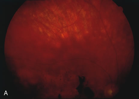
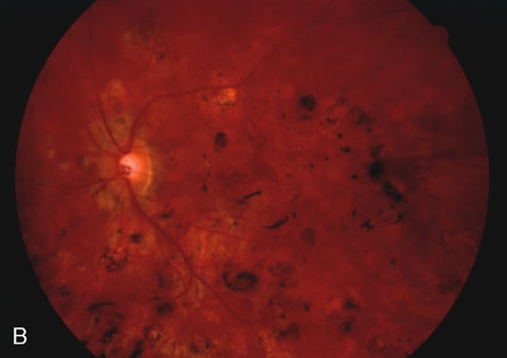
Fig. 9.
A
and
B
. Fundus photographs of the right and left eye showing the classic circular choroidal lesions with marked pigment clumping in a case of multifocal choroiditis.