|
|
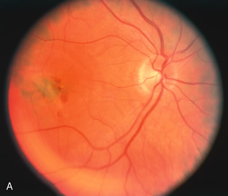
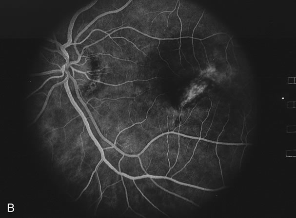
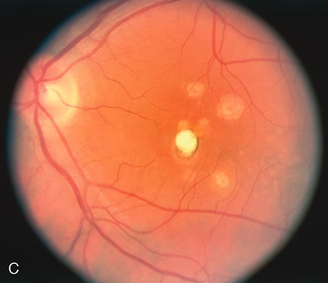
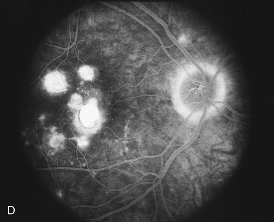
| Fig. 15. A. Right fundus photograph of a patient with punctate inner choroidopathy showing the presence of a choroidal neovascular membrane. B. Fluorescein angiogram of the right eye demonstrating the presence of the choroidal neovascular membrane and perpipapillary window defect. C. Left fundus photograph showing large coalesced spots of inactive choroiditis. D. Fluorescein angiogram of the left eye showing staining of the areas of inactive choroiditis. Note the small areas of fluorescence that could not be seen by color photography. |