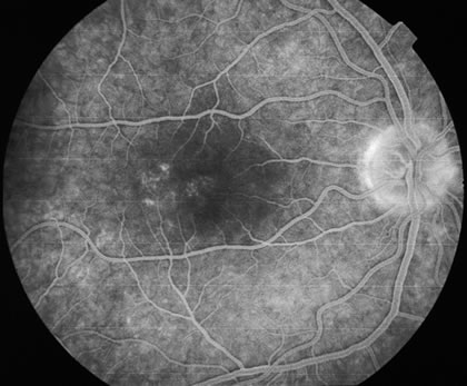
Fig. 3.
Venous phase fluorescein angiogram of fundus shown in Figure 1. It demonstrates the hyperfluorescent dots coalesced near the fovea and in a subtle wreath-like fashion elsewhere.