|
|
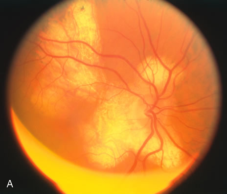
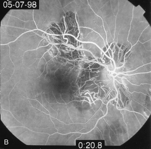
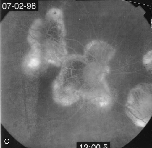
| Fig. 16. A. Color fundus photograph showing the jigsaw pattern of choroidal and retinal atrophy extending from the disc along the arcades. At the inferior edge of the superotemporal lesion there is a recurrence noted by the grayness of the retina. B. Fluorescein angiogram in the laminar venous phase showing a large window defect in the area of inactive choroiditis and blockage of the choroidal fluorescence in the area of active choroiditis. C. In the late phase of the fluorescein angiogram there is staining of the edges of the inactive choroiditis and marked hyperfluorescence in the area of active choroiditis. |