|
|
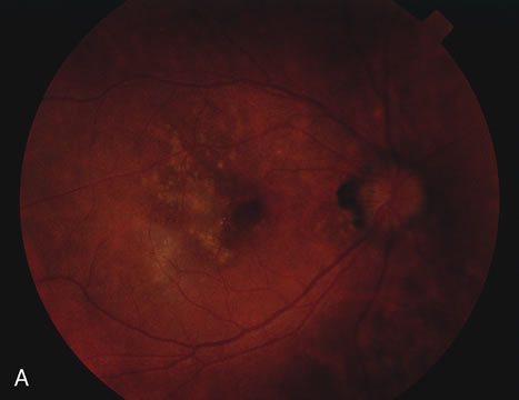
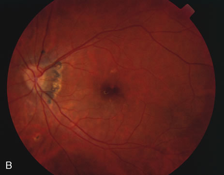
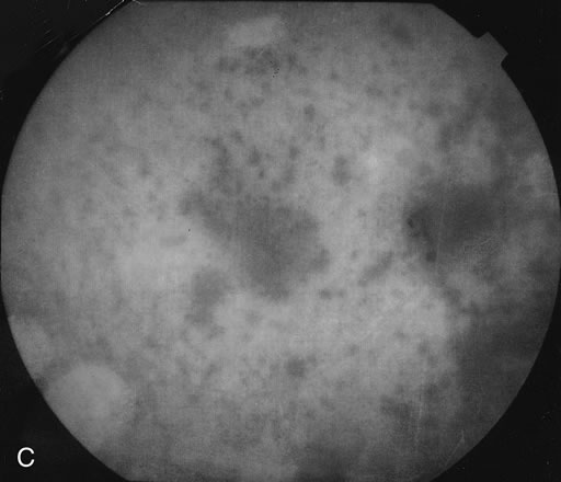
| Fig. 14. A. Right fundus photograph in a patient with multifocal choroiditis showing the small patches of choroiditis. Many have coalesced to form a larger patch. B. Left fundus photograph showing less involvement. C. Indocyanine green angiogram of the fundus shown in Figure 14a showing multiple hypofluorescent spots. |