Chapter 38
Vitreous Biochemistry, Structure, and Clinical Evaluation
J. SEBAG
Main Menu   Table Of Contents

Search

BIOCHEMISTRY
STRUCTURE
CLINICAL EVALUATION OF VITREOUS
REFERENCES

BIOCHEMISTRY

COLLAGENS

As is true for connective tissues everywhere, collagen is an important structural protein in vitreous. Gross1 initially claimed that vitreous collagen fibrils are morphologically distinct from collagen in other connective tissues. However, Swann and coworkers2 demonstrated that the amino acid composition of the insoluble residue of vitreous is similar to that of cartilage collagen and later identified that it is most similar to cartilage collagen composed of alpha 1, type II chains.3 Comparisons of the arthrogenic and immunologic properties of collagens from bovine articular cartilage (type II) and vitreous showed that the two were indistinguishable by these assays.4 Subsequent studies5 demonstrated that, although vitreous collagen contains an alpha 1, type II chain similar to cartilage collagen, there is a lower alanine content. Furthermore, these studies found that vitreous collagen has additional peptides present as uncleaved extension chains containing an amino acid composition different from the alpha chain component. The investigators concluded, however, that the overall similarities in amino acid composition and in the types of cyanogen bromide cleavage peptides indicate that the fibers of the central and posterior peripheral regions of the vitreous are composed of a collagen that should be classified as type II. Linsenmayer and collaborators6 measured in vivo synthesis of types I and II collagen in chick embryo vitreous by radioimmunoprecipitation after tritiated proline labeling and found that more than 90% of the labeled material in the vitreous was type II collagen. Snowden7 provided further physicochemical evidence in support of the similarities between vitreous and cartilage collagens. This may explain why certain clinical phenomena, such as inborn errors of type II collagen metabolism in arthro-ophthalmopathies, manifest phenotypic expression in each of these two tissues.

There are, however, distinct differences in the chemical composition of vitreous and cartilage collagens that are only partly due to the presence of terminal peptide constituents in vitreous collagen.5 Swann and Sotman8 have demonstrated that the carbohydrate content of pepsin-solubilized vitreous alpha chains is significantly greater than cartilage alpha chains, indicating that the carbohydrate side chains of vitreous collagen are largely composed of disaccharide units similar to those found in basement membrane collagen. They proposed that these distinct chemical features are related to the special structure of the mature vitreous fibrils in vivo. Liang and Chakrabarti9 have shown that there are differences between bovine cartilage and vitreous with respect to collagen fibril growth, melting temperature, and fluorescence with a hydrophobic fluorescent probe. These investigators and others10 proposed that vitreous collagen should be considered a “special” type II collagen. Schmut and associates11 employed differential salt precipitation of pepsin-solubilized collagen from bovine vitreous and found that type II collagen is the major component of native vitreous fibers.

Gloor12 pointed out that the collagen content is highest where the vitreous is a gel. Ayad and Weiss13 studied bovine vitreous collagen to determine whether the gel-like structure of vitreous could be explained on the basis of chemical composition. Their findings demonstrated that type II is the major vitreous collagen, but collagens composed of α1, α2, and α3 chains as well as C-PS disulfide-bonded collagen were present in concentrations similar to those in cartilage. In contrast to cartilage, however, vitreous type II collagen was significantly more hydroxylated in the lysine and proline residues. The α1, α2, and α3 collagen chains were interpreted by Van der Rest14 to represent type IX collagen, although Eyre and colleagues15 felt that there was evidence to indicate the presence of type V collagen in vitreous. Furthermore, with respect to the disulfide-bonded collagen, vitreous had three times more C-PS1 and C-PS2 collagens than cartilage, although the molar ratio of C-PS 1 to C-PS2 in both was 1:1, suggesting that in both tissues these collagens are components of a larger molecule. Other studies14 demonstrated that these disulfide-linked triple-helix fragments were actually derivatives of type IX collagen. In this regard vitreous is again similar to cartilage, insofar as both contain type IX collagen,15 although the two tissues differ in the sizes of type IX collagen chains.16 Hong and Davison17 have identified a procollagen in the soluble fraction of rabbit vitreous that was identified as type II by segment-long-spacing banding patterns. Detection of a propeptide extension only at the N-terminus prompted these investigators to conclude that this was a novel type II procollagen. Such distinctive characteristics are possibly related to the unique physiologic roles of vitreous, in particular, its mechanical function.18

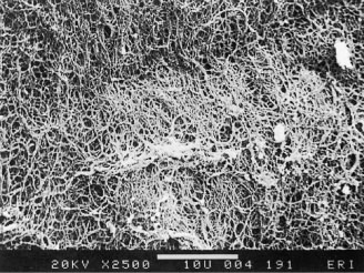
Individual vitreous collagen fibrils are organized as a triple helix of three alpha chains. The major collagen fibrils of the corpus vitreus are heterotypic, consisting of more than one collagen type. Recent studies of pepsinized forms of collagen confirm that the corpus vitreus contains collagen type II, a hybrid of types V/XI, and type IX (Fig. 1).

Fig. 1. Vitreous collagen fibril structure. This schematic diagram of the structure of the major heterotypic collagen fibrils of vitreous is largely speculative but based on the current knowledge of the structure and biophysical attributes of the constituent molecules, particularly in how they would assemble to form fibrils. (From Bishop P: The biochemical structure of mammalian vitreous. Eye 10:64, 1996.)

Type II Collagen

Type II collagen, a homotrimer composed of three identical alpha chains designated as [α1 (II)]3, comprises 75% of the total collagen content in vitreous. When first synthesized as a procollagen and secreted into the extracellular space, type II collagen is highly soluble. The activity of N-proteinase and C-proteinase enzymes reduces the solubility and enables type II collagen molecules to cross-link covalently in a quarter-staggered array. Within this array are likely to be N-propeptides, which probably extend outward from the surface of the forming fibril.11 This may influence the interaction of the collagen fibril with other components of the extracellular matrix. Recent studies19 combined immunolocalization with Western blot analysis of macromolecules extracted from bovine vitreous collagen fibrils and found that the pN-type IIA procollagen is located on the surface of the vitreous collagen fibril. The finding20 that type IIA procollagen propeptides specifically bind transforming growth factor-beta 1 (TGFβ1) and BMP-2 supports the concept that, in certain circumstances, these growth factors and cytokines interact with vitreous fibrils to promote the cell migration and proliferation that result in proliferative diabetic retinopathy and proliferative vitreoretinopathy.

Type IX Collagen

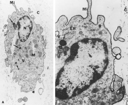
Type IX collagen is a heterotrimer that is disulfide-bonded with an [α1 (IX) α2 (IX) α3 (IX)] configuration. This heterotrimer is orientated regularly along the surfaces of the major collagen fibrils in a “D periodic” distribution, in which it is cross-linked onto the fibril surface. Type IX is not a typical collagen but is a member of the fibrillar-associated collagens with interrupted triple helixes (FACIT) group of collagens. It contains collagenous regions described as COL1, COL2, and COL3 interspersed between noncollagenous regions called NC1, NC2, NC3, and NC4.21,22 In vitreous (as opposed to cartilage) the NC4 domain is small and, therefore, not highly charged and not likely to exhibit extensive interaction with other extracellular matrix components.23 In vitreous, type IX collagen always contains a chondroitin sulfate glycosaminoglycan chain,21,22 which is linked covalently to the α2 (IX) chain at the NC3 domain; this enables the molecule to assume a proteoglycan form. Electron microscopy of vitreous stained with cationic dyes enables visualization of the chondroitin sulfate chains of type IX collagen. In some of these studies sulfated glycosaminoglycans are found distributed regularly along the surface of vitreous collagen fibrils24 and often bridge between neighboring collagen fibrils. Duplexing of glycosaminoglycan chains from adjacent collagen fibrils may result in a ladder-like configuration.25

Type V/XI Collagen

Ten percent of vitreous collagen is a hybrid V/XI collagen, which is believed to comprise the central core of the major collagen fibrils of vitreous.26 Type V/XI is a heterotrimer that contains α1 (XI) and α2 (V) in two chains, but the nature of the third chain is presently not known.15 Along with type II collagen, type V/XI is a fibril-forming collagen. Although the interaction of the fibril with other extracellular matrix components is probably influenced by a retained N-propeptide that protrudes from the surface of the fibril in cartilage,26 it is not known whether this is the case in vitreous.27

Type VI Collagen

Although there are only small amounts of type VI collagen in vitreous, the ability of this molecule to bind both type II collagen and hyaluronan (HA) suggests that it could be important in organizing and maintaining the supramolecular structure of vitreous gel.

GLYCOSAMINOGLYCANS

Glycosaminoglycans are polysaccharides of repeating disaccharide units, each consisting of hexosamine (usually N-acetyl glucosamine or N-acetyl galactosamine) glycosidically linked to either uronic (glucuronic or iduronic) acid or galactose. The nature of the predominant repeating unit is characteristic for each glycosaminoglycan and the relative amount, molecular size, and type of glycosaminoglycan are said to be tissue specific.28 Glycosaminoglycans do not normally occur as free polymers in vivo but are covalently linked to a protein core; the ensemble is called a proteoglycan. A sulfated group is attached to oxygen or nitrogen in all glycosaminoglycans except HA. Balazs29 first documented the presence of sulfated galactosamine-containing glycosaminoglycans in bovine vitreous (less than 5% of total vitreous glycosaminoglycans), and others30,31 identified these as chondroitin-4-sulfate and undersulfated heparan sulfate. Studies in the rabbit32 found a total vitreous glycosaminoglycans content of 58 ng with 13% chondroitin sulfate and 0.5% heparan sulfate.

Hyaluronan

Although HA is present throughout the body, it was first isolated from bovine vitreous. In humans, HA first appears after birth and then becomes the major vitreous glycosaminoglycan. Although it has been proposed that hyalocytes synthesize HA,18 other plausible candidates are the ciliary body and retinal Müller cells. Whereas the synthesis of HA seems to continue at a constant rate in the adult while no extracellular degradation occurs, HA levels are in a steady-state because the molecule escapes via the anterior segment of the eye.29 Laurent and Fraser33 showed that the passage of HA from the vitreous to the anterior segment is strongly molecular-weight dependent, indicating a diffusion-controlled process. In contrast, disappearance of HA from the anterior chamber is independent of molecular weight, suggesting that this is controlled by bulk flow.

HA is a long, unbranched polymer of repeating glucuronic acid β-1,3-N,N-acetylglucosamine disaccharide moieties linked by β 1–4 bonds.5 It is a linear, left-handed, three-fold helix with a rise per disaccharide on the helix axis of 0.98 nm.34 Rotary shadowing electron microscopy of human and bovine vitreous detected lateral aggregates of HA that form an anastomosing three-dimensional network.35 This periodicity, however, can vary depending on whether the helix is in a compressed or extended configuration.36 Changes in the degree of extension of HA could be important in the role vitreous plays in retinal disease. Indeed, the volume of the unhydrated HA molecule is about 0.66cm3/g, whereas the hydrated specific volume is 2000 to 3000 cm3/g.29 Thus, the degree of hydration has a significant influence on the size and configuration of the HA molecular network. Although there is no definitive evidence that adjacent HA chains bind to one another, Brewerton and Mayne37 first proposed such an arrangement. Recent rotary shadowing electron microscopy studies38 of bovine and human vitreous found lateral aggregates of HA that formed three-dimensional lattice-like networks. HA also interacts with the surrounding mobile ions and can undergo changes in its conformation that are induced by changes in the surrounding ionic milieu.39 A decrease in surrounding ionic strength can cause the anionic charges on the polysaccharide backbone to repel one another, resulting in an extended configuration of the macromolecule. An increase can cause contraction of the molecule and, in turn, the entire vitreous body. As a result of HA's entanglement and immobilization within the vitreous collagen fibril matrix, this mechanical force can be transmitted by collagen fibrils to the retina, optic disc, and other structures such as neovascular complexes. In this way, changes in the ionic milieu of vitreous may be converted into mechanical energy via extension or contraction of the HA macromolecule. This can be important in certain pathologic conditions that feature fluctuations in ionic balance and hydration, such as diabetes.40

The sodium salt of HA has a molecular weight of 3 to 4.5 × 10⁶ Da in normal human vitreous.29 Laurent and Granath41 used gel chromatography and found the average molecular weight of rabbit vitreous to be 2 to 3 × 10⁶ Da and of bovine vitreous to be 0.5 to 0.8 × 10⁶ Da. In these studies there were age-related differences in the bovine vitreous, in which HA molecular weight varied from 3 × 10⁶ Da in the newborn calf to 0.5 × 10⁶ Da in old cattle. Furthermore, there may be several species of HA within vitreous that have polysaccharide chains of different lengths42 with a variable distribution in different topographic regions within the corpus vitreous.43

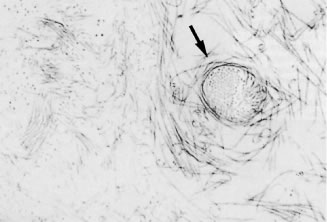
An important property of HA is steric exclusion.44 With its flexible linear chains and random coil conformation, HA occupies a large volume and resists the penetration of this volume by other molecules to a degree dependent on their size and shape.36 This excluded volume effect can influence equilibria between different conformational states of macromolecules and alter the compactness or extension of these molecules.44 Steric exclusion also causes an excess of osmotic pressure when such compounds as albumin and HA are mixed, because the resultant osmotic pressure is greater than the sum of the two components. This could be important in diabetes, in which vascular incompetence can increase vitreous concentrations of serum proteins such as albumin. These osmotic effects can induce contraction and expansion of the corpus vitreus similar to the foregoing description of hydration and ion-induced changes within vitreous and can similarly play an important role in neovascularization and diabetic vitreous hemorrhage.40 An increase in the chemical activity of a compound because of steric exclusion can also cause its precipitation if the solubility limit is reached. This could be important in the formation of pathologic vitreous opacities, such as asteroid hyalosis and amyloid bodies.18

Chondroitin Sulfate

Vitreous contains two chondroitin sulfate proteoglycans. The minor type is actually type IX collagen, which was described above. Most vitreous chondroitin sulfate is in the form of versican.38 This large proteoglycan has a globular N-terminal that binds HA via a 45 kDa link protein.38 Thus, in human, but not bovine vitreous, versican is believed to form complexes with HA, as well as with microfibrillar proteins such as fibulin-1 and fibullin-2.45

Heparin Sulfate

This sulfated proteoglycan is normally found in basement membranes and on cell surfaces throughout the body. It was first detected in bovine vitreous in 197746 and in chick vitreous (as agrin) in 1995.47 However, it is not clear whether heparan sulfate is a true component of vitreous or a contaminant from adjacent basement membranes, such as the internal limiting lamina (ILL) of the retina.48 As pointed out by Bishop45 this may also be the case for nodogen-1, the aforementioned fibulins, and fibronectin.

NONCOLLAGENOUS STRUCTURAL PROTEINS

Fibrillins

Fibrillin-containing microfibrils are more abundant in vitreous than the type VI collagen microfibrils described above. They are the found in vitreous gel, as well as in the zonules of the lens. This fact explains why in Marfan's syndrome the defects in the gene encoding fibrillin-1 (FBN1 on chromosome 15q21) result in both ectopia lentis and vitreous liquefaction.45 The latter probably plays a role in the frequent occurrence of rhegmatogenous retinal detachment in these patients.

Opticin

The major noncollagenous protein of vitreous is a leucine-rich repeat (LRR) protein, which is bound to the surface of the heterotypic collagen fibrils, known as opticin.49 Formerly called vitrican, opticin is believed to be important in collagen fibril assembly and in preventing the aggregation of adjacent collagen fibrils into bundles. Thus, a breakdown in this property or activity may play a role in age-related vitreous degeneration50 (see later discussion below).

VIT 1

Another novel vitreous protein is VIT1, a collagen-binding macromolecule.51 Because of its propensity to bind collagen, this highly basic protein may play an important role in maintaining vitreous gel structure.

MISCELLANEOUS MOLECULAR COMPONENTS

Amino Acids and Proteins

Free amino acids are present in the vitreous, but at one fifth of the levels found in plasma.52 An amino acid concentration gradient is present within the vitreous body, with anterior vitreous concentrations greater than posterior levels. This may be due to uptake and utilization of amino acids by the retina, a consideration that led Reddy53 to propose that the vitreous acts as a metabolic repository for retinal protein metabolism. Other studies54 found that the soluble proteins of vitreous resemble serum proteins with isoelectric points less than 6.0, leading investigators to conclude that the soluble proteins of vitreous derive from plasma and are constantly renewed. Flood and Balazs55 studied 920 human eyes and found the following age-related protein concentrations: ages 10 to 50, 400 to 600 μg/ml; ages 50 to 80, 700 to 800 μg/ml; older than 80 years of age, about 1000 μ/ml. These age-related findings may be due to increased leakage of plasma proteins from the intravascular compartment into the vitreous, resulting from decreased tight-junction integrity with aging of the retinal and ciliary body vasculature and epithelia.

Glycoproteins

Glycoproteins are heteropolysaccharide macromolecules, as opposed to the homogeneous, repeating disaccharide units of glycosaminoglycans that are mostly proteinaceous and contain only a minor carbohydrate component (5% to 10% by weight). According to Balazs29 the most important difference between vitreous and serum proteins is the high content of glycoproteins in the vitreous, because these constitute 20% of the total noncollagenous protein content of vitreous. Sialic acid-containing glycoproteins are believed to be synthesized by hyalocytes.56 Other studies57 have led to the postulate that the inner layer of the ciliary epithelium is responsible for vitreous glycoprotein synthesis. Cartilage oligomeric protein (COMP), an acidic glycoprotein with a characteristic five-armed structure, is present in vitreous, but its function is currently unknown.

Ascorbic Acid

Ascorbic acid concentrations in vitreous are about 0.43 mmol/kg.58 Thus, the vitreous-plasma ratio for ascorbic acid is 9:1. Vitreous levels that are much higher than plasma concentrations are believed to be due to active transport by the ciliary body epithelium.59 The purpose of having high concentrations of ascorbic acid in the vitreous may relate to the abilities of this compound to absorb ultraviolet light and to serve as a free-radical scavenger,60 which may protect not only the retina and lens but also vitreous from the untoward effects of metabolic and light-induced61 singlet oxygen generation.

Lipids

Swann and coinvestigators62 found that the residual fraction of vitreous contained a significant quantity of lipid. Later studies63 found evidence of active lipid metabolism in vitreous of dogs and humans, but in the latter species no significant changes in vitreous lipid composition between the ages of 37 and 82 years.

SUPRAMOLECULAR ORGANIZATION

As described by Mayne,65 vitreous is organized in a dilute meshwork of collagen fibrils interspersed with extensive arrays of long HA molecules. The collagen fibrils provide a scaffold-like structure that is inflated by the hydrophilic HA. If collagen is removed, the remaining HA forms a viscous solution; if HA is removed, the gel shrinks39 but is not destroyed. Early physiologic observations66 suggested the existence of an interaction between HA and collagen that stabilizes collagen. Biomechanical studies67 of vitreous viscoelasticity suggested similar effects on HA. Early investigators68 observed reversible complexes of an electrostatic nature between solubilized collagen and various glycosaminoglycans and suggested that collagen-HA interaction occurs on a physicochemical rather than chemical level. Others69 later demonstrated that the sulfate group of a glycosaminoglycan is largely responsible for such interactions with the guanidino groups of arginine and epsilon-amino groups of lysine in collagen. Comper and Laurent39 proposed that in vitreous electrostatic binding occurs between negatively charged polysaccharides and positively charged proteins. These authors extensively reviewed the existing data characterizing the electrostatic properties of glycosaminoglycans and the factors influencing their electrostatic interactions with different ions and molecules.

Balazs29 hypothesized that the hydroxylysine amino acids of collagen mediate polysaccharide binding to the collagen chain via O-glycosidic linkages. These polar amino acids are present in clusters along the collagen molecule, which may explain why proteoglycans attach to collagen in a periodic pattern.24 Hong and Davison70 identified a type II procollagen in the soluble fraction of rabbit vitreous and proposed a possible role for this molecule in mediating collagen-HA interaction. In cartilage, “link glycoproteins” have been identified that interact with proteoglycans71 and HA.72 Supramolecular complexes of these glycoproteins are believed to occupy the interfibrillar spaces. In Asakura's24 studies of bovine vitreous stained with ruthenium red there are amorphous structures located on collagen fibrils at 55 to 60 nm intervals along the fibrils and filaments connecting these amorphous masses to adjacent collagen fibrils. These filaments may represent “link” structures of either a glycoprotein or proteoglycan nature. HA is known to interact with link proteins, as well as with an HA-binding glycoprotein called hyaluronectin.73 In the cornea, chondroitin sulfate and keratan sulfate bridge the interfibrillar spaces and keep the fibrils at specified distances to achieve transparency.74 The protein cores of these proteoglycans could be the linkage sites to collagen fibrils.

Bishop27 has proposed that an understanding how vitreous gel is organized and stabilized requires an understanding of what prevents collagen fibrils from aggregating and by what means the collagen fibrils are connected to maintain a stable gel structure. Studies75 have shown that the chondroitin sulfate chains of type IX collagen bridge adjacent collagen fibrils in a ladder-like configuration spacing them apart. This arrangement might account for vitreous transparency, because keeping vitreous collagen fibrils separated by at least one wavelength of incident light would minimize light scattering, allowing unhindered transmission of light to the retina for photoreception. However, depolymerizing with chondroitinases does not destroy the gel, suggesting that chondroitin sulphate side chains are not essential for vitreous collagen spacing. Complexed with HA, however, the chondroitin sulfate side chains might space the collagen fibrils,25,74,75 apart but Bishop believes that this form of collagen-HA interaction is “very weak.” Instead, he proposes that the LRR protein opticin is the predominant structural protein in short-range spacing of collagen fibrils. Concerning long-range spacing, Scott25 and Mayne and coworkers77 have claimed that HA plays a pivotal role in stabilizing the vitreous gel via this mechanism. However, studies76 using HA lyase to digest vitreous HA demonstrated that the gel structure was not destroyed, suggesting that HA is not essential for the maintenance of vitreous gel stability, leading to the proposal that collagen alone is responsible for the gel state of vitreous.27

Back to Top
STRUCTURE

HISTORICAL PERSPECTIVE

Duke-Elder78 claimed that the first theories of vitreous structure proposed that the vitreous is composed of “loose and delicate filaments surrounded by fluid,” as conceptualized in 1741 by Demours79 who formulated the alveolar theory. In 1780, Zinn80 proposed that the vitreous is arranged in a concentric, lamellar configuration similar to the layers of an onion. The dissections and histologic preparations of Von Pappenheim81 and Brucke82 provided evidence for the lamellar theory. The radial sector theory was proposed by Hannover in 1845.83 Studying coronal sections at the equator, he described a multitude of sectors approximately radially oriented around the central anteroposterior core that contains Cloquet's canal. Hannover likened this structure to the appearance of a cut orange. In 1848 Bowman84 introduced the fibrillar theory. Employing microscopy, he described fine fibrils that form bundles that Retzius85 described as fibrous structures arising in the peripheral anterior vitreous that assume an undulating pattern similar to a “horse's tail” in the central vitreous but maintain a concentric configuration at the periphery. The elegant studies of Szent-Gyorgi86 in 1917 supported the descriptions of Retzius and introduced the concept that vitreous structure changes with age. Eisner87 studied dissections of human vitreous and found “membranelles,” which he described as funnels packed into one another diverging outward and anteriorly from the prepapillary vitreous. Worst88 has also studied preparations of dissected human vitreous and described that the tracts of Eisner constitute the walls of cisterns within the vitreous body. In Worst's studies these cisterns are visualized by filling with white India ink. Worst has also studied the premacular vitreous in great detail and has proposed the existence of a “bursa premacularis,” which he described as a pear-shaped space that is connected to the cisternal system in front of the ciliary body. Kishi and Shimizu89 recently described a “posterior vitreous pocket” similar to what Worst has reported, which they claim to be an anatomic structure. However, because more than 95% of their study population was age 65 years or older, this is likely to be an age-related phenomenon.90

VITREOUS BODY (CORPUS VITREUS)

There is heterogeneity in the distribution of collagen throughout the vitreous body. Chemical91,92 and light-scattering93,185 studies have shown that the highest density of collagen fibrils is present in the vitreous base, followed by the posterior vitreous cortex anterior to the retina, and then by the anterior vitreous cortex behind the posterior chamber and lens. The lowest density is found in the central vitreous and adjacent to the anterior cortical gel. HA molecules have a different distribution from collagen. They are most abundant in the posterior cortical gel with a gradient of decreasing concentration as one moves centrally and anteriorly.94–96 Balazs96 has hypothesized that this is because vitreous HA is synthesized by hyalocytes in the posterior vitreous cortex and cannot traverse the ILL of the retina. HA leaves the vitreous to enter the posterior chamber by way of the annulus of the anterior vitreous cortex that is not adjacent to a basal lamina. Bound water (non-freezable) has a distribution within the vitreous similar to that of HA,97 presumably resulting from binding by HA.

Laboratory investigations of vitreous structure have long been hampered by the absence of easily recognized landmarks within the vitreous body. Consequently, removal of the vitreous from the eye results in a loss of orientation. The transparency of the vitreous renders observation in conventional diffuse light unrewarding. Attempts to study vitreous structure with opaque dyes88 do visualize the areas filled by dye but obscure the appearance of adjacent structures. The use of histologic contrast-enhancing techniques usually involves tissue fixation, which often includes dehydration of the tissue. Because vitreous is 98% water, dehydration induces profound alteration of the internal morphology. Consequently, any investigation of vitreous structure must overcome these difficulties.

The sclera, choroid, and retina can be dissected and the naked vitreous body can be maintained intact and attached to the anterior segment of the eye (Fig. 2A). This enables study of internal vitreous morphology without a loss of intraocular orientation. However, depending on the person's age and consequently the degree of vitreous liquefaction,55 the dissected vitreous will remain solid and intact in young persons or will be flaccid and collapse in older individuals. Consequently, vitreous turgescence must be maintained to avoid distortion of intravitreal structure. Immersion of a dissected vitreous specimen that is still attached to the anterior segment into a physiologic solution maintains vitreous turgescence and avoids structural distortion (see Fig. 2B). The limitations induced by the transparency of the vitreous were overcome by Goedbloed,98 Friedenwald and Stiehler,99 and Eisner87 who employed darkfield slit illumination of the vitreous body to achieve visualization of intravitreal morphology. Illumination with a slit-lamp beam directed into the vitreous body from the side and visualization of the illuminated portion from above produces an optical horizontal section of the vitreous body.100 The illumination/observation angle of 90 degrees that is achieved by using this technique maximizes the Tyndall effect and, thus, overcomes the limitations induced by vitreous transparency. Furthermore, the avoidance of any tissue fixation eliminates the introduction of many of the artifacts that flawed earlier investigations. Recent studies have used these techniques to investigate human vitreous structure. Within the adult human vitreous there are fine, parallel fibers coursing in an anteroposterior direction18,100–102 (Figs. 3B and C and 4). The fibers arise from the vitreous base (see Figs. 3H and 4) where they insert anterior and posterior to the ora serrata (see Fig. 3H). As the peripheral fibers course posteriorly they are circumferential with the vitreous cortex, whereas central fibers undulate in a configuration parallel with Cloquet's canal.85 The fibers are continuous and do not branch. Posteriorly, these fibers insert into the vitreous cortex (see Fig. 3E and F).

Fig. 2. Human vitreous dissection. A. Vitreous from a 9-month-old child. The sclera, choroid, and retina were dissected off the vitreous body, which remains attached to the anterior segment. A band of gray tissue can be seen posterior to the ora serrata. This is neural retina that was firmly adherent to the vitreous base and could not be dissected. The vitreous body is solid and although situated on a surgical towel exposed to room air maintains its shape; because of the young age of the donor, the vitreous is almost entirely gel. B. Human vitreous dissected of the sclera, choroid, and retina and attached to the anterior segment. The specimen is mounted on a Lucite frame using sutures through the limbus and then immersed in a Lucite chamber containing an isotonic, physiologic solution. This maintains the turgescence of the vitreous and avoids collapse and artifactual distortion of the vitreous structure. (A Courtesy of the New England Eye Bank, Boston, MA. B from Sebag J, Balazs EA: Pathogenesis of C.M.E: Anatomic consideration of vitreo-retinal adhesions. Surv Ophthalmol 28[Suppl]:493, 1984.)

Fig. 3. Human vitreous morphology. Human vitreous structure visualized by dark-field slit illumination. All photographs are oriented with the anterior segment below and the posterior pole above. A. Posterior vitreous in the left eye of a 52-year-old man. The vitreous body is enclosed by the vitreous cortex. There is a hole in the prepapillary (small, to the left) vitreous cortex. Vitreous fibers are oriented toward the premacular region. B. Posterior vitreous in a 57-year-old man. A large bundle of prominent fibers is seen coursing anteroposteriorly and entering the retrocortical space by way of the premacular vitreous cortex. C. Same photograph as B, at higher magnification. D. Posterior vitreous in the right eye of a 53-year-old woman. There is posterior extrusion of vitreous out the prepapillary hole (to the right) and premacular (large extrusion to the left) vitreous cortex. Fibers course anteroposteriorly out into the retrocortical space. E. Horizontal optical section of the same specimen as D, at a different level. A large fiber courses posteriorly from the central vitreous and inserts into the premacular vitreous cortex. F. Same view as E, at higher magnification. The large fiber has a curvilinear appearance because of traction by the vitreous extruding into the retrocortical space (see D). However, because of its attachment to the posterior vitreous cortex, the fiber arcs back to its point of insertion. G. Anterior and central vitreous in a 33-year-old woman. Cloquet's canal is seen forming the retrolental space of Berger. H. Anterior and peripheral vitreous in a 57-year-old man. The specimen is tilted forward to enable visualization of the posterior aspect of the lens and the peripheral anterior vitreous. To the right of the lens there are fibers coursing anteroposteriorly that insert into the vitreous base. These fibers “splay out” to insert anterior and posterior to the ora serrata. (A, E, and F from Sebag J, Balazs EA: Pathogenesis of C.M.E.: Anatomic consideration of vitreo-retinal adhesions. Surv Ophthalmol 28[Suppl]:493, 1984. B, C from Sebag J, Balazs EA: Morphology and ultrastructure of human vitreous fibers. Invest Ophthalmol Vis Sci 30:187, 1989. D, G, and H from Sebag J: The Vitreous: Structure, Function and Pathobiology. New York, Springer-Verlag, 1989. Specimens were courtesy of the New York Bank for Sight and Restoration, New York, NY.)

Fig. 4. A. Vitreous base morphology. Vitreous structure in a 58-year-old woman. Fibers course anteroposteriorly in the central and peripheral vitreous. Posteriorly, fibers orient to the premacular region. Anteriorly, the fibers “splay out” to insert into the vitreous base. B. Fibers of the peripheral anterior vitreous forming the anterior loop. This configuration can provide the scaffold for cell migration and proliferation in the pathophysiology of anterior proliferative vitreoretinopathy. (A from Sebag J, Balazs EA: Pathogenesis of C.M.E.: Anatomic consideration of vitreo-retinal adhesions. Surv Ophthalmol 28[Suppl]:493, 1984. B from Sebag J: The Vitreous: Structure, Function and Pathobiology. New York, Springer-Verlag, 1989. Specimen in A courtesy of the New England Eye Bank, Boston, MA.)

Ultrastructural studies103 have demonstrated that collagen fibrils are the only microscopic structures that could correspond to these fibers. These studies also detected the presence of bundles of packed, parallel collagen fibrils (Fig. 5). It has been hypothesized that visible vitreous fibers form when HA molecules no longer separate microscopic collagen fibrils, resulting in the aggregation of collagen fibrils into bundles from which HA molecules are excluded. Eventually the aggregates of collagen fibrils attain sufficiently large proportions and can be visualized in vitro (see Figs. 3 and 4) and clinically. The areas adjacent to these large fibers have a low density of collagen fibrils separated by HA molecules and, therefore, do not scatter light as intensely as the larger bundles of aggregated collagen fibrils. These adjacent channels probably offer relatively less resistance to bulk flow through the vitreous body and are the areas visualized in studies88,104 using India ink to fill the channels. There are changes occurring in these fibrous structures throughout life,50,100 which probably result from age-related biochemical alterations in the composition and organization of the molecular components that simultaneously result in vitreous liquefaction and fiber formation.

Fig. 5. Ultrastructure of human vitreous. Although specimens were centrifuged to concentrate structural elements, there were no membranes or membranous elements. Only collagen fibrils were detected. There were also bundles of parallel collagen fibrils such as the one shown here in cross section (arrow). (From Sebag J, Balazs EA: Morphology and ultrastructure of human vitreous fibers. Invest Ophthalmol Vis Sci 30:187, 1989.)

VITREOUS BASE

The vitreous base is a three-dimensional zone. It extends 1.5 to 2 mm anterior to the ora serrata, 1 to 3 mm posterior to the ora serrata,105 and several millimeters into the vitreous body.106 The posterior extent of the posterior border of the vitreous base varies with age.50,107 Vitreous fibers enter the vitreous base by splaying out (see Fig. 4A) to insert anterior and posterior to the ora serrata (see Fig. 3H). The anterior-most fibers form the anterior loop of the vitreous base (see Fig. 4B), a structure that is important in the pathophysiology of anterior proliferative vitreoretinopathy.18,108–110 In the posterior portion of the vitreous base, vitreous fibers are closer together than elsewhere. Gartner111 has found that in humans the diameters of collagen fibrils in the vitreous base range from 10.8 to 12.4 nm, with a major period of cross-striations of 50 to 54 nm. Hogan127 demonstrated that just posterior to the ora serrata, heavy bundles of vitreous fibrils attach to the basal laminae of retinal glial cells. Studies by Gloor64 and Daicker112 showed that cords of vitreous collagen insert into gaps between the neuroglia of the peripheral retina. They likened this structure to Velcro (a self-adhesive nylon material) and proposed that this would explain the strong vitreoretinal adhesion at this site. In the anterior vitreous base, fibrils interdigitate with a reticular complex of fibrillar basement membrane material between the crevices of the nonpigmented ciliary epithelium.113 The vitreous base also contains intact cells that are fibroblast-like anterior to the ora serrata and macrophage-like posteriorly.113 Damaged cells in different stages of involution and fragments of basal laminae, presumed to be remnants of the embryonic hyaloid vascular system, are also present in the vitreous base.113

VITREOUS CORTEX

The vitreous cortex is the peripheral shell of the vitreous body that courses forward and inward from the anterior vitreous base to form the anterior vitreous cortex and posteriorly from the posterior border of the vitreous base to form the posterior vitreous cortex. The anterior vitreous cortex, clinically referred to as the anterior hyaloid face, begins about 1.5 mm anterior to the ora serrata. Fine and Tousimis114 described that in this region the collagen fibrils are parallel to the surface of the cortex. Studies by Faulborn and Bowald115 detected dense packing of collagen fibrils in the anterior cortex with looser collagen fibril packing in the subjacent vitreous, giving the impression of lamellae. Rhodes116 studied mouse vitreous and found that the anterior vitreous cortex varied in thickness from 800 to 2000 nm. He also found that there are connections between the loose fibrils in the anterior vitreous and the anterior vitreous cortex.

The posterior vitreous cortex is 100 to 110 μm thick117 and consists of densely packed collagen fibrils (Fig. 6). There is no vitreous cortex over the optic disc (Figs. 3A and 7), and the cortex is thin over the macula because of rarefaction of the collagen fibrils.117 The prepapillary hole in the vitreous cortex can sometimes be visualized clinically when the posterior vitreous is detached from the retina. If peripapillary glial tissue is torn away during posterior vitreous detachment (PVD) and remains attached to the vitreous cortex around the prepapillary hole, it is referred to as Vogt's or Weiss's ring. Vitreous can extrude through the prepapillary hole in the vitreous cortex (see Fig. 3A) but does so to a much lesser extent than through the premacular vitreous cortex (see Figs. 2B and D and 6). Jaffe118 has described how vitreous can extrude into the retrocortical space created following PVD and has proposed that persistent attachment to the macula can produce traction and certain forms of maculopathy.119,120 Although there are no direct connections between the posterior vitreous and the retina, the posterior vitreous cortex is adherent to the ILL of the retina, which is actually the basal lamina of retinal Müller's cells. The exact nature of this adhesion between the posterior vitreous cortex and the ILL is not known but probably results from extracellular matrix molecules.121–123

Fig. 6. Ultrastructure of human vitreous cortex. Scanning electron microscopy demonstrates the dense packing of collagen fibrils in the vitreous cortex. To some extent this arrangement is exaggerated by the dehydration that occurs during specimen preparation for scanning electron microscopy.

Fig. 7. Posterior human vitreous structure. Posterior vitreous in the left eye of a 59-year-old man. The vitreous cortex envelops the vitreous body and multiple, small, very bright points, which are hyalocytes (see Figs. 8, 9), are seen to scatter light intensely. There are two areas in the posterior vitreous cortex through which vitreous extrudes into the retrohyaloid space. The prepapillary hole is smaller (to the left, see arrows at nasal edge) and has a small amount of extruding vitreous. Larger amounts of vitreous extrude by way of the premacular vitreous cortex and fibers course from the central vitreous into the retrohyaloid space. (From Sebag J: Age-related differences in the human vitreoretinal interface. Arch Ophthalmol 109:966, 1991.)

Hyalocytes

Reeser and Aaberg106 consider the vitreous cortex to be the “metabolic center” of the vitreous because of the presence of hyalocytes (Figs. 7, 8, and 9). These mononuclear cells are embedded in the vitreous cortex (see Figs. 7 and 9A), widely spread apart in a single layer situated 20 to 50 μm from the ILL of the retina posteriorly and the basal lamina of the ciliary epithelium at the pars plana and vitreous base. Quantitative studies of cell density in the bovine124 and rabbit125 vitreous found the highest density of hyalocytes in the region of the vitreous base, followed next by the posterior pole, with the lowest density at the equator. Hyalocytes are oval or spindle-shaped; are 10 to 15 μm in diameter; and contain a lobulated nucleus, a well-developed Golgi complex, smooth and rough endoplasmic reticula, and many large periodic acid-Schiff (PAS)-positive lysosomal granules and phagosomes.117,126 Hogan and colleagues127 described that the posterior hyalocytes are flat and spindle-shaped, whereas anterior hyalocytes are larger, rounder, and at times star-shaped. Saga and coworkers128 have described that different ultrastructural features can be present in different individual cells of the hyalocyte population in a single eye. Whether this relates to different origins for the different cells or different states of cell metabolism or activity is not clear. Balazs129 pointed out that hyalocytes are located in the region of highest HA concentration and suggested that these cells are responsible for vitreous HA synthesis. There is experimental evidence in support of this hypothesis.130–135 Swann5 claimed that there is no evidence that hyalocytes are responsible for the synthesis of vitreous HA. There is, however, evidence to suggest that hyalocytes maintain ongoing synthesis and metabolism of glycoproteins within the vitreous.136,137 Hyalocytes have also been shown to synthesize vitreous collagen138 and enzymes.139 The phagocytic capacity of hyalocytes has been described in vivo140 and demonstrated in vitro.141,142 This activity is consistent with the presence of pinocytic vesicles and phagosomes126 and the presence of surface receptors that bind IgG and complement.142 Balazs96 has proposed that, in their resting state, hyalocytes synthesize matrix glycosaminoglycans and glycoproteins and that the cells internalize and reutilize these macromolecules by means of pinocytosis. They become phagocytic cells in response to inducting stimuli and inflammation. HA may have a regulatory effect on hyalocyte phagocytic activity.143,144

Fig. 8. Human hyalocytes. Phase-contrast microscopy of flat mount preparation of hyalocytes in the vitreous cortex from an 11-year-old girl. No stains or dyes were used in this preparation. These mononuclear cells are round and distributed in a single layer within the vitreous cortex, with pseudopodia in some cells. (Courtesy of New England Eye Bank, Boston, MA.)

Fig. 9. Ultrastructure of human hyalocyte. A. A mononuclear cell is seen embedded within the dense collagen fibril (C) network of the vitreous cortex. There is a lobulated nucleus (N) with dense marginal chromatin (white C). In the cytoplasm there are mitochondria (M), dense granules (arrows), vacuoles (V), and microvilli (Mi). (× 11,670). B. Higher magnification view (× 33,000) demonstrates dense granules (arrow), rough endoplasmic reticulum (ER), vacuoles (V), ribosomes (R), surface microvilli (Mi), and adjacent conical collagen fibrils (C). (Photographs courtesy of JL Craft and DM Albert, Cogan Laboratory of Ophthalmic Pathology, Harvard Medical School, Boston.)

Fibroblasts

There is a second population of cells in the vitreous cortex, which in some cases may be mistaken for hyalocytes. These cells constitute less than 10% of the total vitreous cell population. Several investigations113,145 have determined that these represent fibroblasts, which are present in the vitreous cortex at the vitreous base and adjacent to the optic disc and ciliary processes. The argument for a role in normal vitreous collagen synthesis is mostly by analogy to studies of fibrillogenesis in tendon in which investigations146 have found that secreted collagen molecules are assembled into fibrils within invaginations of secreting fibroblasts. The locations of fibroblasts in the anterior peripheral vitreous (vitreous base and near the ciliary processes) and posterior vitreous may explain how vitreous fibers become continuous structures spanning the distance between these locations. Balazs and associates147 have found that, near the pars plana, vitreous fibroblasts decrease in number with age. Gartner111 has suggested that changes in these cells are responsible for aging changes in the collagen network of the vitreous base.

VITREORETINAL INTERFACE

The interface between vitreous and the retina posteriorly, as well as the ciliary body and lens anteriorly, consists of a complex formed by the vitreous cortex and the basal laminae of adjacent cells. These basal laminae are firmly attached to their cells,127,148 and vitreous cortex collagen fibrils purportedly insert into the basal laminae.114,145 The only region not adjacent to a basal lamina is the annulus of the anterior vitreous cortex, which is directly exposed to the zonules and the aqueous humor of the posterior chamber. Balazs96 has pointed out the structural similarities between this zone and the surface of articular cartilage, which in joints is exposed to synovial fluid. The significance of such an arrangement in the vitreous is not known, although it probably enables aqueous to enter the vitreous and allows various substances (red blood cells, HA, growth factors, etc.) to exit the vitreous anteriorly.

The basal laminae surrounding the vitreous are composed of type IV collagen closely associated with glycoproteins.122,123,149 At the ciliary body, the basal lamina of the pars plicata is a meshwork of lamina densa that is 0.05 to 0.1 μm thick and is organized in a reticular, multilayered structure that is 2 to 6 μm thick and fills the spaces between the crevices of the ciliary epithelium. At the pars plana, the basal lamina has a true lamina densa with insertions of vitreous collagen fibrils. The basal lamina posterior to the ora serrata is actually the basement membrane of retinal Müller's cells, known as the ILL of the retina. Immediately adjacent to the Müller's cells is the lamina rara, which is 0.03 to 0.06 μm thick and demonstrates neither species variations nor changes with topography or age. The lamina densa is thinnest at the fovea (0.01 to 0.02 μm). It is thicker elsewhere in the posterior pole (0.5 to 3.2 μm) than at the equator or vitreous base. At the rim of the optic nerve head the retinal ILL ceases, although the basement membrane continues as the inner limiting membrane of Elschnig.150 This membrane is 50 nm thick and is believed to be the basal laminae of the astroglia in the optic nerve head. At the central-most portion of the optic disc the membrane thins to 20 nm, follows the irregularities of the underlying cells of the optic nerve head, and is composed only of glycosaminoglycans and no collagen.150 This structure is known as the central meniscus of Kuhnt. Balazs96 has stated that the Müller cell's basal lamina prevents the passage of cells, as well as molecules larger than 15 to 20 nm. Consequently, the thinness and chemical composition of the central meniscus of Kuhnt and the membrane of Elschnig may account for, among other effects, the frequency with which abnormal cell proliferation arises from or near the optic nerve head.108,110 Zimmerman and Straatsma151 claimed that there are fine, fibrillar attachments between the posterior vitreous cortex and the ILL and proposed that this was the source for the strong adhesion between vitreous and retina. The composition of these fibrillar structures is not known and their presence has never been confirmed. It is more likely that an extracellular matrix “glue” exists between the vitreous and retina in a fascial, as opposed to focal, apposition.122,123 These and other studies152 have demonstrated the presence of fibronectin, laminin, and other extracellular matrix components. Certain extracellular matrix components that were found at the vitreoretinal interface were not present at the interface between the peripheral vitreous and the basal lamina of the ciliary epithelium at the pars plana. For example, N-acetylgalactosamine was present in the lamina rara externa of the retinal ILL but not in the basal lamina of the ciliary epithelium, suggesting a specific role in mediating vitreoretinal adhesion. Of particular interest is the finding that there was intense reactivity of numerous antibodies directed against chondroitin sulfate glycosaminoglycan. The pattern of immunoreactivity was most intense at the vitreous base and optic disc margin, which are sites of strong vitreoretinal adhesion. Western blots of separated proteins derived from dissected vitreoretinal tissues identified two chondroitin sulfate-containing proteoglycans of approximately 240-kDa molecular weight that are believed to function as adhesive molecules at the vitreoretinal interface. These findings formed the rationale for experimental and clinical studies on pharmacologic vitreolysis153 using avidin-biotin complex (ABC) chondroitinase.

The vitreous is known to be firmly adherent to the vitreous base, optic disc and macula, and over retinal blood vessels. In the posterior pole, vitreoretinal adhesion is not focal but extends as a sheet encompassing the disc, peripapillary region, and macula.121 So-called attachment plaques between the Müller cells and the ILL have been described in the basal and equatorial regions of the fundus but not in the posterior pole, except for the fovea. It has been hypothesized that these develop in response to vitreous traction on the retina. It has also been proposed that the thick ILL in the posterior pole dampens the effects of vitreous traction except at the fovea where the ILL is thin. The thinness of the ILL and the purported presence of attachment plaques at the perifoveal central macula could explain the predisposition of this region to changes induced by traction.

There is an unusual vitreoretinal interface overlying retinal blood vessels. Kuwabara and Cogan154 described “spider-like bodies” in the peripheral retina that coil around blood vessels and connect with the ILL. Pedler155 found that the ILL was thin over blood vessels, whereas Wolter156 noted the existence of pores in the ILL along blood vessels and found that vitreous strands inserted where the pores were located. Mutlu and Leopold157 described that these strands extend through the ILL to branch and surround vessels in what they termed “vitreo-retino-vascular bands.” Physiologically, such structures may provide a shock-absorbing function, damping arterial pulsations. However, pathologically, this structural arrangement could also account for the hemorrhagic and proliferative events associated with vitreous traction on retinal blood vessels during posterior vitreous detachment18,158 and proliferative diabetic vitreoretinopathy.40

Back to Top
CLINICAL EVALUATION OF VITREOUS

CLINICAL ANATOMY

The vitreous body of an emmetropic human eye is approximately 16.5 mm in axial length with a depression anteriorly just behind the lens (patellar fossa). Various structures and regions within the vitreous body are named after the anatomists and histologists who first described them (Fig. 10). The hyaloideocapsular ligament (of Wieger) is the annular region 1 to 2 mm in width and 8 to 9 mm in diameter where the vitreous is attached to the posterior aspect of the lens. Erggelet's or Berger's space is at the center of the hyaloideocapsular ligament. Arising from this space and coursing posteriorly through the central vitreous is the canal of Cloquet (see Figs. 3G and 10), which is the former site of the hyaloid artery. Posteriorly, Cloquet's canal opens into a funnel-shaped region anterior to the optic disc known as the area of Martegiani.

Fig. 10. Clinical anatomy of vitreous. Schematic diagram of human vitreous. (From Schepens CL, Neetens A: The Vitreous and Vitreo-retinal Interface. New York, Springer-Verlag, 1987:20.)

PHYSICAL EXAMINATION

Examination of the vitreous essentially consists of visualizing a structure intended to be virtually invisible.158 The clarity of the vitreous and its position within the inner recesses of the eye make clinical examination difficult. Optical transparency necessitates maximizing the Tyndall effect for visualization. Although this can be achieved in vitro (see previous discussion), there are limitations in the illumination/observation angle that can be achieved in vivo. This is even more troublesome in the presence of meiosis, corneal and lenticular opacities, and poor patient compliance. Essential to the success of achieving an adequate Tyndall effect are maximizing pupil dilation in the patient, because the Tyndall effect increases with an increasingly subtended angle between the axis of illumination and the line of observation (up to a maximum of 90 degrees) and dark adaptation in the examiner.159 Some observers160 propose that green light further enhances the Tyndall effect.

Direct Ophthalmoscopy

With direct ophthalmoscopy, light rays emanating from a point in the patient's fundus emerge as a parallel beam that is focused on observer's retina and an image is formed. However, incident light reaches only the part of the fundus onto which the image of the light source falls, and only light from the fundus area onto which the observer's pupil is imaged reaches that pupil. Thus, the fundus can be seen only where the observed and the illuminated areas overlap and where the light source and the observer's pupil are aligned optically. This restricts the extent of the examined area and also because of a limited depth of field, this method is rarely used to assess vitreous structure.

Indirect Ophthalmoscopy

Indirect ophthalmoscopy extends the field of view by using an intermediate lens to gather rays of light from a wider area of the fundus. Although binocularity provides stereopsis, the image size is considerably smaller than with direct ophthalmoscopy and only significant alterations in vitreous structure, such as the hole in the prepapillary posterior vitreous cortex (see Figs. 3D and 7), vitreous hemorrhage, or asteroid hyalosis, are reliably diagnosed by indirect ophthalmoscopy. There is also difficulty in examining the peripheral vitreous resulting from a loss of stereopsis because when examining the periphery the circular pupil becomes an elliptic aperture, making it difficult to obtain an adequate view with both of the observer's eyes. This is more of a problem in the horizontal meridians than in the vertical, because the examiner's two eyes are positioned horizontally. Scleral indentation during indirect ophthalmoscopy decreases this difficulty, but more so for peripheral fundus examination than for the vitreous.

Slit-Lamp Biomicroscopy

The anterior vitreous is easily examined at the slit lamp without preset or contact lenses. In the absence of a crystalline or artificial intraocular lens, vitreous prolapse into the anterior chamber could be important in vitreocorneal touch and the risks of corneal endothelial cell dysfunction.161 Vitreous adhesions to a cataract wound or to the iris may be important in the pathogenesis of postoperative cystoid macular edema.162 Particulate opacities in the anterior vitreous can be seen at the slit lamp and can give important clues as to the possible presence of posterior pathology, such as retinitis pigmentosa.163 Cells can be the augury of retinal infection, inflammation, tears, and/or detachments. Lacqua and Machemer164 described that an increase in the number and size of pigmented cells in the vitreous of patients with retinal detachment (preoperatively or postoperatively) heralds the development of proliferative vitreoretinopathy. Bleeding can be associated with red blood cells in the anterior vitreous. Various neoplastic diseases, for example, endophytic retinoblastoma, choroidal melanoma, and reticulum cell sarcoma, can result in anterior vitreous cells. Anterior vitreous structures such as Mittendorf's dot, a remnant of embryonic hyaloid vessel regression, can be seen at the slit lamp and should alert the examiner to the possibility of other developmental disorders, such as persistent hyperplastic primary vitreous in the fellow eye.165 Effectively using slit-lamp biomicroscopy to overcome vitreous transparency necessitates maximizing the Tyndall effect. Although this can be achieved in vitro, as described previously (and for the anterior vitreous), there are limitations to the illumination/observation angle that can be achieved clinically in the mid- and posterior vitreous. This is even more troublesome in the presence of meiosis, corneal and/or lenticular opacities, and limited patient cooperation. Pupil dilation in the patient should be maximized to achieve an adequate Tyndall effect, because the Tyndall effect increases with an increasingly subtended angle between the plane of illumination and the line of observation (up to a maximum of 90 degrees) and dark adaptation in the examiner. Some observers purport that green light enhances the Tyndall effect, although this has never been explained or tested scientifically.

Preset lens biomicroscopy attempts to increase the available illumination/observation angle, offers dynamic inspection of vitreous in vivo, and provides the capability of recording the findings in real-time.166 Initially introduced on a wide scale for use with a Hruby lens and currently practiced by using a hand-held 90.00 diopter (D) lens at the slit lamp, this technique can be performed either with a plano concave lens (e.g., -55.00 to -58.60 D Hruby lenses) or various convex lenses (e.g., +32.00, +58.60, +60.00, or +90.00 D). Plano concave lenses produce a highly magnified, narrow-field, erect image and enable visualization of the posterior pole, although it is difficult to achieve an adequate illumination/observation angle to examine the posterior vitreous. Peripheral examination can be performed only by varying the position of the fixation point of the eye(s), and the quality of the image is reduced by optical distortions. El-Bayadi167 first proposed the use of a +55.00 D preset lens and advised maintaining at least a 10 degree illumination/observation angle. The resultant image is inverted and can be photographed. The advantage offered by this form of posterior vitreous biomicroscopy is that the avoidance of a contact lens facilitates the “ascension/descension” examination technique,168 whereby eye movements are used to displace the vitreous. This can be helpful in visualizing structures such as an operculum or Vogt's (or Weiss's) ring, which may have descended inferiorly in the presence of a PVD with vitreous syneresis.108 It is principally this feature that makes the approach superior to contact lens systems for examination of the posterior vitreous.169 A +90.00 D double aspheric lens can similarly be used in a “preset” manner, also with photographic capabilities.170 To examine the peripheral vitreous, Schepens168 suggests reducing the illumination/observation angle, rotating the slit beam to the axis of the meridian being observed, and reducing the interpupillary distance of the slit-lamp eyepieces as ways of minimizing the loss of stereopsis.

Peripheral vitreous examination has been traditionally performed with the various mirrors of the Goldmann lens. Both Schepens168 and Jaffe171 give excellent detailed accounts of the procedure to be followed for peripheral vitreous examination. Each describes the use of the “oscillation” technique of “rocking” the slit-lamp joystick to alternate between direct and retroillumination for visualization of particulate or cellular opacities in the posterior vitreous. Schepens168 further describes the use of a tilted slit-lamp column to enhance visualization of the peripheral vitreous. Eisner87 has devised a cone-shaped apparatus that fits onto a three-mirror Goldmann lens and enables peripheral scleral indentation during slit-lamp biomicroscopy with a contact lens. Binocular indirect ophthalmoscopy with scleral indentation can also permit such examination. The recent development of inverted-image contact lenses with various-size fields has greatly enhanced stereoscopic examination of the vitreous and fundus, and they are now routinely used for laser photocoagulation therapy of the fundus. The Mainster retina laser lens172 is a +61.00 D convex aspheric contact lens that produces a real inverted image of about 45 degrees of the posterior fundus with excellent stereopsis. The “panfunduscopic” lens is a +85.00 D convex spheric lens that provides less magnification and image clarity but offers a wider angle view. It is useful for peripheral examination to 60 or 70 degrees with a 15-degree tilt.

DIAGNOSTIC TECHNOLOGIES

Scanning Laser Ophthalmoscopy

The Scanning laser ophthalmoscope (SLO) was developed at the Schepens Eye Research Institute in Boston. This technique features tremendous depth-of-field and offers real-time recording of findings. Monochromatic green, as well as other wavelengths of light, are available for illumination.173 To date, however, the SLO has really only improved the ability to visualize details in the prepapillary posterior vitreous, such as Weiss's ring (see Figs. 3D and 7). Unfortunately, despite the dramatic depth-of-field possible with this technique, SLO does not adequately image the vitreous body and an attached posterior vitreous cortex, probably because of limitations in the level of resolution. Thus, PVD, by far the most common diagnosis to be entertained when imaging vitreous clinically, is not reliably determined by SLO. Indeed, there is an increasing awareness among vitreous surgeons that the reliability of the clinical diagnosis of total PVD by any existing technique is woefully inadequate. This awareness arises because vitreous surgery following clinical examination often reveals findings that are contradictory to preoperative assessments.

Ultrasonography

When examination of the vitreous is made difficult by opacification of the cornea, lens, or vitreous, there can, nevertheless, be worthwhile information garnered from careful study. As pointed out by Charles,174 much can be learned from studying the geometric configuration of an opaque or semi-opaque vitreous. When opacification is advanced, however, ultrasonography can be helpful in defining the nature of the opacification, the three-dimensional configuration of the opaque vitreous, and the presence or absence of structural pathology behind the vitreous.

Ultrasound is an inaudible acoustic wave that has a frequency of more than 20 kHz. The frequencies used in ophthalmology are generally in the range 8 to 10 MHz. Although these very high frequencies produce wavelengths as short as 0.2 mm, these are not short enough to adequately assess normal internal vitreous structure such as the fibers described previously. Even the posterior vitreous cortex, about 100 μm at its thickest point in the normal state, is below the level of resolution of conventional ultrasonography. The utility of this technique results from strong echoes produced at acoustic interfaces found at the junctions of media with different densities and sound velocities, and the greater the difference in density between the two media that create the acoustic interface, the more prominent the echo. Thus, age-related or pathologic phase alterations within the vitreous body are detectable by ultrasonography.

Oksala, among the first to employ B-scan ultrasonography to image vitreous in the late 1950s and early 1960s, summarized his findings of aging changes in 1978.175 In that report of 444 “normal” subjects, Oksala defined the presence of acoustic interfaces within the vitreous body as evidence of vitreous aging and determined that the incidence of such interfaces was 5% between the ages of 21 to 40 years and was 80% in individuals older than 60 years of age. In clinical practice, however, only profound entities such as asteroid hyalosis, vitreous hemorrhage, and intravitreal foreign bodies (if sufficiently large) are imaged by ultrasonography. At the vitreoretinal interface, the presence of a PVD is often suspected on the basis of B-scan ultrasonography but can never be definitively established, because the level of resolution is not sufficient to reliably image the posterior vitreous cortex, which is only a little more than 100 μm thick at its thickest portion. However, recent studies have successfully used this technique to determine the presence of a split (Fig. 11) in the posterior vitreous cortex, called vitreoschisis, in patients with proliferative diabetic vitreoretinopathy.176 The success achieved in using ultrasound to identify this important pathologic entity probably results because in advanced cases this tissue is significantly thickened by nonenzymatic glycation of vitreous collagen and other proteins177 in the posterior vitreous cortex. Future studies should determine if vitreoschisis can be identified by ultrasound in other conditions, especially premacular membranes with pucker and macular holes. However, it may turn out that the thickness of these tissue planes is below the level of resolution presently available with ultrasonography. Thus, it follows that the diagnosis of complete PVD cannot be reliably established by ultrasonography.

Fig. 11. Vitreoschisis. B-scan ultrasonography of vitreoschisis demonstrates splitting of the vitreous cortex (arrow) that can mimic posterior vitreous detachment. In diabetic patients, blood can be present in the vitreoschisis cavity.176 (I, inner wall; P, posterior wall of vitreoschisis cavity within the posterior vitreous cortex) (Photograph courtesy of Dr. Ronald Green. From Green RL, Byrne SF: Diagnostic ophthalmic ultrasound. In Ryan SJ (ed). Retina. St Louis: CV Mosby, 1989.)

Optical Coherence Tomography

Introduced in 1991, optical coherence tomography (OCT) is a new technique for high-resolution cross-sectional imaging of ocular structure.178 OCT is based on the principle of low-coherence interferometry, in which the distances between and sizes of structures in the eye are determined by measuring the “echo” time it takes for light to be backscattered from the different structures at various axial distances. The resolution of all echo-based instrumentation (such as ultrasound and OCT) is based on the ratio of the speed of the incident wave to that of the reflected wave. As described previously, clinical ultrasonography is performed with a frequency of approximately 10 MHz and has a 150 μm resolution. Although recently introduced ultrasound biomicroscopy has increased the frequency (up to 100 MHz), and, thus, has a spatial resolution of 20 microns, penetration into the eye is no more than 4 to 5 mm. Light-based devices, such as the OCT, use an incident wavelength of 800 nm and have increased axial resolution to 10 μm, providing excellent imaging of retinal architecture, although vitreous applications are less useful. The limitations of OCT include the inability to obtain high-quality images through media opacities such as dense cataract or vitreous hemorrhage. The use of OCT is also limited to cooperative patients who are able to maintain fixation for the full acquisition time of 2.5 seconds per section. Thus, to date OCT has primarily been used to image, and to some extent quantitate, retinal laminar structure. Some vitreous applications have been useful, especially those that involve imaging the vitreomacular interface in patients with macular holes.179 However, the exact nature and molecular composition of these preretinal tissue planes cannot be definitively deduced using OCT, and little information can be garnered about structures within the vitreous body.

Spectroscopy

NUCLEAR MAGNETIC RESONANCE SPECTROSCOPY.

The nuclear magnetic resonance (NMR) phenomenon is based on the fact that, when placed in a magnetic field, magnetic nuclei such as water protons tend to orient their magnetic vectors along the direction of the magnetic field. The time constant for this orientation, known as the longitudinal relaxation time T1, reflects the thermal interactions of protons with their molecular environment. Magnetic vectors that have previously been induced to be in phase with each other undergo a dephasing relaxation process that is measured by the transverse relaxation time T2. It is the transverse relaxation time T2 that reflects inhomogeneities within the population of protons. Protons oriented by a magnetic field absorb radio waves of the appropriate frequency to induce transactions between their two orientations. This absorption is the basis of the NMR signal used to index relaxation times. Relaxation times in biologic tissues vary with the concentration and mobility of water within the tissue. Because the latter is influenced by the interaction of water molecules with macromolecules in the tissue, this noninvasive measure can assess the gel to liquid transformation that occurs in vitreous during aging180 and in disease states, such as diabetic vitreopathy.50,181 These considerations led Aguayo et al182 to use NMR in studying the effects of pharmacologic vitreolysis153 of bovine and human vitreous specimens and intact bovine eyes in vitro. Collagenase induced measurable vitreous liquefaction, more so than hyaluronidase. Thus, this noninvasive method could be used to evaluate age and disease-induced synchisis (liquefaction) of the vitreous body, although it is not clear that this technique would adequately evaluate the vitreoretinal interface. There have, curiously, been few recent studies that have employed NMR spectroscopy in research or clinical applications on vitreous.

RAMAN SPECTROSCOPY.

This form of spectroscopy was first described in 1928 by C.V. Raman in India. Raman spectroscopy is an inelastic light scattering technique wherein the vibrational mode of molecules in the study specimen absorb energy from incident photons, causing a downward frequency shift, which is called the Raman shift. Because the signal is relatively weak, current techniques employ laser-induced stimulation with gradual increases in the wavelength of the stimulating laser, to detect the points at which the Raman signal becomes apparent as peaks superimposed on the broad background fluorescence. The wavelengths at which these peaks are elicited are characteristic of the chemical bonds, such as aliphatic C-H (2939 cm-1), water O-H (3350 cm-1), and C-C and C-H stretching vibrations in π-conjugated and aromatic molecules (1604 cm-1 and 3057 cm-1). To date, most applications of this technique in the eye have been for analysis of lens structure and pathology.183 The use of near-infrared (IR) excitation wavelengths is particularly effective in the lens, because these wavelengths have better penetration through cataractous lenses.

Studies184 of samples of excised human vitreous obtained during surgery used near-IR excitation at 1064 nm provided by a diode-pumped neodymium:yttrium-aluminum-garnet (Nd:YAG) CW laser with a diameter of 0.1 mm and a power setting of 300 mW. Back-scattering geometry with an optical lens collected scattered light that was passed through a Rayleigh light rejection filter into a spectrophotometer. The results showed that this technique was able to detect peaks at 1604 cm-1 and 3057 cm-1 in vitreous of diabetic patients that were not present in controls. Further research and development is needed to reliably interpret such results and refine the methodology for noninvasive use in situ. Although this has already been achieved in the lens, it is not clear that this will be possible for vitreous applications.

Dynamic Light Scattering

Dynamic light scattering (DLS) is an established laboratory technique to measure average size (or size distribution) of microscopic particles as small as 3 nm in diameter that are suspended in a fluid medium in which they undergo random Brownian motion. Light scattered by a laser beam passing through such a dispersion will have intensity fluctuations in proportion to the Brownian motion of the particles. Because the size of the particles influences their Brownian motion, analysis of the scattered light intensity yields a distribution of the size(s) of the suspended particles. Visible light from a laser diode (power 50 μW) is focused into a small scattering volume inside the specimen (excised lens or vitreous, autopsy or living eye). The detected signal is processed via a digital correlator to yield a time autocorrelation function (TCF). For dilute dispersions of spherical particles, the slope of the TCF provides a quick and accurate determination of the particle's translational diffusion coefficient, which can be related to its size via a Stokes-Einstein equation, provided that the viscosity of the suspending fluid, its temperature, and its refractive index are known. For the lens and vitreous, a viscosity of η = 0.8904 centipoise, a refractive index of n = 1.333, and a temperature of 25°C for in vitro studies and 37°C for in vivo studies were used to determine macromolecule sizes.

Studies185–187 in the lens and vitreous employ DLS instrumentation that was developed by Dr. Rafat Ansari at National Aeronautics and Space Administration (NASA) to conduct fluid physics experiments on-board the space shuttle and space station orbiters. The input beam from a semiconductor laser (670-nm wavelength) at 50 μW power was projected into the specimens, and the scattered signal was collected by the DLS probe for a duration of 10 seconds. The signal was then detected by an avalanche photodiode detector system. A TCF was constructed using a digital correlator card. The slope of the TCF provides a measure of particle sizes in the selected measurement sites (volume = 50 μm3). Studies186 in the lens found that DLS was significantly more sensitive that the Scheimpflug photography in detecting early changes in the lens. When the DLS probe was used to obtain measurements from the entire vitreous body, scanning was performed in conjunction with a micropositioning assembly, which controlled detector position in the X, Y, and Z planes. This enabled semi-automated measurements from a sufficient number of sites within the bovine vitreous body to create a three-dimensional map of the distribution of the average particle sizes of vitreous macromolecules. Furthermore, in studies187 of autopsy human eyes, DLS was able to detect the structural changes181 resulting from diabetic vitreopathy.40

Back to Top
REFERENCES

1. Gross J: Comparative biochemistry of collagen. In Florkin M, Mason HS (eds). Comparative Biochemistry. New York: Academic Press, 1963:307–347

2. Swann DA, Constable IJ, Harper E: Vitreous structure. III. Composition of bovine vitreous collagen. Invest Ophthalmol 11:735, 1972

3. Swann DA, Caulfield JB, Broadhurst JB: The altered fibrous form of vitreous collagen following solubilization with pepsin. Biochem Biophys Acta 427:365, 1976

4. Stuart JM, Cremer MA, Dixit SN et al: Collagen-induced arthritis in rats: Comparison of vitreous and cartilage-derived collagens. Arthritis Rheum 22:347, 1979

5. Swann DA: Chemistry and biology of vitreous body. Int Rev Exp Pathol 22:1, 1980

6. Linsenmeyer TF, Gibney E, Little CD: Type II collagen in the early embryonic chick cornea and vitreous-immunoradiochemical evidence. Exp Eye Res 34:371, 1982

7. Snowden JM: The stabilization of in vivo assembled collagen fibrils by proteoglycans/glycosaminoglycans. Biochem Biophys Acta 703:21, 1982

8. Swann DA, Sotman SS: The chemical composition of bovine vitreous humour collagen fibres. Biochem J 185:545, 1980

9. Liang JN, Chakrabarti B: Stereoscopic studies on pepsin-solubilized vitreous and cartilage collagens. Curr Eye Res 1:175, 1981

10. Von der Mark K: Localization of collagen types in tissues. Int Rev Connect Tissue Res 9:265, 1981

11. Schmut O, Mallinger R, Paschke E: Studies on a distinct fraction of bovine vitreous body collagen. Graefes Arch Clin Exp Ophthalmol 221:286, 1984

12. Gloor BP: The Vitreous. In Moses RA (ed). Adler's Physiology of the Eye. St Louis: CV Mosby, 1975:246–267

13. Ayad S, Weiss JB: A new look at vitreous humour collagen. Biochem J 218:835, 1984

14. Van der Rest M: Type IX collagen. In Structure and Function of Collagen Types. New York: Academic Press, 1987:195–221

15. Eyre DR, Apon S, Wu JJ et al: Collagen type IX: Evidence for covalent linkages to type II collagen in cartilage. Fed Eur Biochem Soc 220:337, 1987

16. Wright DW, Mayne R: Vitreous humor of chicken contains two fibrillar systems: An analysis of their structure. J Ultrastr Mol Struct Res 100:224, 1988

17. Hong BS, Davison DF: Identification of type II procollagen in rabbit vitreous. Ophthalmic Res 17:162, 1985

18. Sebag J: The Vitreous: Structure, Function and Pathobiology. New York: Springer-Verlag, 1989:63–64

19. Reardon A, Sandell L, Jones CJP et al: Localization of pN-type IIA procollagen on adult bovine vitreous collagen fibrils. Matrix Biol 19:169, 2000

20. Zhu Y, Oganesian A, Keene DR et al: Type IIA procollagen containing the cysteine-rich amino propeptide is deposited in the extracellular matrix of prechondrogenic tissue and binds to TGFbeta-1 and BMP-2. J Cell Biol 144:1069, 1999

21. Bishop PN, Crossman MV, McLeod D: Extraction and characterization of the tissue forms of collagens type II and IX from bovine vitreous. Biochem J 299:497, 1994

22. Bishop PN, Reardon AJ, McLeod D et al: Identification of alternatively spliced variants of type II procollagen in vitreous. Biochem Biophys Res Commun 203:289, 1994

23. Brewton RG, Ouspenskaia MV, Van der Rest M et al: Cloning of the chicken alpha 3 (IX) collagen chain completes the primary structure of type IX collagen. Eur J Biochem 205:443, 1992

24. Asakura A: Histochemistry of hyaluronic acid of the bovine vitreous body as studied by electron microscopy. Acta Soc Ophthalmol Jpn 89:179, 1985

25. Scott JE: The chemical morphology of the vitreous. Eye 6:553, 1992

26. Zhidkova NI, Justice S, Mayne R: Alternative in RNA processing occurs in the variable region of the pro-peptide. J Biol Chem 270:9485, 1995

27. Bishop PN: Structural macromolecules and supramolecular organization of the vitreous gel. Prog Retinal Eye Res 19:323, 2000

28. Toledo DMS, Dietrich CP: Tissue specific distribution of sulfated mucopolysaccharides in mammals. Biochem Biophys Acta 498:114, 1977

29. Balazs EA: The vitreous. In Davson H (ed). The Eye. Vol 1a. London: Academic Press, 1984:533–589

30. Breen M, Bizzell JW, Weinstein MG: A galactosamine-containing proteoglycan in human vitreous. Exp Eye Res 24:409, 1977

31. Allen WS, Otterbein EC, Wardi AH: Isolation and characterization of the sulfated glycosaminoglycans of the vitreous body. Biochem Biophys Acta 498:167, 1977

32. Kamei A, Totani A: Isolation and characterization of minor glycosaminoglycans in the rabbit vitreous body. Biochem Biophys Res Comm 109:881, 1982

33. Laurent UBG, Fraser JRE: Turnover of hyaluronate in aqueous humor and vitreous body of the rabbit. Exp Eye Res 36:493, 1983

34. Sheehan JK, Atkins EDT, Nieduszynski IA: X-ray diffraction studies on the connective tissue polysaccharides. Two dimensional packing scheme for threefold hyaluronic chains. J Mol Biol 91:153, 1975

35. Atkins EDT, Phelps CF, Sheehan JK: The conformation of the mucopolysaccharides—Hyaluronates. Biochem J 128:1255, 1972

36. Chakrabarti B, Park JW. Glycosaminoglycans structure and interaction. CRC Crit Rev Biochem 8:225, 1980.

37. Brewton RG, Mayne R: Mammalian vitreous humor contains networks of hyaluronan molecules. Exp Eye Res 198:237, 1992

38. Reardon A, Heinegard D, McLeod D et al: The large chondroitin sulphate proteoglycans versican in mammalian vitreous. Matrix Biol 17:325, 1998

39. Comper WD, Laurent TC: Physiological functions of connective tissue polysaccharides. Physiol Rev 58:255, 1978

40. Sebag J: Diabetic vitreopathy. Ophthalmology 103:205, 1996

41. Laurent UBG, Granath KA: The molecular weight of hyaluronate in the aqueous humour and vitreous body of rabbit and cattle eyes. Exp Eye Res 36:481, 1983

42. Laurent TC, Ryan M, Pietruszkiewiecz A: Fractionation of hyaluronic acid: The polydispersity of hyaluronic acid from the vitreous body. Biochem Biophys Acta 42:476, 1960

43. Berman ER: Studies on mucopolysaccharides in ocular tissues. 1. Distribution and localization of various molecular species of hyaluronic acid in the bovine vitreous body. Exp Eye Res 2:1, 1963

44. Ogston AG, Phelps CF: The partition of solutes between buffer solutions containing hyaluronic acid. Biochem J 78:827, 1961

45. Bishop PN: Structural macromolecules and supramolecular organization of the vitreous gel. Prog Retinal Eye Res 19:323, 2000

46. Allen WS, Ottenbein E, Wardi AH: Isolation and characterization of the sulphated glycosaminoglycans of the vitreous body. Biochim Biophys Acta 498:167, 1977

47. Tsen G, Halfter W, Kroger S et al: Agrin is a heparan sulphate proteoglycans. J Biol Chem 270:3392, 1995

48. Kroger S: Differential distribution of agrin isoforms in the developing and adult avian retina. Mol Cell Neurosci 10:149, 1997

49. Reardon AJ, LeGoff M, Briggs MD et al: Identification in vitreous and molecular cloning of opticin, a novel member of the family of leucine-rich repeat proteins of the extracellular matrix. J Biol Chem 275:2123, 2000

50. Sebag J: Ageing of the vitreous. Eye 1:254, 1987

51. Mayne R, Ren Z-X, Liu J et al: VIT1—The second member of a new branch of the von Willebrand A domain superfamily. Biochem Soc Trans 27:832,1999

52. Reddy DVN, Rosenberg C, Kinsey VE: Steady state distribution of free amino acids within aqueous humour, vitreous body and plasma of the rabbit. Exp Eye Res 1:175, 1961

53. Reddy VN: Dynamics of transport systems in the eye. Invest Ophthalmol Vis Sci 18:1000,1979

54. Chen CH, Chen SC: Studies on soluble proteins of vitreous in experimental animals. Exp Eye Res 32:381, 1981

55. Balazs EA, Denlinger JL: Aging changes in the vitreous. In Aging and Human Visual Function. New York: Alan R Liss, 1982:45–57

56. Jacobson B: Degradation of glycosaminoglycans by extracts of calf vitreous hyalocytes. Exp Eye Res 39:373, 1984

57. Haddad A, Almeida JC, Laicine EM et al: The origin of the intrinsic glycoproteins of the rabbit vitreous body: An immunohistochemical and autoradiographic study. Exp Eye Res 51:555, 1990

58. McGahan MC: Ascorbic acid levels in aqueous and vitreous humors of the rabbit: Effects of inflammation and ceruloplasmin. Exp Eye Res 41:291, 1985

59. Reddy DV, Kinsey VE: Composition of the vitreous humor in relation to that of plasma and aqueous humors. Arch Ophthalmol 63:715, 1960

60. Balazs EA: Studies on structure of vitreous body: Absorption of ultraviolet light. Am J Ophthalmol 38:21, 1954

61. Ueno N, Sebag J, Hirokawa H et al: Effects of visible-light irradiation on vitreous structure in the presence of a photosensitizer. Exp Eye Res 44:863, 1987

62. Swann DA, Constable IJ, Caulfield JB: Vitreous structure. IV. Chemical composition of the insoluble residual protein fraction from the rabbit vitreous. Invest Ophthalmol 14:613, 1975

63. Reddy TS, Birkle DL, Packer AJ et al: Fatty acid composition and arachidonic acid metabolism in vitreous lipids from canine and human eyes. Cur Eye Res 5:441, 1986

64. Gloor BP: The Vitreous. In Moses RA (ed). Adler's Physiology of the Eye. St Louis: CV Mosby, 1975:246–267.

65. Mayne R: The eye. In Connective Tissue and Its Heritable Disorders. Wiley-Liss, 2001:131–141

66. Jackson DS: Chondroitin sulphuric acid as a factor in the stability of tendon. Biochem J 54:638, 1953.

67. Tokita M, Fujiya Y, Hikichi K: Dynamic viscoelasticity of bovine vitreous body. Biorheology 21:751, 1984

68. Mathews MB: The interaction of collagen and acid mucopolysaccharides: A model for connective tissue. Biochem J 96:710, 1965

69. Podrazky V, Stevens FS, Jackson DS et al: Interaction of tropocollagen with protein-polysaccharide complexes: An analysis of the ionic groups responsible for interaction. Biochem Biophys Acta 229:690, 1971

70. Hong BS, Davison DF: Identification of type II procollagen in rabbit vitreous. Ophthalmic Res 17:162, 1985

71. Baker J, Caterson B: The isolation of “link proteins” from bovine nasal cartilage. Biochem Biophys Acta 532:249, 1978

72. Hardingham TE: The role of link-protein in the structure of cartilage proteoglycan aggregates. Biochem J 177:237, 1979

73. Delpech B, Halavent C: Characterization and purification from human brain of a hyaluronic acid-binding glycoprotein, hyaluronectin. J Neurochem 36:855, 1981

74. Scott JE: Proteoglycan-collagen interactions and corneal ultrastructure. Biochem Soc Trans 19:1991, 1992

75. Scott JE, Chen Y, Brass A: Secondary and tertiary structures involving chondroitin and chondroitin sulphate in solution, investigated by rotary shadowing electron microscopy and computer simulation. Eur J Biochem 209:675, 1992

76. Bishop PN, McLeod D, Reardon A: The role of glycosaminoglycans in the structural organization of mammalian vitreous. Invest Ophthalmol Vis Sci 40:2173, 1999

77. Mayne R, Brewerton RG, Ren Z-H: Vitreous body and zonular apparatus. In Harding JJ (ed). Biochemistry of the Eye. London: Chapman and Hall, 1997:135–143

78. Duke-Elder WS: The nature of the vitreous body. Br J Ophthalmol 14(Suppl):6, 1930

79. Demours P: Observations anatomiques sur la structure cellulaire du corps vitre. Memoires de Paris, 1741

80. Zinn JG: Descriptio anatomica oculi humani. Goettingen, 1780

81. Von Pappenheim A: Die spezielle gewebelehie des auges. 1842:179–184; as cited by Redslob E: Le Corps Vitre. Paris: Masson et Cie, 1932:160–217

82. Brucke E: Uber den innern bau des glaskorpers. Arch Anal Physiol Wissensch Med (Mueller) 1843; as cited by Redslob E: Le Corps Vitre. Paris: Masson et Cie, 1932:160–217

83. Hannover A: Endeckung des baues des glaskorpers. Müller Archiv 1845:467–477

84. Bowman W: Observations on the structure of the vitreous humour. Dublin Q J Med Sci 6:102, 1848

85. Retzius R: Om membrana limitans retinae interna. Norkdiskt Archiv III(2):1, 1871

86. Szent-Gyorgi A: Untersuchungen ueber die struktur des glaskoerpers des menschen. Archiv Mikroscop Anat 89:324, 1917

87. Eisner G: Biomicroscopy of the Peripheral Fundus. New York: Springer-Verlag, 1973

88. Worst JGF: Cisternal systems of the fully developed vitreous body in the young adult. Trans Ophthalmol Soc U K 97:550, 1977

89. Kishi S, Shimizu K: Posterior precortical vitreous pocket. Arch Ophthalmol 108:979, 1990

90. Sebag J: Letter to the editor. Arch Ophthalmol 190:1059, 1991

91. Balazs EA: The vitreous. Int Ophthalmol Clin 15:53, 1973

92. Balazs EA, Laurent TC, Laurent UBG et al: Studies on the structure of the vitreous body. VIII. Comparative biochemistry. Arch Biochem Biophys 81:464, 1959.

93. Bettelheim FA, Balazs EA: Light-scattering patterns of the vitreous humor. Biochem Biophys Acta 158:309, 1968

94. Osterlin SE, Balazs EA: Macromolecular composition and fine structure of the vitreous in the owl monkey. Exp Eye Res 7:534, 1968

95. Berman ER, Michaelson IC: The chemical composition of the human vitreous body as related to age and myopia. Exp Eye Res 3:9, 1964

96. Balazs EA: Functional anatomy of the vitreous. In Duane TD, Jaeger EA (eds). Biomedical Foundations of Ophthalmology. Vol 1. Philadelphia: Harper & Row, 1982:6–12

97. Ali S, Bettelheim FA: Distribution of freezable and nonfreezable water in bovine vitreous. Curr Eye Res 3:1233, 1984

98. Goedbloed J: Studien am Glaskorper. I. Die Struktur des Glaskorpers. Graefes Arch Clin Exp Ophthalmol 323, 1934

99. Friedenwald IF, Stiehler RD: Structure of the vitreous. Arch Ophthalmol 14:789, 1935

100. Sebag J: Age-related changes in human vitreous structure. Graefes Arch Clin Exp Ophthalmol 225:89, 1987

101. Sebag J, Balazs EA: Pathogenesis of C.M.E.: Anatomic consideration of vitreo-retinal adhesions. Surv Ophthalmol 28(Suppl):493, 1984

102. Sebag J, Balazs EA: Human vitreous fibres and vitreoretinal disease. Trans Ophthalmol Soc U K 104:123, 1985

103. Sebag J, Balazs EA: Morphology and ultrastructure of human vitreous fibers. Invest Ophthalmol Vis Sci 30:1867, 1989

104. Jongebloed WL, Worst JFG: The cisternal anatomy of the vitreous body. Doc Ophthalmol 67:183, 1987

105. Hogan MJ: The vitreous: Its structure in relation to the ciliary body and retina. Invest Ophthalmol 2:418, 1963

106. Reeser FH, Aaberg T: Vitreous Humor. In Records PE (ed). Physiology of the Human Eye and Visual System. Hagerstown, MD: Harper & Row, 1979:1–31

107. Teng CC, Chi HH: Vitreous changes and the mechanism of retinal detachment. Am J Ophthalmol 44:335, 1957

108. Sebag J: Vitreous pathobiology. In Tasman W, Jaeger EA (eds). Clinical Ophthalmology. Philadelphia: JB Lippincott, 1992

109. Sebag J: Anatomy and pathology of the vitreo-retinal interface. Eye 6:541, 1992

110. Sebag J: Surgical anatomy of vitreous and the vitreo-retinal interface. In Tasman W, Jaeger EA (eds). Clinical Ophthalmology. Philadelphia: JB Lippincott, 1994

111. Gartner J: Electronmicroscopic study on the fibrillar network and fibrocyte-collagen interactions in the vitreous cortex at the ora serrata of human eyes with special regard to the role of disintegrating cells. Exp Eye Res 42:21, 1986

112. Gloor BP, Daicker BC: Pathology of the vitreo-retinal border structures. Trans Ophthalmol Soc U K 95:387, 1975

113. Gartner J: The fine structure of the vitreous base of the human eye and the pathogenesis of pars planitis. Am J Ophthalmol 71:1317, 1971

114. Fine BS, Tousimis AJ: The structure of the vitreous body and the suspensory ligaments of the lens. Arch Ophthalmol 65:95, 1961

115. Faulborn J, Bowald S: Combined macroscopic, light microscopic, scanning and transmission electron microscopic investigation of the vitreous body. II, The anterior vitreous cortex. Ophthalmic Res 14:117, 1982

116. Rhodes RH: An ultrastructural study of complex carbohydrates in the posterior chamber and vitreous base of the mouse. Histochem J 17:291, 1985

117. Streeten BA: Disorders of the vitreous. In Garner A, Klintworth GK (eds). Pathobiology of Ocular Disease: A Dynamic Approach. Part B. New York: Marcel Dekker, 1982:1381–1419

118. Jaffe NS: Vitreous traction at the posterior pole of the fundus clue to alterations in the vitreous posterior. Trans Am Acad Ophthalmol Otolaryngol 71:642, 1967

119. Jaffe NS: Macular retinopathy after separation of vitreoretinal adherence. Arch Ophthalmol 78:585, 1967

120. Schachat AP, Sommer A: Macular hemorrhages associated with posterior vitreous detachment. Am J Ophthalmol 102:647, 1986

121. Sebag J: Age-related differences in the human vitreo-retinal interface. Arch Ophthalmol 109:966, 1991

122. Sebag J, Hageman GS: Interfaces. Rome: Fondazione G.B. Bietti, 2000:47–50

123. Sebag J, Hageman GS: Interfaces. Eur J Ophthalmol 10:1, 2000

124. Balazs EA, Toth LZ, Eckl EA et al: Studies on the structure of the vitreous body. XII. Cytological and histochemical studies on the cortical tissue layer. Exp Eye Res 3:57, 1964

125. Gloor BP: Cellular proliferation on the vitreous surface after photocoagulation. Graefes Arch Clin Exp Ophthalmol 178:99, 1969

126. Bloom GD, Balazs EA: An electron microscope study of hyalocytes. Exp Eye Res 4:249, 1965

127. Hogan MJ, Alvarado JA, Weddel JE: Histology of the Human Eye: An Atlas and Textbook. Philadelphia: WB Saunders, 1971:607

128. Saga T, Tagawa Y, Takeuchi T et al: Electron microscopic study of cells in vitreous of guinea pig. Jpn J Ophthalmol 28:239, 1984

129. Balazs EA: Structure of vitreous gel. Acta XVII Concilium Ophthalmologicum 11:1019, 1954

130. Balazs EA, Sundblad L, Toth LZJ: In vitro formation of hyaluronic acid by cells in the vitreous body and by comb tissue. Abstr Red Proc 17:184, 1958

131. Jacobson B, Osterlin S, Balazs EA: A soluble hyaluronic acid synthesizing system from calf vitreous. Proc Fed Am Soc Exp Biol 25:588, 1966

132. Osterlin SE: The synthesis of hyaluronic acid in the vitreous. III. In vivo metabolism in the owl monkey. Exp Eye Res 7:524, 1968

133. Osterlin SE: The synthesis of hyaluronic acid in the vitreous. IV. Regeneration in the owl monkey. Exp Eye Res 8:27, 1969

134. Berman ER, Gombos GM: Studies on the incorporation of U-14 C-glucose into vitreous polymers in vitro and in vivo. Invest Ophthalmol 18:521, 1969

135. Bleckmann H: Glycosaminoglycan metabolism of cultured fibroblasts from bovine vitreous. Graefes Arch Clin Exp Ophthalmol 222:90, 1984

136. Rhodes RH, Mandelbaum SH, Minckler DS et al: Tritiated glucose incorporation in the vitreous body, lens and zonules of the pigmented rabbit. Exp Eye Res 34:921, 1982

137. Jacobson B: Identification of sialyl and galactosyl transferase activities in calf vitreous hyalocytes. Curr Eye Res 3:1033, 1984

138. Newsome DA, Linsemayer TF, Trelstad RJ: Vitreous body collagen. Evidence for a dual origin from the neural retina and hyalocytes. J Cell Biol 71:59, 1976

139. Hoffmann K, Baurwieg H, Riese K: Uber gehalt und vertailang niederund hoch molekularer substanzen in glaskorper. II. Hock molekulare substanzen (LDH, MDH, GOT). Graefes Arch Clin Exp Ophthalmol 191:231, 1974

140. Teng CC: An electron microscopic study of cells in the vitreous of the rabbit eye. Part I. The macrophage. Eye Ear Nose Throat Monthly 48:91, 1969

141. Szirmai JA, Balazs EA: Studies on the structure of the vitreous body. 111. Cells in the cortical layer. Arch Ophthalmol 59:34, 1958

142. Grabner G, Baltz G, Forster O: Macrophage-like properties of human hyalocytes. Invest Ophthalmol Vis Sci 19:333, 1980

143. Forrester JV, Balazs EA: Inhibition of phagocytosis by high molecular weight hyaluronate. lmmunology 40:435, 1980

144. Sebag J, Balazs EA, Eakins KE et al: The effect of Na-hyaluronate on prostaglandin synthesis and phagocytosis by mononuclear phagocytes. Invest Ophthalmol Vis Sci 20:33, 1981

145. Gartner J: Vitreous electron microscopic studies on the fine structure of the normal and pathologically changed vitreoretinal limiting membrane. Surv Ophthalmol 9:291, 1964

146. Birk DE, Zycbard EI: Collagen fibrillogenesis in situ: Fibril segments are intermediates in matrix assembly. Proc Natl Acad Sci U S A 86:4549, 1989

147. Balazs EA, Toth LZ, Ozanics V: Cytological studies on the developing vitreous as related to the hyaloid vessel system. Graefes Arch Clin Exp Ophthalmol 213:71, 1980

148. Cohen AI: Electron microscopic observations of the internal limiting membrane and optic fiber layer of the retina of the rhesus monkey. Am J Anat 108:179, 1961

149. Kefalides NA: The biology and chemistry of basement membranes. In Kefalides NA (ed). Proceedings of the First International Symposium on the Biology and Chemistry of Basement Membranes. New York: Academic Press, 1978:215–228

150. Heergaard S, Jensen OA, Prause JU: Structure of the vitreal face of the monkey optic disc (Macacca mulatta): SEM on frozen resin-cracked optic nerve heads supplemented by TEM and immunohistochemistry. Graefes Arch Clin Exp Ophthalmol 226:377, 1988

151. Zimmerman LE, Straatsma BR: Anatomic relationships of the retina to the vitreous body and to the pigment epithelium. In Schepens CL (ed). Importance of the Vitreous Body in Retina Surgery with Special Emphasis on Reoperation. St Louis: CV Mosby, 1960:15–28

152. Russell SR, Shepherd JD, Hageman GS: Distribution of glycoconjugates in the human internal limiting membrane. Invest Ophthalmol Vis Sci 32:1986, 1991

153. Sebag J: Pharmacologic vitreolysis. Retina 18:1, 1998

154. Kuwabara T, Cogan DG: Studies of retinal vascular patterns. I. Normal architecture. Arch Ophthalmol 64:904, 1960

155. Pedler C: The inner limiting membrane of the retina. Br J Ophthalmol 45:423, 1961

156. Wolter JR: Pores in the internal limiting membrane of the human retina. Acta Ophthalmol Copenh 42:971, 1964

157. Mutlu F, Leopold IH: Structure of the human retinal vascular system. Arch Ophthalmol 71:93, 1964

158. Sebag J: Guest editorial: Classifying posterior vitreous detachment—a new way to look at the invisible. Br J Ophthalmol 81:521, 1997

159. Packer AJ, Newsome DA: Practical guidelines for posterior segment biomicroscopy. In Newsome DA (ed). Retinal Dystrophies and Degenerations. New York: Raven Press, 1988:1–4

160. Jalkh AE, Trempe CL: Clinical methods of vitreous examination. In Schepens CL, Neetens AE (eds). The Vitreous and Vitreoretinal Interface. New York: Springer-Verlag, 1987:73–83

161. Boruchoff SA: Corneo-vitreal contact. Trans Ophthalmol Soc U K 95:417, 1975

162. Jaffe NS: The Vitreous in Clinical Ophthalmology. St Louis: CV Mosby, 1968

163. Pruett RC: Retinitis pigmentosa—Clinical observations and correlations. Trans Am Ophthalmol Soc 81:693, 1983

164. Lacqua H, Machemer R: Clinico-pathologic correlation in massive preretinal proliferation. Am J Ophthalmol 80:913, 1975

165. Awan KJ, Thurmayan M: Changes in the contralateral eye in uncomplicated persistent hyperplastic primary vitreous. Am J Ophthalmol 99:122, 1985

166. Schepens Cl, Trempe CL, Takahashi M: Atlas of Vitreous Biomicroscopy. Boston: Butterworth Heinemann, 1999

167. El Bayadi G: A new method of slit-lamp micro-ophthalmoscopy. Br J Ophthalmol 37:625, 1953

168. Schepens CL: Methods of examination. In Retinal Detachment and Allied Diseases. Philadelphia: WB Saunders, 1983:126–133

169. Buzney SM, Welter JJ, Furukawa H et al: Examination of the vitreous: Comparison of biomicroscopy using the Goldmann and El Bayadi-Kajiura lenses. Ophthalmology 92:1745, 1985

170. Takehashi A, Akiba J, Trempe CL: Vitreous photography with a +90 diopter double aspheric preset lens vs the El Bayadi-Kajiura preset lens. Arch Ophthalmol 109:962, 1991

171. Jaffe NS: Methods of examination of the vitreous and fundus. In Jaffe NS (ed). The Vitreous in Clinical Ophthalmology. St Louis: CV Mosby, 1968:75–82

172. Mainster MA, Grossman JL, Erickson PJ et al: Retinal laser lenses: Magnification, spot size, and field of view. Br J Ophthalmol 74:177, 1990

173. Mainster MA, Timberlake GT, Webb RH et al: Scanning laser ophthalmoscopy—Clinical applications. Ophthalmology 89:852, 1982

174. Charles S: Vitreous Microsurgery. Baltimore: Williams & Wilkins, 1981:7–8

175. Oksala A: Ultrasonic findings in the vitreous body at various ages. Graefes Arch Clin Exp Ophthalmol 207:275, 1978

176. Chu T, Lopez PF, Cano MR et al: Posterior vitreoschisis—an echographic finding in proliferative diabetic retinopathy. Ophthalmology 103:315, 1996

177. Sebag J, Buckingham B, Charles MA et al: Biochemical abnormalities in vitreous of humans with proliferative diabetic retinopathy. Arch Ophthalmol 110:1472, 1992

178. Fujimoto JG, Brezinski ME, Tearney GJ et al: Optical biopsy and imaging using optical coherence tomography. Nature Med 1:970, 1995

179. Gaudric A, Haouchine B, Massin P et al: Macular hole formation—New data provided by optical coherence tomography. Arch Ophthalmol 117:744, 1999

180. Sebag J: Age-related changes in human vitreous structure. Graefes Arch Clin Exp Ophthalmol 225:89, 1987

181. Sebag J: Abnormalities of human vitreous structure in diabetes. Graefes Arch Clin Exp Ophthalmol 231:257, 1993

182. Aguayo J, Glaser B, Mildvan A et al: Study of vitreous liquefaction by NMR spectroscopy and imaging. Invest Ophthalmol Vis Sci 26:692, 1985

183. Nie S, Bergbauer KL, Kuck JFR Jr et al:Near infrared Fourier transform Raman spectroscopy in human lens research. Exp Eye Res 51:619, 1990

184. Sebag J, Nie S, Reiser KA et al: Raman spectroscopy of human vitreous in proliferative diabetic retinopathy. Invest Ophthalmol Vis Sci 35:2976, 1994

185. Ansari RR, Dunker S, Suh K et al: Quantitative molecular characterization of bovine vitreous and lens with non-invasive dynamic light scattering. Exp Eye Res 73:859–866, 2001

186. Ansari RR, Datiles MB III, King JF et al: Measuring lens opacity: Combining quasi-elastic light scattering with Scheimpflug imaging system. SPIE 3246:35, 1998

187. Sebag J, Ansari RR, Dunker S et al: Dynamic light scattering of diabetic vitreopathy. Diabetes Technol Ther 1:169, 1999

Back to Top