|
|
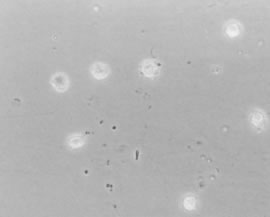
| Fig. 8. Human hyalocytes. Phase-contrast microscopy of flat mount preparation of hyalocytes in the vitreous cortex from an 11-year-old girl. No stains or dyes were used in this preparation. These mononuclear cells are round and distributed in a single layer within the vitreous cortex, with pseudopodia in some cells. (Courtesy of New England Eye Bank, Boston, MA.) |