|
|
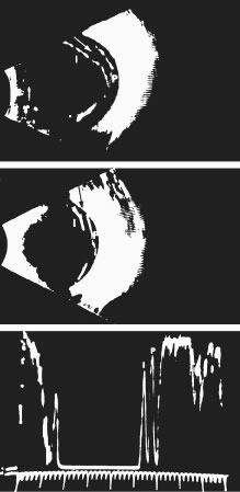
| Fig. 11. Vitreoschisis. B-scan ultrasonography of vitreoschisis demonstrates splitting of the vitreous cortex (arrow) that can mimic posterior vitreous detachment. In diabetic patients, blood can be present in the vitreoschisis cavity.176 (I, inner wall; P, posterior wall of vitreoschisis cavity within the posterior vitreous cortex) (Photograph courtesy of Dr. Ronald Green. From Green RL, Byrne SF: Diagnostic ophthalmic ultrasound. In Ryan SJ (ed). Retina. St Louis: CV Mosby, 1989.) |