|
|
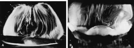
| Fig. 4. A. Vitreous base morphology. Vitreous structure in a 58-year-old woman. Fibers course anteroposteriorly in the central and peripheral vitreous. Posteriorly, fibers orient to the premacular region. Anteriorly, the fibers “splay out” to insert into the vitreous base. B. Fibers of the peripheral anterior vitreous forming the anterior loop. This configuration can provide the scaffold for cell migration and proliferation in the pathophysiology of anterior proliferative vitreoretinopathy. (A from Sebag J, Balazs EA: Pathogenesis of C.M.E.: Anatomic consideration of vitreo-retinal adhesions. Surv Ophthalmol 28[Suppl]:493, 1984. B from Sebag J: The Vitreous: Structure, Function and Pathobiology. New York, Springer-Verlag, 1989. Specimen in A courtesy of the New England Eye Bank, Boston, MA.) |