|
|
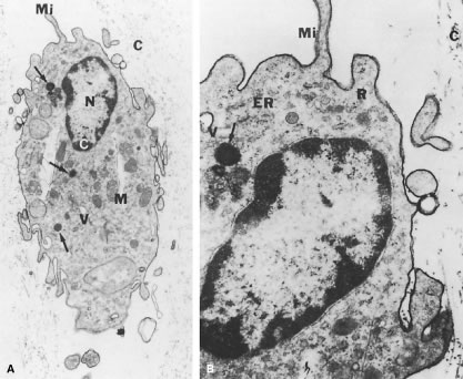
| Fig. 9. Ultrastructure of human hyalocyte. A. A mononuclear cell is seen embedded within the dense collagen fibril (C) network of the vitreous cortex. There is a lobulated nucleus (N) with dense marginal chromatin (white C). In the cytoplasm there are mitochondria (M), dense granules (arrows), vacuoles (V), and microvilli (Mi). (× 11,670). B. Higher magnification view (× 33,000) demonstrates dense granules (arrow), rough endoplasmic reticulum (ER), vacuoles (V), ribosomes (R), surface microvilli (Mi), and adjacent conical collagen fibrils (C). (Photographs courtesy of JL Craft and DM Albert, Cogan Laboratory of Ophthalmic Pathology, Harvard Medical School, Boston.) |