|
|
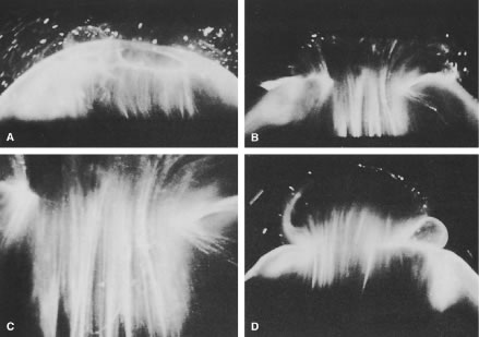
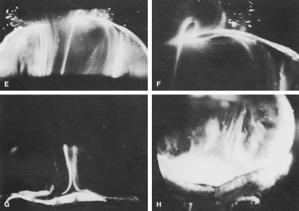
| Fig. 3. Human vitreous morphology. Human vitreous structure visualized by dark-field slit illumination. All photographs are oriented with the anterior segment below and the posterior pole above. A. Posterior vitreous in the left eye of a 52-year-old man. The vitreous body is enclosed by the vitreous cortex. There is a hole in the prepapillary (small, to the left) vitreous cortex. Vitreous fibers are oriented toward the premacular region. B. Posterior vitreous in a 57-year-old man. A large bundle of prominent fibers is seen coursing anteroposteriorly and entering the retrocortical space by way of the premacular vitreous cortex. C. Same photograph as B, at higher magnification. D. Posterior vitreous in the right eye of a 53-year-old woman. There is posterior extrusion of vitreous out the prepapillary hole (to the right) and premacular (large extrusion to the left) vitreous cortex. Fibers course anteroposteriorly out into the retrocortical space. E. Horizontal optical section of the same specimen as D, at a different level. A large fiber courses posteriorly from the central vitreous and inserts into the premacular vitreous cortex. F. Same view as E, at higher magnification. The large fiber has a curvilinear appearance because of traction by the vitreous extruding into the retrocortical space (see D). However, because of its attachment to the posterior vitreous cortex, the fiber arcs back to its point of insertion. G. Anterior and central vitreous in a 33-year-old woman. Cloquet's canal is seen forming the retrolental space of Berger. H. Anterior and peripheral vitreous in a 57-year-old man. The specimen is tilted forward to enable visualization of the posterior aspect of the lens and the peripheral anterior vitreous. To the right of the lens there are fibers coursing anteroposteriorly that insert into the vitreous base. These fibers “splay out” to insert anterior and posterior to the ora serrata. (A, E, and F from Sebag J, Balazs EA: Pathogenesis of C.M.E.: Anatomic consideration of vitreo-retinal adhesions. Surv Ophthalmol 28[Suppl]:493, 1984. B, C from Sebag J, Balazs EA: Morphology and ultrastructure of human vitreous fibers. Invest Ophthalmol Vis Sci 30:187, 1989. D, G, and H from Sebag J: The Vitreous: Structure, Function and Pathobiology. New York, Springer-Verlag, 1989. Specimens were courtesy of the New York Bank for Sight and Restoration, New York, NY.) |