Chapter 14
Arterial Obstructive Disease of the Eye
GEORGE E. SANBORN and LARRY E. MAGARGAL
Main Menu   Table Of Contents

Search

ACUTE ARTERIAL OBSTRUCTIVE DISEASE
CHRONIC ARTERIAL OBSTRUCTIVE DISEASE
REFERENCES

Arterial obstructive disease of the eye is frequently encountered in clinical practice. It usually occurs suddenly and presents dramatically but there is also a chronic form of presentation that is less frequently seen. In this chapter, we first discuss the acute presentations of arterial obstructive disease and then the chronic forms; usually, the chronic forms are due to some manifestation of carotid artery disease.
Back to Top
ACUTE ARTERIAL OBSTRUCTIVE DISEASE

CENTRAL RETINAL ARTERY OBSTRUCTION

Central retinal artery obstruction is one of the most sudden and dramatic events seen by ophthalmologists and was described as early as 1859.1 Although there have been numerous clinical and experimental studies of the pathophysiology of central retinal artery obstruction and although these studies have been augmented by the introduction of intravenous fluorescein angiography in the 1960s, the disease still has a relatively poor visual prognosis.

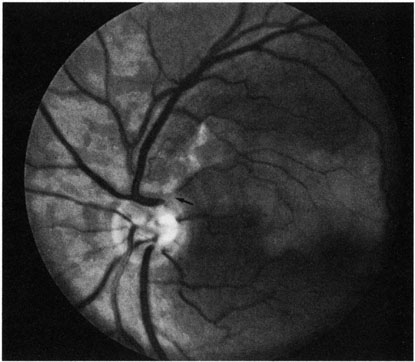
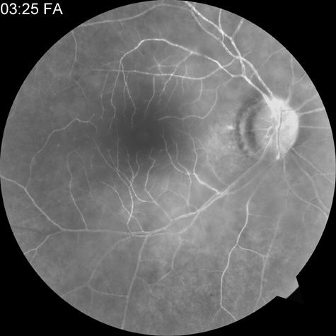
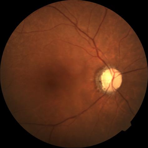
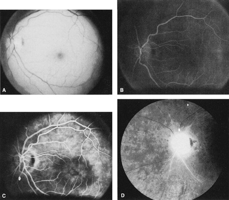
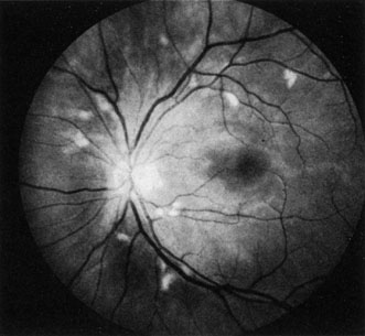
The clinical picture is typically striking. Patients present with a sudden painless loss of vision. The appearance of a cherry-red spot in the fundus is characteristic (Fig. 1).2 The cherry-red spot appears because soon after obstruction of the blood flow to the inner retina, the normally transparent retina becomes opaque and blocks the brownish-red color from the underlying choroid, which is still supplied by blood. Because the retina overlying the foveola is relatively thin, however, the normal color of the choroid is still visible in this area.3,4 Although characteristic, the cherry-red spot is not pathognomonic for central retina artery obstruction.5 Sometimes the characteristic cherry-red spot does not develop; there may be only a slight accentuation of the brownish-red color in the foveola.4 It is not known how long it takes this cherry-red spot to appear, but in a primate model, it has appeared as early as 30 minutes after obstruction.6 An afferent pupil defect is usually present.2

Fig. 1. A: Acute central retinal artery obstruction with a cherry-red spot. B and C: Intravenous fluorescein angiography. There is a delay of dye appearance in the central retinal artery, and when it does appear, it does not fill the arteries completely. D: Ocular coherence tomography (OCT) at the time of occlusion showing the increase in retinal thickness and reflectivity of the inner layers of the retina. E: Seven months later there is significant optic atrophy(G), and the OCT (F) shown now has marked thinning of the retina.

With obstruction, virtually all eyes have narrowing and irregularity in the arteries and there is frequently an irregularity in the caliber of the retinal veins.2,4,6,7 Segmentation (boxcarring) of the blood column frequently develops; this segmentation is particularly noticeable in the veins and can be seen as a to-and-fro movement of the blood elements.3,4 Retinal hemorrhages, however, are not characteristic of central retinal artery obstruction.7

With time, the retinal opacity diminishes, generally leaving an optic nerve that is atrophic (Fig. 1). Frequently, thinned retinal arteries and veins also remain.7,8 No foveolar light reflex is evident, and a finely pigmented appearance of the macula is typical.7 In some cases, arterial collaterals develop at the optic disc.9–11 Rarely, anatomoses that exist between the central retinal artery and the ciliary arteries become visible as preretinal loops (Nettleship collaterals) after an occlusion at the edge of the disc.12,13,378In approximately 20% of patients, an embolus is evident somewhere in the arterial system.14 Emboli are discussed later.

Intravenous fluorescein angiography is useful in showing the details of the abnormal circulation of a central retinal artery obstruction (Fig. 1). The principal abnormality is the delay in the appearance of the dye in the central retinal artery and its branches.2 Rather than the central retinal artery and its branches filling rapidly, considerable time may elapse before the entire arterial system is filled.15 The filling of the retinal arteries is often abnormal, with the fluorescein partially filling an artery (a dye front) or hugging the vessel wall, as in normal venous filling.15

Segmentation of the blood column is often well defined on fluorescein angiography.15 Venous filling is usually slowed and occasionally the dye does not progress beyond laminar flow during the study.14 In approximately 10% of eyes that have central retinal artery obstruction, there are abnormal choroidal filling defects, reflecting posterior circulation obstruction. These defects can occur even in patients who appear to have a typical central retinal artery obstruction.14 Leakage of dye from the vessel walls is not normally seen except at the site where an embolus lodges within a retinal artery.16 With time, the flow within the artery and its branches is reestablished and the appearance of the intravenous fluorescein angiogram may return to normal.

The electroretinogram (ERG) is usually abnormal in central retinal artery obstruction. The b-wave is reduced, indicating a marked abnormality in the circulation of the inner retina.17,18 The a-wave, indicating normal photoreceptor function, is preserved or accentuated. There does not appear to be a good correlation, however, between the extent of b-wave abnormality and the degree of retinal function,3 although Yotsukura and Adachi-Usami420 reported that the b-wave amplitude increased with visual improvement. The photopic negative response (PhNR) shows severe depression in this condition reflecting the significant loss of the ganglion cells and their axons.377 The electrooculogram may be either normal or abnormal. Visual field defects are usually profound but either occasionally a small portion of the temporal peripheral visual field remains or the patient has a large central scotoma.3 The onset of obstruction occurs most often between the hours of midnight and 6 AM, with the second most common period being between 6 AM and noon.2

Most patients with central retinal artery obstruction complain of a sudden loss of visual acuity in the affected eye; most of these patients present with visual acuity ranging from finger-counting to light perception.14 Only about 5% of patients have visual acuity of no light perception and most of these have involvement of the posterior ciliary circulation, which can be seen on either intravenous fluorescein angiography or electrophysiology.14 Without a partial central retinal artery obstruction or a spared cilioretinal artery (to be discussed), it is unlikely that any patient will have initial visual acuity better than finger-counting.14 With or without treatment, most eyes retain visual acuity of finger-counting to light perception. Patients with a spared cilioretinal artery do not seem to have a follow-up visual acuity that is significantly different from those without a spared cilioretinal artery.14

Central retinal artery obstruction is caused by the disruption or cessation of blood flow to the central retinal artery, its branches, and the retinal layers supplied by those vessels. The inner two thirds of the retina derives its blood supply from the central retinal artery and its branches; the outer third of the retina is supplied from the choroidal circulation.19–21 The central retinal artery and its branches function as an end artery, and with the exception of a few anastomotic connections between the retina and the ciliary circulation at the optic disc, there are no other anastomoses.19 No intrinsic retinal vessels are present in the fovea because the inner retinal layers are not present in this area.19

Histopathologically, when a central retinal artery obstruction occurs, the retina initially becomes edematous. With time, all the retinal layers supplied by the central retinal artery and the artery's branches, including the nerve fiber layer, ganglion cells, inner plexiform layers, and the inner portion of the inner nuclear layer, are lost.20 In trypsin digest preparations after obstruction of the central retinal artery, the greatest change is in the capillary bed,22 where extensive ischemic changes take place. Usually, little attempt is made to repair the retina with gliosis or neovascularization.20 The most common site of obstruction of the central retinal artery is at the level of the lamina cribrosa.20

Clinically, it is possible to examine the blood flow velocity in the central retinal artery in a noninvasive manner, using a variety of techniques.21 The most useful appears to be color Doppler imaging, providing simultaneous Doppler and B-scan ultrasound images of the optic nerve head, which primarily represents flow in the central retinal artery.23 Studies using this technique demonstrate a marked reduction of or no blood flow in the central retinal artery with an acute central retinal artery occlusion that returns to normal with time.24

Characteristics of the retinal blood flow can be measured clinically with retinal Doppler velocimetry.385 This is an instrument that can measure the blood column diameter, the centerline blood velocity, and the blood flow in major retinal vessels. However, when evaluating the blood flow in the optic nerve, choroidal, and retina, the Doppler laser flowmetry is more sensitive to the blood flow in the superficial layers than in the deeper layers.410

Masaoka and colleagues384 studied the hemodynamic changes in the retinal circulation with a scanning laser ophthalmoscope and intravenous fluorescein angiography. This technique allows the assessment of erythrocyte velocities and movement with high resolution continuous images and is a useful technique for the evaluation of hemodynamic changes in vascular occlusions.

Analysis of the retinal nerve fiber layer by one of the confocal laser scanning tomography techniques can provide information about a central retinal artery occlusion. Foroozan and co-workers383 studied patients with a central retinal artery occlusion within 1 week of occlusion with a scanning laser polarimeter. Diffuse attenuation of the retardation of the nerve fiber layer surrounding the optic nerve was seen in all eyes studied. Four patients were scanned in follow-up at intervals ranging from 1 week to 6 weeks; all had unchanged studies.383

Optic coherence tomography (OCT) of an acute central retinal artery occlusion shows a slight increase in the thickening of the retina and reflectivity of the inner layers of the retina that correspond to the opacity seen clinically (Fig. 1).409 With time, the disc becomes atrophic and the retina thinner.

Experimental studies of central retinal artery obstruction in the cat25 and the rhesus monkey26–28 have increased our understanding of arterial obstruction. The most clinically relevant finding is that there is a period in which a total lack of blood flow (ischemia) to the inner retina can be tolerated; that is, if the blood flow is not obstructed longer than this period, visual function may return to normal. This critical time in the cat eye is 90 minutes,25 and in the young, healthy rhesus monkey it is 100 minutes26–28—any longer than this and the histologic changes are irreversible and the animal has permanent visual loss. Hayreh and co-workers379,380 have repeated this experiment to determine how long the retina can survive central retinal artery occlusion in middle-aged and elderly rhesus monkeys, a situation that might be more applicable to the human situation where most occlusions occur in older patients. That study showed that an occlusion of less than approximately 100 minutes produced no permanent damage and that an occlusion of longer than 240 minutes produced massive and irreversible retinal damage. It is not known why the older monkeys had a longer retinal survival time with occlusion of the central retinal artery than younger monkeys.

Because there is no evidence that the monkey and the human have different retinas,379 the studies by Hayreh and co-workers have implications for studies that attempt to treat central retinal artery occlusion. Any treatment of this condition will almost certainly have to be initiated within 100 to 240 minutes, assuming the occlusion is total, or the patient will likely have permanent damage to the retina, regardless of treatment.

On average, central retinal artery obstruction occurs in patients who are in the fifth or sixth decades of life, with the age range varying from 17 to 84 years. Fewer than 10% of patients are younger than 30 years of age.14,29,30

In some cases, the cause of central retinal artery obstruction is relatively clear, such as when due to a visible embolus. In other cases, however, especially when found in association with a systemic disorder such as diabetes, the association is less clear. Table 1 lists some of the conditions associated with central retinal artery occlusion. Arteriosclerosis is probably the most commonly associated systemic condition. With the progressive buildup of subendothelial atheromatous material, the lumen of the central retinal artery becomes narrowed over time and eventually may become occluded.20 This association is inferred because most patients with central retinal artery obstruction are in the fifth to sixth decades of life, when atherosclerotic changes are expected.

TABLE 1. Conditions Associated With Retinal Arterial Obstructions


Systemic
  1. Conditions associated with embolus formation
    1. Arterial hypertension30,31
    2. Carotid artery disease
      1. Atherosclerosis30,32–34,254
      2. Fibromuscular dysplasia6,433
      3. Surgical obstruction30
      4. Moyamoya disease255
      5. Irradiation256
    3. Cardiac or valvular disease30,240
    4. Myocardial infarction (mural thrombus)257
    5. Tumors
      1. Cardiac myxoma258–261,431
      2. Metastatic tumors262,263,430
      3. Aortic valve papillary fibroelastoma432
    6. Postretinal detachment surgery
    7. Radiologic procedures
      1. Lymphangiography264
      2. Hysterosalpingography265
      3. Arteriography266,267
      4. Cardiac catheterization268,269
      5. Interventional radiology270–272,427
    8. Pancreatitis273,274
    9. Purtscher's retinopathy275,276
    10. Intravenous drug abuse29,277–280
    11. Scalp, nasal, facial, or orbital corticosteroid injection281–288
    12. Subacute bacteiral endocarditis31
    13. Heart surgery289
    14. Atrial fibrillation
    15. Cardiac arrhythmias
    16. Amniotic fluid426
    17. Systemic cholesterol microembolization syndrome290
  2. Coagulopathies
    1. Platelet and factor abnormalities29,31,45,291–298,425
    2. Pregnancy29,31,299,300
    3. Oral contraceptives29,31,301,302,441
    4. Sickle cell disease30,302–307
    5. Homocysteinuria308–310
    6. Use of nasal spray311
    7. Hyperglobulinemia in hair cell leukemia312
    8. Anticardiolipin antibodies55,313,314,428,429,446
    9. Lupus anticoagulant antibodies31,313
    10. Thrombocytopanic purpura
    11. Dysplasminogenemia315
    12. Ulcerative colitis316
    13. Homocystenemia46
    14. Antiphospholipid antibodies422–425
  3. Other
    1. Artherosclerosis317,413
    2. Arterial hypertension14,31
    3. Diabetes mellitus30,31
    4. Migraine28,31,318–323
    5. Fabry's disease324
    6. Loiasis325
    7. Sydenham's chorea326
    8. Hypotension327
    9. Systemic lupus erythematosus30,328–331,446
    10. Polyarteritis nodosa332,444
    11. Syphilis30
    12. Giant cell (temporal) arteritis30,333–336,415
    13. Herpes zoster337–339
    14. Sneddon's disease340
    15. Mediterranean spotted fever341
    16. Inflammatory bowel disease342
    17. Ventriculography343
    18. Wegener's granulomatosis440
    19. Lyme Disease344,345
    20. Acute myclogenous leukemia31
    21. Plasma cell dyscrasias346
    22. Intranasal cocaine and methamphetamines44,347
    23. Varicella348
    24. Radiation retinopathy349
    25. Chickenpox
    26. Chiropractic neck manipulation350
    27. T-cell lymphoma351
    28. Cat-scratch disease
    29. Monoclonal antibody therapy434
    30. Acquired immunodeficiency syndrome and central nervous system lymphoma439
    31. Internal carotid artery dissection438
    32. Internal carotid artery aneurym437
    33. Endophthalmitis435
    34. Wada test442
    35. Crohn's disease443
    36. Melanocytoma of the optic disc445
Ocular and Periocular
  1. Blunt trauma304,352
    1. Compression56,147,353–355
    2. Penetrating injury
    3. Retrobulbar injection303,356–359
    4. Orbital fracture repair360
    5. Belpharotplasty361
    6. Common carotid dissection362
  2. Increased intraocular pressure305
  3. Prepapillary arterial loops363–369
  4. Optic nerve drusen323,366,367
  5. Optic neuritis368
  6. Orbital mucomycosis369,370
  7. Toxocara canis infection371
  8. Toxoplasmosis retinochoroditis372
  9. Papilledema373,374
  10. Carotid artery disease31,375,376

The next most common systemic condition associated with central retinal artery obstruction is arterial hypertension; it can be found in as many as two thirds of all patients with central retinal artery obstruction.14,30,31 Carotid artery disease has been found in about 45% of patients who have central retinal artery obstruction,30 and hemodynamically significant stenosis occurs in approximately 20%.32 O'Farrell and Fitzgerald33 found ipsilateral carotid artery disease in 88% of patients presenting with retinal ischemia but neither study defined retinal ischemia nor indicated the ages or method of selection for patients studied. No difference was demonstrated between stenosis in the ipsilateral and the contralateral carotid artery33; this suggests that the cause of retinal artery occlusions may not be carotid artery stenosis—it may be simply associated with these occlusions or whatever process causes the carotid artery disease. Merchut and co-workers34 found no relation between the types of arterial occlusion—branch or central—and the type of underlying carotid artery disease. Wijman and colleagues35 found a high-grade internal carotid stenosis of 70% or more in 73% of patients with retinal ischemia that included transient ischemic attacks (TIAs) and arterial occlusions, both acute and chronic.

Diabetes mellitus14,28 and valvular heart disease28 are probably the next two most common associated conditions.

A variety of hematology parameters have been associated with central retinal artery occlusive disease.291–293 Walsh and co-workers291 found significant elevation of platelet coagulant activities (associated with the early stages of intrinsic coagulation) were elevated in patients with a central or branch retinal artery occlusion compared to controls. Kiesewetter and co-workers292 studied blood parameters in six patients with a central retinal artery occlusion before treatment was instituted. They found that compared to controls, the plasma viscosity, the extent of erythrocyte aggregation, and the plasma fibrinogen was significantly increased, while the erythrocyte deformation was reduced.

Sagripanti and associates389 found elevation of the prothrombin fragment (F1+2), the formation of thrombin-antithrombin III complex, and D-dimer (a stable degradation product of cross-linked fibrin) in acute central and branch retinal artery occlusions compared to controls. On follow-up examinations, the elevated F1+2 and the thrombin-antithrombin III complex significantly decreased while the D-dimer did not. The investigators conclude that increased thrombin generation may have a role in artery occlusions. However, the patients studied were treated with intravenous heparin and methylprednisolone, and the manuscript does not indicate whether their blood samples were taken prior to treatment or whether treatment could have affected the studies performed.

Greiner and co-workers388 studied 13 patients with a central retinal artery occlusion and 7 patients with a branch retinal vein occlusion for deficiencies in the protein C pathway. They concluded that the incidence of protein C deficiency in the artery occlusions they studied was not statistically significant from their control group.

Two research groups have shown a higher plasma level of homocyst(e)ine in patients with a central retinal artery occlusion, but not branch retinal artery occlusion.390,391 Wenzler and co-workers392 found hyperhomocyst(e)inemia in 2 patients: 1 patient with a central retinal artery occlusion in one eye and a branch retinal artery occlusion in the opposite eye, and 1 patient with bilateral branch retinal artery occlusions. However, the studies in the literature in regard to plasma homocyst(e)ine do not provide an answer as to whether elevated plasma homocyst(e)ine levels are an independent risk factor for retinal artery occlusive disease or are related to other vascular risk factors. Hyperhomocyst(e)inemia is an independent risk factor for atherosclerosis and atherothrombosis.416

The difference in the etiology of central retinal artery obstruction depends on the age of patients presenting with an obstruction. Central retinal artery obstruction in patients 30 years of age or younger tends to be associated with migraine, coagulant disorders, intraocular abnormalities, and trauma.29 Systemic diseases such as systemic arterial hypertension, atherosclerotic disease, and diabetes mellitus are not found in this younger age group.28,29 Some patients in the younger age group do not have a known cause for the obstruction; this was the case in as many as 12% of patients in one large series29 and in 40% of patients in a smaller series.30

Greven and associates31 studied 27 eyes in 21 patients with a retinal artery occlusion; all patients were younger than 40 years of age; emboli were identified in only 7 patients (33%) who had cardiac valvular disease, the most common cause in 4 patients (19%). The authors were able to identify various associated factors leading to a hypercoagulable state or embolic condition in 19 patients (91%).31 Despite their findings, the authors believe that the percentage of patients with ophthalmoscopically visible emboli is not indicative of the percentage of patients whose etiology for obstruction is embolic, and that most central retinal artery occlusions are embolic in origin.36

Two hundred fifty-six consecutive patients with acute unilateral retinal artery obstruction were studied using carotid Doppler ultrasonography; 18.8% had more than 60% stenosis of the internal carotid artery.37 Of those who had significant carotid stenosis, only 19 (40%) had visible retinal emboli. Thus, if ultrasound testing had been based only on visible retinal emboli, almost 60% of significant carotid artery stenotic lesions would have been missed.37 All patients at risk for extracranial carotid artery disease with acute retinal artery occlusion—not only those with visible emboli—should undergo duplex carotid Doppler ultrasonography.37

Evaluation of a patient with an arterial obstruction should be tailored somewhat to the age of the patient. A patient 20 years of age likely does not have extra-cranial carotid artery disease, whereas a 70-year-old patient clearly should be evaluated for such disease. Likewise, a 70-year-old patient with central retinal artery obstruction may have temporal arteritis, whereas this diagnosis in a 30-year-old patient is not likely.

There is probably little value, however, in determining the type of emboli if one is observed in the fundus. Sharma and co-workers38 found poor intraobserver and interobserver agreement on the quantitative assessment of retinal emboli when assessing them into groups of cholesterol, calcific, or both. Sharma and co-workers386 performed a retrospective multicenter chart review of 104 patients with both embolic and nonembolic acute retinal artery occlusion on whom transthoracic echocardiography was performed to determine the accuracy of visible retina emboli as a diagnostic test for anticoagulation or cardiac surgery. In this study, 41 patients had visible retinal emboli. The presence of a visible embolus in this review had no effect on whether a patient received anticoagulation or cardiac surgery, based on transthoracic echocardiography.

In some patients, the source of emboli from the heart can be determined with transthoracic echocardiography.39,40,412 When transthoracic echocardiography does not demonstrate the source of emboli, transesophageal echocardiography may be of more diagnostic value.41–43,381,412 Kramer and co-workers381,382 studied 18 patients with a retinal artery occlusion with transthoracic and transesophageal echocardiography. Seven of these patients had a central retinal artery occlusion. Sixty-one percent of the patients studied had at least one cardiac or aortic source of emboli detected by transesophageal echocardiography that was missed by transthoracic echocardiography. The authors recommend performing transthoracic echocardiography first, and then if the findings suggest cardiac or aortic conditions, performing the study via the esophagus: these findings include mitral valve disease, other valvular calcifications, enlargement of the left atrium, and severe left ventricular dysfunction.386

For all patients with a central retinal artery occlusion, the authors recommend an evaluation by a primary care physician or internist, to rule out systemic disease, and suggest as part of the evaluation, a Doppler study of the extracranial carotid arteries as well as a transthoracic echocardiogram. For patients with a visible embolus who have a negative carotid Doppler and normal transthoracic echocardiogram the authors generally suggest a transesophageal echocardiogram (depending on the patient's age as this is an invasive procedure). In older patients (however that is defined) with a central retinal artery occlusion we obtain a Westergren sedimentation rate and C-reactive protein to help eliminate the possibility that a patient may have temporal arteritis. Because elevated homocyst(e)ine has been associated with this disease and is an independent risk factor for atherosclerosis,416 the authors recommend testing the plasma homocyst(e)ine. Bilateral simultaneous central retinal artery occlusions413 are extremely rare and should prompt a careful search for a cause.

Thew authors' approach is more aggressive with younger patients. If a careful physical examination and search for emboli (including transesophageal echocardiography) are unrevealing and no obvious systemic cause exists for the obstruction, it may be useful to screen for hemotologic abnormalities; the most common finding is protein S deficiency.44,45 Whether this abnormality is responsible for or simply associated with retinal occlusions is unknown.

Treatment of either central retinal artery obstruction or branch retinal artery obstruction is unsatisfactory. No method has been proved to be more effective than the natural course of the disease.393,394 Although anecdotal cases abound of dramatic improvement in vision after some therapeutic intervention, no evidence is available that such intervention changes the natural course of the disease. The authors have seen one patient who presented on two different occasions with typical central retinal artery obstruction and severely decreased vision, who regained normal vision with no therapeutic intervention. Other clinicians have seen similar spontaneous recovery.399

The theoretical goal of treatment is to restore the retinal blood flow as soon as possible. Lowering intraocular pressure is one of the principal methods of treatment because the pressure load may perfuse the retina or dislodge an embolus.6,47 Paracentesis may be the fastest way to accomplish this.48,49 There is no documented evidence, however, that it is possible to improve arterial circulation with paracentesis or that paracentesis dislodges emboli in humans. Experimental studies in animals failed to show that paracentesis results in a significant increase in retinal perfusion.50–52 At least one case has been reported of endophthalmitis after paracentesis for central retinal artery obstruction.53 Reduction of intraocular pressure can also be accomplished with intravenous acetazolamide47 or topical glaucoma medications. Some investigators advocate continuation of acetazolamide for up to 2 weeks.54

Ocular massage also appears to be a useful technique for treatment of central retinal artery obstruction.395 Raising intraocular pressure for 30 seconds by pressure on the eye can result in both dilation of retinal arteries55–57 and increased blood flow volume.47,50 The change in arterial diameter with decreased perfusion (retinal arterial reactivity) decreases with the age of the patient.58 Digital massage also acts to lower intraocular pressure; it is easy to perform and can be done by the patient while awaiting other treatment.

A variety of gases have been suggested for use in treatment of central retinal artery obstruction, including oxygen (either delivered at standard atmospheric pressure or hyperbaric oxygen)59 and a mixture of 95% oxygen and 5% carbon dioxide (carbogen).54 Theoretically, it may be possible to deliver enough oxygen to perfuse the outer retina of a patient with central retinal artery obstruction by delivering 100% oxygen at atmospheric pressure. It has been found, however, that administration of 100% oxygen leads to vasoconstriction and decreased blood flow.60 The rationale for the addition of carbon dioxide to oxygen is that carbon dioxide leads to vasodilation,61 and the combination may allow delivery of the 100% oxygen to the retina without causing vasoconstriction.

Experimentally, the results of oxygenization to the retina have been variable and in some cases contradicting. Flower and Patz62 were not able to restore retinal function by delivering 100% oxygen at 1 atm of pressure to either the cat or monkey retina after retinal artery obstruction. Landers,63 however, was able to show in similar experiments that ventilation with 100% oxygen at 1 atm was capable of restoring inner retinal function, even though retinal circulation was occluded. Berkowitz64 showed in the adult and newborn rat that breathing carbogen oxygenates the inner retina better than 100% oxygen does. Birol and co-workers405 found that hyperoxia was preferable to room air in the cat model of arterial occlusion for preserving the b-wave amplitude of the electroretinogram. Deutsch and associates65 demonstrated in young, healthy human volunteers that there is no significant difference between vasoconstriction of retinal arteries with either 100% oxygen or a mixture of 95% oxygen and 5% carbon dioxide.

Experiments in healthy subjects have shown that carbogen breathing increases the retinal blood flow when studied with the blue field entopic phenomena66 and the scanning laser ophthalmoscope,68 but the retinal blood flow is not increased when tested with laser Doppler velocimetry. Schmetterer and colleagues studied the effect of different mixtures of O2 and CO2 on ocular pulsations in healthy volunteers using a laser interferometer coupled to a fundus camera.417 They concluded that the addition of 5% CO2 to oxygen is appropriate to maintain pulsatile ocular blood flow in the macula and optic disc at a level comparable to breathing air.417

Harino and co-workers421 demonstrated in young healthy volunteers that rebreathing into a bag for two minutes significantly increased the leucocyte velocity in the macular capillaries.

There is no evidence that any of these treatments are efficacious in restoring vision in patients with any type of arterial obstruction. Atebara and co-workers69 examined the records of 89 consecutive Wills Eye Hospital patients who had acute central retinal artery obstruction and received both anterior chamber paracentesis and carbogen therapy, which were compared with the records of those patients who had not been treated. No statistically significant difference was demonstrated between treated and untreated groups after adjusting for initial visual acuity.69

Although it is theoretically possible for choroidal circulation to supply adequate oxygen to the inner retina with the use of hyperbaric oxygen, the quantity of oxygen required appears to be toxic to the retina in a relatively short time.70 This may be true even when 100% oxygen under standard atmospheric pressure is delivered for prolonged periods of time.71 Two separate groups, however, have shown experimentally in animals that it is possible to supply oxygen to the inner retina after vascular obstruction by means of intravitreal perfusion.72,73

A variety of drugs to induce vasodilation have been injected into the retrobulbar space in patients with central retinal artery obstruction47 but no experimental or clinical evidence is available to suggest that any of the agents delivered by retrobulbar injection are of value in restoring blood flow. In addition, it is theoretically possible that such an injection could produce a retrobulbar hemorrhage, which would further decrease retinal blood flow.47

A few patients have been treated with a technique involving surgical cannulation of the supraorbital artery and perfusion of this artery with heparin, papaverine, streptokinase, or a combination of these agents.74,75 Watson74 reported complete restoration of vision in the eyes of 4 patients in a series of 14 with central retinal artery obstruction; he also reported 2 patients who had considerable improvement of visual acuity and visual fields. No control group of patients was used in this study, however. Schmidt and colleagues76,77 treated 46 patients with either urokinase or recombinant tissue plasminogen activator (tPA) by inserting a microcatheter into the proximal segment of the ophthalmic artery. They were able to show that visual acuity improved with this treatment in patients who have “slight edema of the retina,” no progression, well-perfused perimacular arterioles, and who were treated within 14 hours of occlusion.77 It is notable, however, that there was no standardized protocol or control group, and intravenous fluorescein angiography was performed in only about three-fourths of the patients.77

Richard and co-workers treated 46 patients with a central retinal artery occlusion and seven patients with branch retinal artery occlusion treated with rTPA infused into the ophthalmic artery via a transfemoral incision and reported that vision improved in 66% of the patients.396 However, this was an uncontrolled study that did not include pre and posttreatment fluorescein angiography to either confirm the diagnosis or to evaluate the effect of treatment on retinal circulation,397–399 and the final results of treatment may not differ from the natural history of this disease.397

Seventeen patients were treated by Weber and associates78 with intraarterial urokinase placed into the proximal segment of the ophthalmic artery; these investigators believed that complete or partial improvement of visual acuity was achieved in approximately one-third. This was not a clinical trial, however, and some patients also received paracentesis or carbogen therapy.

Rumelt and co-workers400 reported the treatment of 11 patients with a central retinal artery occlusion using an arbitrary, nonsystematic regimen of ocular massage, sublingual isosorbide dinitrate, intravenous acetazolamide, intravenous mannitol, oral glycerol, anterior chamber paracentesis, intravenous methylprednisolone and streptokinase, and retrobulbar tolazoline; although not all patients received all treatments. Improvement of blood flow was determined at the end of treatment by three-mirror contact lens biomicroscopy. The authors report that 73% had improved visual acuity after these treatments.

Kattah and co-workers401 treated 12 patients with a central retinal artery or ophthalmic artery occlusion with a stroke protocol involving the intravenous injection of rTPA and reported 10 patients had improved visual acuities after treatment. Some but not all patients were treated before injection of rTPA with a paracentesis of the anterior chamber, and four of the patients treated developed neovascular glaucoma within 3 to 4 weeks of the arterial occlusion. No results of vascular perfusion pretreatment or posttreatment with intravenous fluorescein angiography were reported in this study and there was no control group.

It has been demonstrated in rats that tPA is effective when given intravenously for lysing experimental retinal arterial thrombi.79 The use of tPA in acute obstruction has probably been increased by its proved value when given intravenously as a treatment for acute ischemic stroke.13 In the case of acute ischemic stroke, tPA is administered within 3 hours of onset; the benefits of such treatment are observed 3 months after the stroke. The systemic administration of tPA is not innocuous, and whether this risk can be justified in a patient who has another normal eye has not been demonstrated.

Beatty and Au Eong418 performed a meta-analysis of the published reports of local intra-arterial fibrinolysis to treat central retinal artery occlusion. They studied 16 reports, all retrospective and nonrandomized, and concluded that there may be a marginal visual acuity benefit of local intraarterial fibrinolysis compared to conventional management, but there is currently insufficient evidence to justify the use of this intervention outside of a randomized controlled clinical trial.418

Zheng and co-workers80 treated 245 cases of retinal artery obstruction—including 168 cases of central retinal artery obstruction—by daily acupuncture treatments delivered over several 10-day courses. They reported marked visual improvement in 25% of patients and good visual improvement in 30%. They compared these results to a retrospective group of untreated patients from their clinic but the study was not performed as a controlled clinical trial or in a prospective fashion.80

Hausmann and Richard81 treated 4 patients with high-dose bolus intravenous steroids and observed that intravenous fluorescein angiography demonstrated improved circulation. These patients were also treated patients with plasma expanders, heparin, and nicoumalone, however, and no visual acuities were reported.

It is technically possible to cannulate the central retinal artery or its branches402,404 or even remove emboli from retinal arteries.111,403 Whether this will result in improvement in vision after this surgery is unknown. An animal model of transient retinal ischemia has been developed in the rat retina407 that may allow experiments with thrombolytic agents to be conducted prior to human testing.

There are no randomized, prospective clinical trials of large enough size to determine whether any of the forms of treatment of central retinal artery obstruction are better than no treatment.393 Augsburger and Magargal54 reported the visual outcome in 34 consecutive cases of treated acute central retinal artery obstruction. The treatment in their series included paracentesis, controlled ocular massage, inhalation of 95% oxygen and 5% carbon dioxide, and the administration of oral acetazolamide and aspirin. They reported that 35% of the patients had final visual acuity equal to or greater than 20/100. No control group was available in their series, however. Hayreh and Podhajsky,82 in a detailed study of 64 eyes with central retinal artery obstruction, state that no treatment is effective for central retinal artery obstruction, although they provide no details to support this statement. Some patients recover good visual acuity after central retinal artery obstruction with no treatment,83 whereas others may reestablish normal blood flow and visual acuity several days after treatment.84 The study by Atebara and co-workers,69 which did not find anterior chamber paracentesis or carbogen therapy effective for treatment of arterial occlusive disease, has been mentioned.

Two experimental studies to protect the retina from vascular obstruction after ischemia should be mentioned. Faberowski and associates85 showed that an ice pack applied to the rat eye protected it against ischemic injury after ligation of the major vessels and the optic nerve. Yoon and Marmor86 showed in the rabbit that intravenous dextromethorphan protected against retinal ischemic damage from vascular obstruction when the obstruction was produced by elevation of intraocular pressure.

Neovascularization is probably the most serious complication that can develop after central retinal artery obstruction. Duker and Brown87,88 studied 168 cases of central retinal artery obstruction and found neovascularization of the disc developed in 1.8% of eyes; neovascularization of the iris occurred in 16.6% of eyes. Hayreh and Podhajsky82 reported neovascular glaucoma in 16% of 64 eyes with central retinal artery obstruction. These authors believe that in most patients, central retinal artery obstruction is actually caused by neovascular glaucoma secondary to severe carotid artery disease.82 There is, however, at least one case reported in which neovascular glaucoma resulted from central retinal artery obstruction in a patient who did not have carotid artery obstructive disease.89

CENTRAL RETINAL ARTERY OBSTRUCTION WITH CILIORETINAL ARTERY SPARING

A cilioretinal artery90 is an anatomic variant that is found in approximately 30% of eyes.91 In patients with central retinal artery obstruction, approximately 25% have some degree of macular sparing as a result of a patent cilioretinal artery.92 When the cilioretinal artery makes a major contribution to perfusion of the macula, these patients may have some degree of visual preservation. Brown and Magargal14 reported on 73 eyes with acute central retinal artery obstruction in which 48 eyes had no cilioretinal sparing. None of these affected patients presented with visual acuity better than finger-counting. Of the 25 eyes with some degree of cilioretinal sparing, however, 16% had an acuity of 20/200 or better. Brown and Shields92 analyzed 107 cases of central retinal artery obstruction, of which 26% showed some degree of macular sparing because of a patent central retinal artery. They believed that with rare exceptions, patients with central retinal artery obstruction but no cilioretinal sparing had poor vision and that patients with a patent cilioretinal artery did relatively well—with or without treatment in terms of visual acuity—but were usually left with a severe peripheral visual field defect.

BRANCH RETINAL ARTERY OBSTRUCTION

In one large series of retinal artery obstructions, 57% of obstructions involved the central retinal artery, 5% involved the cilioretinal artery and the remaining 38% involved one of the branch retinal arteries.92 Branch retinal artery obstructions usually occur at bifurcations.19,93

When a branch retinal artery is obstructed, there is usually whitening of the retina in the area supplied by the artery (Fig. 2). In most eyes (62% in one series), emboli are responsible for the obstruction.94 The temporal branch arteries are involved in almost all cases that have been reported,92,94 but whether this is because nasal branch retinal artery obstructions are rare or because such obstructions are simply asymptomatic is unknown. The characteristics of a branch retinal artery obstruction on intravenous fluorescein angiography are similar to those for central retinal artery obstruction.

Fig. 2. Multiple branch retinal artery obstructions in a young woman with encephalopathy and hearing loss. (Photograph courtesy of Jack W. Pierce, M.D.)

The patient describes either a sudden loss of vision if the branch retinal artery involves the macula or a visual field defect or both. Initial visual acuities can range from 20/15 to hand movements, but in one series, more than three-fourths of all patients had an initial visual acuity of 20/40 or better.94 Most patients who are seen with have branch retinal artery obstruction have unilateral obstructions, but about 8% later develop branch retinal artery obstruction in the opposite eye.94

Occasionally, patients present with a bilateral branch retinal artery obstruction.94 Some have obstructive retinal arteries without evidence of systemic abnormalities,95,96 whereas others have accompanying symptoms such as deafness.97,98 Some younger patients—usually women—have been reported to have an idiopathic syndrome of branch retinal artery obstruction, encephalopathy, and hearing loss (Susac syndrome).99–104,406 The eyes on these patients are characterized by significant systemic thromboembolic events; recurrent bilateral retinal arterial occlusions; segmental arteriolar staining by fluorescein angiography, occasionally associated with periarteriolar retinal whitening or mild vitreous cells; and a predilection for vestibuloauditory or transient sensorimotor symptoms.104 Some patients, however, present with only recurrent bilateral retinal arteriolar occlusions and never develop the associated vestibuloauditory or sensorimotor systems; whether these patients have a partial manifestation of Susac syndrome is unknown. The etiology of branch retinal artery obstruction in most cases is similar to that of central retinal artery obstruction (see Table 1). Retinal diseases (such as toxoplasmosis) can also cause branch retinal artery obstruction.

Hasegawa and co-workers411 have reported on the multifocal electroretinogram in patients with a branch retinal vein occlusion. They found that both the first and second order multifocal electroretinogram amplitudes decreased in the effected area of the retina compared with the normal retina, but the second order multifocal electroretinogram was more affected by a branch retinal artery occlusion, suggesting that the second order multifocal electroretinogram is a more sensitive indicator of inner retinal dysfunction.411

The complications of branch retinal artery obstruction can include retinal neovascularization, although this is rare.105 When neovascularization develops, it may be related to decreased blood flow to the eye, such as that due to carotid artery disease106 or diabetes mellitus.107 Neovascularization has been reported as a sequel to obstructive retinal arteriolitis.95 Vitreous hemorrhage can occur after neovascularization.87,107 Occasionally, retinal breaks occur after branch retinal artery obstruction.108

Dutton and Craig109 treated a retinal embolus causing branch retinal artery occlusion with the argon laser after other therapy had failed; they were able to make the embolus break up and disappear but without improvement in visual function.109 Ciulla and colleagues,110 however, were able to only disrupt one retinal emboli out of four in a rabbit model of artery occlusion using human atherosclerotic material treated with the argon laser. They also failed to dislodge emboli in a human with branch retinal artery obstruction, and no clinical improvement resulted.110 Peyman and Gremillion111 removed a branch retinal artery embolus in a patient with branch retinal artery occlusion using a 30-gauge needle and a pars plana vitrectomy; the patient regained vision to 20/200.

Most clinicians do not treat branch retinal artery obstruction, and there are no published clinical trials that suggest any treatment is better in terms of a final visual result than no treatment at all. An investigation into associated systemic abnormalities is important, and treatment of any systemic abnormalities that may be found is necessary in most cases. In those patients who present with retinal neovascularization, the treatment of choice is scatter photocoagulation delivered to the area of ischemic retina, with delivery similar to that used for neovascularization secondary to branch retinal vein obstruction.106,112

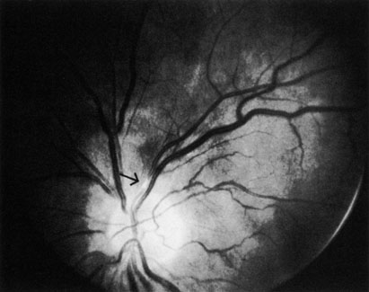
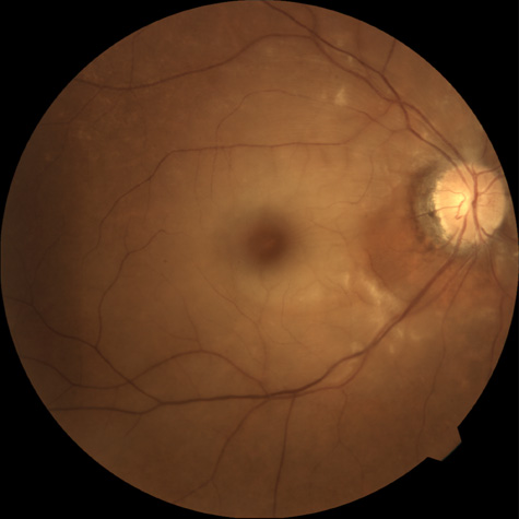
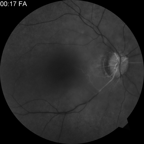
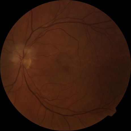
CILIORETINAL ARTERY OBSTRUCTION

Cilioretinal arteries can be selectively obstructed (Fig. 3). More commonly, however, they occur in association with central retinal vein obstruction.113 When they supply a significant portion of the macula, decreased visual acuity is common. Prognosis for visual outcome in such patients is good. Brown and co-workers114 studied 10 eyes having an isolated cilioretinal artery obstruction. Of those, 60% had an initial visual acuity of 20/40 or better and on follow-up examination, all eyes but one had a best-corrected visual acuity of 20/40 or better.114

Fig. 3. A: Cilioretinal artery obstruction showing opacity of the retina in the area of obstruction. B: The cilioretinal artery has begun to fill after the branches of the central retinal artery but is still not completely filled at this point in the angiogram.

OBSTRUCTION OF THE OPHTHALMIC ARTERY, POSTERIOR CILIARY ARTERIES, OR ARTERIES SUPPLYING THE CHORIOCAPILLARIS

Symptoms that involve obstruction of the ophthalmic artery, posterior ciliary arteries, or arteries supplying the choriocapillaris depend on which artery is affected. In some patients, the location of the obstruction must be inferred because it is not always possible to visualize it, even with intravenous fluorescein angiography. The ophthalmic artery is a branch of the carotid artery that branches into the central retinal artery and the posterior ciliary arteries.115 There are usually two main ciliary arteries; each one supplies about half of the choroid. The medial posterior ciliary artery supplies the nasal half, and the lateral posterior ciliary artery supplies the temporal half, with a watershed zone in between. The branches of the medial and lateral posterior ciliary arteries (and occasionally a superior ciliary artery) become the long or short posterior ciliary artery.116,117 Each lobule of the choriocapillaris supplied by the terminal choroidal arteriole forms one of the branches of the posterior ciliary artery, and each lobule is drained by a venule.116,118There is no anastomotic communication between lobules.

OPHTHALMIC ARTERY OBSTRUCTION

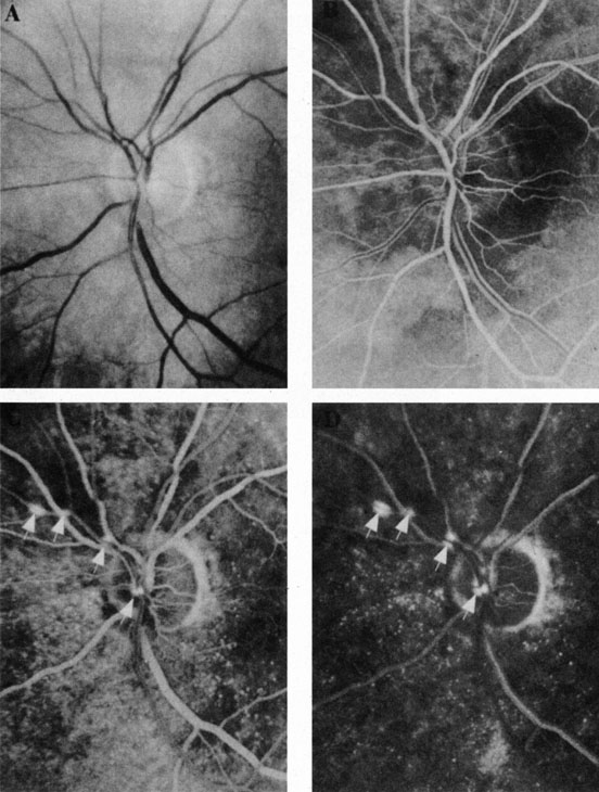
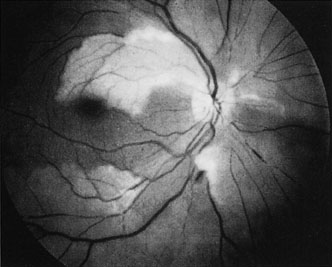
Ophthalmic artery obstruction is characterized by an initial opacification of the entire retina in a manner similar to that of central retinal artery obstruction (Fig. 4A). The cherry-red spot may or may not be present, however.119 The difference between an ophthalmic artery obstruction and central retinal artery obstruction is that in an ophthalmic artery obstruction, with time, optic atrophy develops, as does varying amounts of pigmentation due to the lack of perfusion of the retinal pigment epithelium (see Fig. 4D). This pigment is generally diffusely scattered throughout the posterior pole but it may also be seen in greater amounts in the periphery.

Fig. 4. A: Ophthalmic artery obstruction showing a cherry-red spot: the visual acuity was no light perception. B: At 26.2 seconds, the retinal vessels are filled, but no dye is seen in the choroidal circulation. C: By 37.7 seconds, the choroidal circulation is more visible, and a doughnut-shaped area of hypofluorescence is noted. D: Six months after the initial photographs, optic atrophy is present, as is atrophy of the retinal vessels. Diffuse pigment disturbance is evident.

Intravenous fluorescein angiography generally shows moderate to marked abnormalities in the filling of the choroid, in addition to a delay in filling or even nonfilling of retinal vessels (see Fig. 4B and 4C). Staining of the retinal pigment epithelium can also be seen; this staining either can occur locally in the macular area or it can be diffuse.119 The electroretinogram shows abnormalities of both a- and b-waves, reflecting ischemia to both the inner and outer retina. Almost all reported patients have had an initial visual acuity of no light perception; virtually no patients can be expected to have a final visual acuity better than that.119 An acuity of no light perception is a clue to the presence of an ophthalmic artery obstruction. Because relatively few patients with central retinal artery obstruction have an initial visual acuity of no light perception, no light perception usually suggests the likelihood of some obstruction of the choroidal circulation.14

The causes of ophthalmic artery obstruction vary. In the 10 eyes reported by Brown and co-workers,119 the cause in 2 patients was carotid artery disease. In 1 patient each, the following causes were found: embolization from an arterial myxoma, atrial fibrillation, external compression of the orbit and globe, trauma (a knife wound), orbital mucormycosis, and one of unknown cause.119 Because the ophthalmic artery makes an acute bend in the orbit, it is likely that emboli could lodge at that location.115,120 The site of this type of obstruction must be speculative, however, because there is no way to know that a simultaneous obstruction of the retinal and the choroidal circulation has not occurred distal to the ophthalmic artery.

Two of the cases reported by Brown and coworkers119 were bilateral. One occurred in a patient with bilateral mucormycosis and the other was in a patient experiencing atrial fibrillation with accompanying thyroid fever.119 Patients with a presumed ophthalmic artery obstruction require careful and complete systemic evaluation in the search for possible causes.

POSTERIOR CILIARY ARTERY OBSTRUCTION

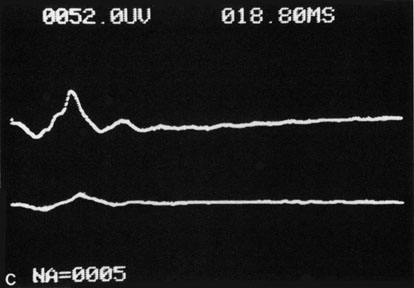
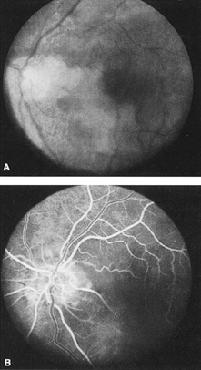
Obstruction of one of the posterior ciliary arteries may result in a whitening of the area of the retina supplied by the involved posterior ciliary artery and the subsequent appearance of patchy pigmentation in the same area. However, in patients with anterior ischemic optic neuropathy secondary to temporal arteritis the retina may appear normal (Fig. 5). In the case of either the medial posterior ciliary artery or the lateral posterior ciliary artery, the area involved is the size of approximately half of the choroid.121–123 In the case of a long posterior ciliary artery, the temporal side of the eye including the macula is involved.121

Fig. 5. A: Ischemic optic neuropathy in a patient with giant cell arteritis. B and C: Intravenous fluorescein angiography demonstrates delayed filling of the lateral posterior ciliary artery.

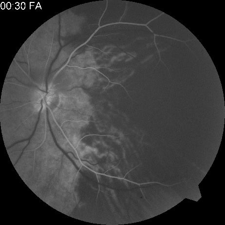
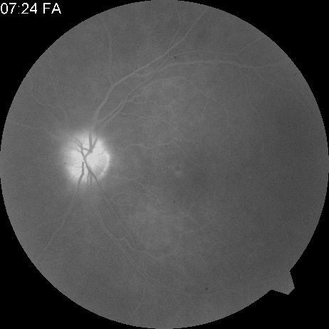
We have seen several patients with anterior ischemic optic neuropathy with suspected temporal arteritis where the Westergren sedimentation rate and C-reactive protein were normal and the intravenous fluorescein angiogram helped confirm the diagnosis (Fig. 5).

CHOROIDAL OBSTRUCTION

The appearance of a lesion involving the arterial supply to the choroid varies, depending on the size of the vessel occluded. Obstruction of a larger choroidal artery produces a whitening of the retina in a wedge shape, with the apex positioned toward the posterior pole.124–126 With time, pigmentation occurs in this area.

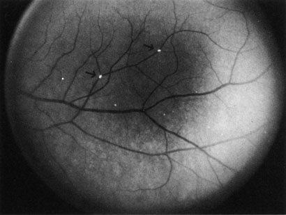
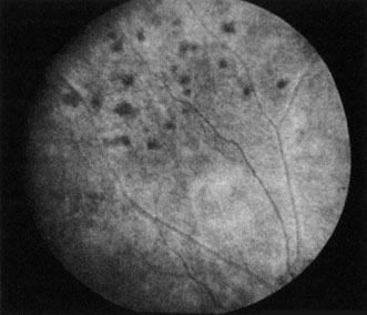
Obstruction of smaller terminal choroidal arterioles produces only small patchy areas of whitening, with overlying pigmentation (Elschnig spots, Fig. 6).127 The obstruction of smaller arterioles may occur in multiple sites in some cases.128

Fig. 6. Elschnig spots. (Courtesy of W.B. Benson, M.D.)

The syndrome of serous retinal detachments in various systemic diseases, such as arterial hypertension,129 eclampsia (toxemia of pregnancy),130 Goodpasture's syndrome,131 thrombotic thrombocytopenic purpura,132 periarteritis nodosa,133 and disseminated intravascular coagulation,134 are probably due to obstruction of the choroidal vessels affecting the pigment epithelium, with breakdown of the chorioretinal barrier but without frank infarction.135 Experimental work involving the injection of microspheres into the posterior ciliary arteries supports this concept.136 Some patients have resolution of the serous retinal detachment in addition to their systemic disease; depending on the severity of the ischemia produced, they may be left with decreased visual function and scarring, whereas others may be left with normal vision.135 Some investigators believe that acute multifocal posterior placoid pigment epitheliopathy (which Hayreh calls multifocal acute ischemic choroidopathy) and geographic (helicoid or serpiginous) choroidopathy are both manifestations of acute choroidal ischemia.135–137

A syndrome that appears to affect the choroidal and outer retinal circulation has been reported in a few patients after phacoemulsification138,139 and has been termed the outer retinal ischemic infarction syndrome.139,140 After uneventful phacoemulsification, such patients are observed to have loss of central and paracentral vision and diffuse and patchy whitening of the outer retinal layer in the posterior fundus. They also have an unusual polygonal pattern of fluorescein angiography and staining of the retinal pigment epithelium and outer retinal layers. They experience clearing of the retinal whitening that is later replaced by mottled pigmentation of the retinal pigment epithelium and partial recovery of the central visual field.139 The optic nerve does not become pale, and the vessel caliber is normal. The cause is postulated to be an elevation of the intraocular pressure that temporarily obstructs the blood flow to the choroid (and probably also to the retina). This obstruction then causes a temporary obstruction of the blood supply of the retinal pigment epithelium and outer retinal layers but results in sparing of both the macula and the periphery.139,140 This syndrome has been reported to occur also in patients undergoing pars plana vitrectomy with a scleral buckling procedure.141

OBSTRUCTION OF A RETINAL ARTERIOLE

Obstruction of a retinal arteriole produces cotton-wool spots, which have also been called white patches or soft exudates (Fig. 7). Cotton-wool spots are caused by focal hypoxia, which obstructs axoplasmic flow, resulting in accumulations of axoplasmic debris in the nerve fiber layers of the retina.142–144 Cotton-wool spots may be either single or multiple. On intravenous fluorescein angiography, they correspond to areas of capillary nonperfusion.6 The patients are often asymptomatic but may experience severe visual loss if the areas of arteriolar obstructions are extensive (Fig. 8).145

Fig. 7. Cotton-wool spots in a patient with acquired immunodeficiency syndrome.

Fig. 8. A: Extensive cotton-wool spots and (B) corresponding large areas of vascular nonperfusion in a patient with Purtscher's-like retinopathy associated with systemic lupus erythematosus. The condition was bilateral.

There are multiple causes for cotton-wool spots. Brown and co-workers146 studied 24 consecutive patients who presented with a cotton-wool spot; any patient with known diabetes mellitus was excluded. In this series, 23 of the 24 patients were found to have one or more associated systemic diseases. The disease entities associated with cotton-wool spots are listed in Table 2.

TABLE 2. Conditions Associated with Cotton-Wool Spots


Diabetes mellitus
Systemic arterial hypertension
Collagen vscular diseases
Systemic lupus erythematosus
Dermatomyositis
Polyarteritis nodosa
Scleroderma
Giant cell arteritis
Cardiac valvular disease
Mitral valve prolapse
Rheumatic heart disease
Endocarditis
Acquired immunodeficiency syndrome (AIDS)
Leukemia
Trauma
Radiation retinopathy
Central retinal artery obstruction
Retinal vein obstruction
Metastatic carcinoma
Leptospirosis
Rocky Mountain spotted fever
High altitude retinopathy
Severe anemia
Acute blood loss
Papilledema
Papillitis
Carotid artery obstruction
Dysproteinemias
Septicemia
Aortic arch syndrome (pulseless disease)
Intravenous drug abuse
Acute pancreatitis
Onchocerciasis

Adapted from Brown GC, Brown MM, Hiller T, et al: Cotton-wool spots. Retina 5:206, 1985.


The most common cause of cotton-wool spots is diabetes mellitus, and most of these patients are already aware of their disease. The second most likely cause is either acquired immunodeficiency syndrome or systemic arterial hypertension. Occasionally, a healthy patient has a cotton-wool spot and on systemic evaluation is found to be entirely normal. This was the case with one of Brown and co-workers146 patients, and we have encountered similar patients. However, we have also seen an occasional patient with no history of systemic disease who presented with a single cotton wool spot and subsequently was diagnosed with diabetes.

COMBINED CENTRAL RETINAL ARTERY AND CENTRAL RETINAL VEIN OBSTRUCTION

Occasionally, patients present who have an obstruction of both the central retinal artery and the central retinal vein.56 Such patients have fundus characteristics of both conditions. Generally, there is a whitening of the retina associated with a cherry-red spot and in addition, optic disc edema, retinal hemorrhages, and venous engorgement. On follow-up examination, these patients develop optic atrophy accompanied by extreme narrowing of the retinal vessels. Subsequent visual acuity tends to be poor, although rare patients have been reported to have a good return of visual function.147 In the largest series reported in the literature, one-third of patients developed neovascular glaucoma.56

Back to Top
CHRONIC ARTERIAL OBSTRUCTIVE DISEASE

CAROTID ARTERY DISEASE

The principal cause of chronic arterial obstruction is usually some kind of carotid artery disease. Atherosclerosis is the most common cause of carotid artery disease in the United States but there are other causes, such as moyamoya disease,148 fibromuscular dysplasia,149 and irradiation.150 Atherosclerosis is a disease affecting medium-sized arteries in humans. The primary symptoms of carotid artery disease are caused by the atheromatous or fibromuscular plaque.151,152 Atherosclerosis may result in narrowing (obstruction) or occlusion of the artery or in embolization. The bifurcation of the common carotid artery into the external and internal carotid artery is one of the sites at particular risk for atheromatous disease.153 The eye is frequently involved in atheromatous disease of the carotid artery bifurcation because the ophthalmic artery is one of the first branches of the internal carotid artery in the neck. Carotid artery disease can be manifest by a variety of ocular and non ocular symptoms. The ocular manifestations are important—not only because they occur in many patients with carotid insufficiency154 but also because they often are the first indication that carotid artery disease exists.

There are many ways that patients with carotid artery disease present to the ophthalmologist.8 Some present with a TIA. If the TIA involves the carotid system, the symptoms include hemiparesis, hemisensory loss, aphasia, and transient monocular blindness (amaurosis fugax).155 Another group of patients have symptoms that are purely ocular. These patients present with one of several symptoms, such as (i) partial or complete visual loss due to an artery obstruction, (ii) decreased visual acuity, or (iii) pain resulting from the ocular ischemic syndrome. Other patients are asymptomatic and any ocular findings consistent with carotid artery disease are incidental.

Amaurosis Fugax

The type of TIA known as amaurosis fugax is the most common symptom of carotid artery disease.154,156 Amaurosis fugax is characterized by a sudden, painless, monocular loss of vision that typically lasts between 2 and 10 minutes. The patient frequently describes his or her vision as being “dark” or “dim.” The return of vision can be sectorial or altitudinal and is occasionally described as a “curtain rising.” Generally, the vision returns to normal immediately after an attack. The frequency of these attacks varies from 1 or 2 attacks per month to 10 or 20 per day.

The retina—if observed during an amaurotic attack—may be normal155 or it may show obstruction (such as in central retinal artery obstruction). It may exhibit migratory white retinal emboli (within the retinal arterioles and in association with disruption of the arterial circulation)157 or it may show cholesterol emboli moving through the arterial system.155 It is important to recognize amaurosis fugax as a TIA because it is frequently caused by microembolization from an atheromatous ulcerative lesion in the ipsilateral extracranial carotid artery, at least in older patients.158,159 Approximately one-third of all patients with an untreated TIA can be expected to have a stroke; this rate is about four times greater than that of an age-matched population.160

It must be noted, however, that not all amaurosis fugax and other forms of TIA are caused by carotid artery disease. Other causes of amaurosis fugax include temporal arteritis,161 pseudotumor cerebri,161 migraine,161 structural cardiac defects,162 ophthalmic artery stenosis,163 ophthalmic artery aneurysms, hematology disorders,164,165 ocular hypertension,160 arterial hypotension, and vasospasm.165 A group—usually younger patients—may have no identifiable disease.161,162,167

Ocular Ischemia

There are several forms of chronic arterial obstructions that occur in carotid artery disease. These typically result from hypoperfusion of the retina caused by either stenosis or the complete obstruction of either the extracranial internal or the common carotid artery. One form of chronic arterial occlusion is a unilateral retinopathy that is ipsilateral to the carotid artery lesion and is characterized by cotton-wool patches, dilated arterioles and veins, an absence of hemorrhages, and normal vision. This retinopathy was first recognized as a sign of carotid artery disease by Hollenhorst154 in 1958, although such changes had been previously noted after ligation of the common and external carotid arteries in the treatment of traumatic carotid cavernous fistula.168 In a milder form of this retinopathy, the only abnormality may be larger retinal arterioles on the side of a ligated carotid artery in patients with hypertension169 or asymmetric hypertensive retinal vascular changes, in which the eye ipsilateral to the carotid lesion has the fewer changes.8

Another type of chronic ophthalmic artery obstruction produced by hypoperfusion caused by stenosis or occlusion of the ipsilateral extracranial carotid artery was first reported by Hedges170 and termed venous stasis retinopathy by Kearns and Hollenhorst.171 Venous stasis retinopathy is probably not a good term for this condition, however, because the basic mechanism involves reduction of the arterial inflow, not the venous outflow. Even more confusion has resulted from the use of the term to describe the features of the nonischemic form of central retinal vein obstruction.172 A better term is hypoperfusion or hypotensive retinopathy.172 Klijin and co-workers414 found this type of chronic ocular ischemia in approximately one-third of patients studied with symptomatic carotid artery occlusive disease.

Hypoperfusion retinopathy is characterized by dot and blot hemorrhages in the midperiphery of the fundus, by venous tortuosity and engorgement, by microaneurysms, and by the occasional sludging of blood within the veins. Fluorescein angiography may show areas of capillary nonperfusion; it may also show microaneurysms in the midperiphery and slow arm to retina or arteriovenous transit time (Fig. 9A and 9B).174 The condition does not usually affect the posterior pole, and patients typically have normal visual acuity, although an occasional patient may have macular edema.174 The entire periphery of the eye is usually affected, although there may be more hemorrhages in one quadrant than another. Patients occasionally experience ocular discomfort or eye pain despite normal intraocular pressure. Reduction of the ophthalmic arterial pressure is a pathognomonic feature. Disc edema and disc collaterals are generally not present. The electroretinogram shows abnormalities in both the a- and b-waves (see Fig. 9C).

Fig. 9. Intravenous fluorescein angiogram of a patient with hypotensive or hypoperfusion retinopathy. A: There is a marked delay in the choroidal and retinal filling. B: In the recirculation of the angiogram, there is a characteristic staining of both arteries and veins. C: Electroretinogram shows normal a- and b-waves in the normal right eye (upper tracing) and marked redirection of the a- and b-waves in the affected left eye (lower tracing).

Histopathologic examination of eyes with hypotensive retinopathy demonstrates intraretinal hemorrhages and numerous microaneurysms, particularly in the midperiphery and periphery, and a decrease of the pericyte to endothelial cell ratio.175 The ophthalmoscopic appearance and effect on the retina and optic nerve has been reproduced in an animal model using mature rats after carotid artery ligation176,177,447–451. Unilateral carotid artery ligation has produced inconsistent and less severe changes.176 Symptoms do not occur until the blood flow in the carotid artery distal to a stenosis is reduced, which is only when the stenosis is more than 60% to 65%.177

The third type of chronic arterial occlusion caused by carotid artery disease is the ocular ischemic syndrome.178–181 Ocular ischemic syndrome is frequently associated with bilateral high-grade stenosis or occlusion of the common or internal carotid artery, although the syndrome usually affects only one eye.181 It has been reported to occur after carotid artery dissection.182 The basic abnormality is reduced flow in the ophthalmic artery. In patients with stenosis or occlusion of more than one artery, the collateral flow through the circle of Willis is insufficient, and the blood flow in the ophthalmic artery reverses and the artery functions as an external-internal carotid shunt.183,184

Ocular ischemic syndrome is characterized posteriorly by hypoperfusion retinopathy and choroidal perfusion defects, with some patients exhibiting neovascularization of either the disc or the retina elsewhere. Anteriorly, there may be episcleral vascular congestion and signs of anterior segment ischemia. This ischemia may consist of corneal edema, anterior chamber cells and flare (ischemic uveitis), neovascularization of the iris, elevated intraocular pressure (neovascular glaucoma),185 a poorly reactive pupil, and severe orbital pain (ocular angina). It is not unusual for a patient with ocular ischemic syndrome to present with neovascularization of the iris and complete angle closure but with normal intraocular pressure. The ischemic pain is different from pain secondary to elevated pressure caused by neovascular glaucoma. Before its recognition by Knox,178 patients with this syndrome were generally believed to have uveitis.

Some patients with carotid artery obstructive disease complain of blurring of vision when going from a darkened area to a bright area.186 This blurring generally lasts only a few minutes and has been associated with an abnormal visually evoked response during the attack.187 This phenomenon appears to be reversible after vascular surgery but not after anticoagulant therapy.186,187

Another group of patients with carotid artery disease are asymptomatic; signs of the disease are an incidental finding. Generally, this incidental finding involves the discovery of retinal microemboli, although these emboli can also be found in association with arterial obstructions and can produce ocular symptoms (Table 3). Microemboli are sometimes observed after or during an episode of amaurosis fugax.157

TABLE 3. Retinal Arterial Emboli


Emboli Appearance Source
Endogenous   
CholesterolGlinting orange, mobile, usually at bifurcations, often without evident infarctionUlceration of atheromata in extra cranial artery or other major vessel
CalciumWhite, nonmobile, frequently with infarction of retinaFragments of calcium from cardiac valves
Platelet-fibrinLong, dull, white, slowly mobile plugs that break up and clear; associated with transient ischemic attack of retina and brain; can produce branch artery obstructionsSurface of atheromas, especially in carotid stenosis
Lipid/fatMultiple cotton-wool spots from microemboli (Purtscher's retinopathy) macromoleculesBone marrow in fractures or soft tissue injury with release of fat
PlateletProbably not visible in fundusFloppy mitral valve
Intravascular aggregation of blood elementsMultifocal, embolic, retinal artery obstructions resembling Purtscher's retinopathy, milky white material can fill arteriolesVarious diseases including disseminated intravascular coagulopathy, collagen vascular disease, renal dialysis, nomotensive obstetric patients after precipitous delivery induced with intravenous oxytocin, trauma, and pancreatitis
BacteriaWhite centered hemorrhages (Roth's spots)Acute and subacute bacterial endocarditis and drug addiction
TumorCentral retinal artery obstruction, usually left eyeAtrial myxoma or bronchogenic carcinoma
Amniotic fluidRetinal edema and pallor with late degeneration of pigment epitheliumComplicated delivery or pregnancy
Parasites and fungiAppearance varies greatly according to organismCysticercus, Angiostrongylus cantonensis, Schistosom mansoni, Mucor, and others
Exogenous  
SiliconeMultiple small emboliHeart valve prosthesis; plastic surgery of nose and face
Air and mercuryMultiple small emboliAccidental emboli at surgery
OilGlistening dotsHysterosalpingography contrast media
Talc and cornstarchRed-yellow glistening particles near macula, cotton-wool patchesIntravenous drug users
Glass beadsRetinal artery obstructionsInjected into carotid to close cerebral aneurysms
Cloth particlesVariable; can appear as exudate on arterioles or shiny emboliCloth covered artificial cardiac valves


Two types of microemboli are associated with carotid artery disease. The first is cholesterol emboli, which are a bright yellow or orange color, often irregular in shape, and appear slightly larger than the blood column; these are called Hollenhorst or bright plaques (Fig. 10). They tend to lodge at the bifurcation of medium and small vessels but can also change position, moving downstream from one bifurcation to another. They generally do not produce complete retinal artery obstruction but can produce localized partial obstruction of the arteriole, which is sometimes accompanied by dilation of the adjacent vein.188 Transient obstructions producing amaurosis fugax are common.189 Some of these emboli remain unchanged for long periods, whereas others disappear. In some patients, an inflammatory segmental perivascular cuff gradually appears at the site of cholesterol lipid embolus, sometimes obscuring the embolus (Fig. 11).188,190 This periarteriolar sheathing is probably due to endothelial cell damage and sometimes disappears with time, leaving a narrowed blood column.189 Digital pressure on the eye occasionally causes the embolus to “flash” and become more apparent191; further pressure sometimes causes the embolus to move toward the optic disc as the central retinal artery partially collapses. At times, after the embolus has ophthalmoscopically disappeared, fluorescein angiography provides evidence of microembolization in the form of a focal area of arteriolar leakage (Fig. 12).192

Fig. 10. Cholesterol emboli (arrows) in a patient with carotid artery disease.

Fig. 11. Perivascular cuff or sheathing (arrow) after cholesterol mbolus.

Fig. 12. A: Ophthalmoscopy shows no emboli in the left eye of a patient who had experienced an episode of amaurosis fugax. B: No abnormality is seen during the arteriolar phase of the angiogram. C and D: In the venous and recirculation plates, fluorescein leakage is seen at arteriolar bifurcation, indicating endothelial damage caused by microemboli (arrows). (Courtesy of William F. Hoyt, M.D.)

Cholesterol emboli are caused by ulceration of an atheromatous lesion in the extracranial carotid artery. Although such emboli have generally been thought to be present only when the extracranial carotid artery is not entirely occluded, the occurrence of embolization distal to a completely occluded carotid artery has been documented. This embolization originates from an ulcerative lesion of the patent external carotid artery, from the “stump” of the occluded internal carotid artery, or from both.193,194

Platelet-fibrin emboli are the second type of emboli associated with carotid artery disease. These emboli are grayish-white amorphous structures that are approximately the same size as the blood column. They are highly mobile, tend to move rapidly from one bifurcation to another, and tend to fragment rapidly. They can be asymptomatic but may produce either an amaurosis fugax attack157 or branch retinal artery obstruction (Fig. 13).189 Because of the fleeting nature of platelet-fibrin emboli, they are seen less often than cholesterol emboli.189 These emboli are composed predominantly of platelet aggregates and often originate from the surface of a thrombotic occlusion of the extracranial carotid artery.195,196

Fig. 13. Platelet-fibrin embolus (arrow) with branch retinal artery obstruction in a patient with carotid artery disease.

These two types of emboli are not the only ones seen within the retinal circulation. A list of retinal emboli is shown in Table 3. The one most likely to be confused with emboli of carotid origin is the calcific embolus, which typically originates from aortic or valvular heart disease.197 Calcific emboli are grayish white, are the same size or larger than the arterial lumen, and are usually found near the optic disc. They frequently produce a high-grade sustained retinal artery occlusion.

Another type of intraluminal plaque within the arterial system is not an embolus; however, it is sometimes confused with one of the emboli described previously. This plaque is an atheroma involving the retinal arterioles. In the patient documented histopathologically, one eye was normal, but the other eye showed multiple yellow-white plaques that involved all the retinal arteriole branches.198 The patient showed no evidence of extracranial carotid artery disease.

The importance of the finding of either cholesterol emboli or platelet-fibrin emboli as a sign of carotid artery disease has been well established.199 One study showed that 69% of patients with cholesterol emboli experienced either a TIA or a stroke within 10 days of being examined.200 Patients with cholesterol or platelet-lipid emboli have increased death rates, compared with age-matched normal patients201; however, the largest single cause of death in these patients is not stroke but cardiovascular disease.202 The Beaver Dam Eye Study found that persons with a retinal embolus are an increased risk of stroke-related death.408

DIAGNOSIS

After a complete history and ocular examination, the status of the extracranial carotid artery should be ascertained.203 Each carotid should be palpated and the strength of each pulse judged. This procedure is important because the carotid pulse is often diminished or absent in the presence of a complete or nearly complete occlusion. Auscultation of the carotid arteries should also be performed. The occurrence of a cervical bruit is related to the degree of extracranial carotid artery stenosis.204 The absence of a cervical bruit does not exclude a significant stenotic lesion, however.204,205 A bruit may be present in patients with high-grade carotid obstruction but may disappear when the carotid artery becomes occluded.

Invasive Tests

The standard reference test for carotid artery disease is selective carotid film-screening arteriography (conventional), using a selective arterial injection of contrast material.206 This test is expensive, time-consuming, and has a low but definite morbidity and mortality.206,207 It was once hoped that intravenous venous digital subtraction would be a safer alternative in the diagnosis of carotid artery disease.208,209 In this technique, contrast material is injected through a venous catheter. The images are produced on an image intensifier and are computer-enhanced, with bone and soft-tissue structures subtracted; the images are then stored on videotape or videodisc for replaying and filming.

Venous digital subtraction angiography has some disadvantages, however. Because all the neck vessels are imaged at the same time, it is difficult to obtain a true lateral view of the carotid bifurcation without the opposite carotid arteries being superimposed (Fig. 14). Spatial resolution often is not optimal, and motion artifacts can be a problem. Moreover, the volume of contrast required is large and may be a problem for patients with heart disease or impaired renal function.210 Because of these problems, intravenous digital subtraction angiography is no longer recommended for evaluation of patients with carotid artery disease.206

Fig. 14. Color Doppler imaging of the ophthalmic artery of a patient with severe occlusive carotid artery disease who underwent carotid endarterctomy; images obtained before surgery (A), 1 week (B), and 1 month (C) after surgery. Observe that before surgery, the ophthalmic artery flow was reversed, whereas after surgery it was anterograde. (From Costa V, Kuzniec S, Molnar L, et al: The effects of carotid endarterectomy on the retrobulbar circulation of patients with severe occlusive carotid artery disease: An investigation by color Doppler imaging. Ophthalmology 106:306, 1999.)

In the last few years, digital subtraction angiography has been performed with an intraarterial injection of contrast material by the femoral route.208 This technique can selectively show an individual artery, with better image quality than intravenous injection and almost equal to that of film-screen angiography (Fig. 15). A smaller volume of contrast material is required than for intravenous injections—indeed, approximately half as much as in film-screen angiography. Intraarterial digital subtraction angiography is probably as accurate as conventional carotid angiography,211 is less expensive, has fewer complications, and can be performed on an outpatient basis.206

Fig. 15. Intraarterial digital subtraction angiogram of the patient in Figure 13. The high-grade stenosis of the right internal carotid artery (A, large arrow) and the complete obstruction of the left internal carotid artery (B, small arrow) are well seen.

The magnetic resonance equipment can also be used to assess the status of the carotid arteries; this is termed magnetic resonance arteriography.

Noninvasive Tests

To avoid many of the problems inherent in conventional selective arteriography, a variety of noninvasive techniques have been developed or modified for use in carotid artery disease testing. Generally, these tests are used as screening tests.

The ideal screening test should be noninvasive, inexpensive, easy to perform (ideally without requiring highly trained personnel), easy to interpret, and should have a high degree of sensitivity and specificity. The noninvasive tests can be divided into two basic groups: indirect and direct.

The indirect tests are based on the measurement or assessment of the carotid blood flow or pressure. The most common indirect tests used in the past are ophthalmodynamometry,212 oculoplethysmography,213 oculopneumoplethysmography,214 carotid Doppler ultrasonography,206 and periorbital Doppler ultrasonography.215 All have one common problem: they cannot be used to detect a carotid artery stenosis the area of which is less than approximately 80% of the lumen. Experimental studies, as predicted by fluid mechanics,216 verify that there is no significant pressure drop in the carotid artery distal to an acute obstruction until the area of lumen of the vessel is reduced by at least 80%, the “critical stenosis.”217–219 In a chronic obstruction produced by carotid artery disease, the drop in pressure may be even less because of the extensive collateral anastomosis between the external carotid artery and the ophthalmic artery.220,221

Because this limitation has been known for some time,159 several studies describe only how accurate a particular technique is for identifying vessels that have a high degree of stenosis; typically, these examine the accuracy of detecting a reduction in diameter of more than 50%,222 more than 75%,223,224 or 90% or more.225,226

There is little evidence that the degree of stenosis of the carotid artery correlates with symptoms, however. Several clinical studies have shown that ischemic events are not correlated with the degree of obstruction,205,227,228 although one study showed an increased risk of stroke in patients with a complete occlusion of the carotid, in comparison with lesser degrees of stenosis or ulceration.229 Patients with ulcerative but nonstenotic lesions of the carotid appear as likely to develop a stroke as those who have significant stenotic lesions. Therefore, “techniques that detect only lesions that reduce pressure and flow, even if 100% accurate, will identify only half the patients at risk.”227 For this reason, noninvasive tests such as ophthalmodynamometry, oculoplethysmography, and oculopneumoplethysmography are not recommended when other noninvasive tests are available.206

Carotid Doppler ultrasonography detects blood flow velocity changes that are associated with carotid artery stenosis.207 Doppler ultrasonography is safe, inexpensive, and an alternative noninvasive test when duplex ultrasonography is not available.206 Periorbital Doppler ultrasonography is a safe and inexpensive test that assesses the blood flow in the orbital circulation.215 Color Doppler imaging has been particularly useful in assessment of the ophthalmic, central retinal artery, and posterior ciliary arteries in carotid disease.228–231 It probably has no place as the sole noninvasive test but appears to have some value when combined with carotid Doppler ultrasonography.206

The second group of tests is those that image the carotid bifurcation directly. Theoretically, tests that image the carotid directly should overcome the limitations of indirect tests. They do not depend on blood flow or pressure and can provide information about ulcerative (nonstenotic) lesions of the carotid in addition to stenotic lesions. These tests provide information about only a small portion of the extracranial carotid arteries and no information about the intracranial circulation.

Duplex ultrasonography is the preferred noninvasive test.206 It embodies two different ultrasound techniques. A pulsed-echo or b-mode ultrasonogram gives an anatomic image of the vessel studies, and a pulsed Doppler ultrasonogram assesses changes in blood flow.206,234 Although complex and expensive, duplex ultrasonography can detect ulceration, plaque, and plaque hemorrhages; it may actually surpass the accuracy of carotid arteriography for that purpose.235

An article by Fuessner and Matchar236 and an accompanying position paper by the American College of Surgeons206 appear to be the most complete and concise sources of information about the evaluation of carotid artery disease

TREATMENT

A detailed review of vascular surgery for carotid artery disease is beyond the scope of this chapter. A variety of invasive procedures have been developed to treat extracranial carotid occlusive disease.237–247 Less invasive treatments for this condition include carotid angioplasty423,424 and carotid artery stenting.425

However, in general, large randomized, controlled clinical trials have shown that carotid endarterectomy is effective in preventing a stroke in patients with carotid stenosis and a recent transient ischemic attack or cerebral infarction. In asymptomatic patients with carotid stenosis the degree of stroke prevention is less than among symptomatic patients, but studies suggest that asymptomatic patients with a carotid stenosis of 60% or greater have a lower risk of future cerebral infarction with carotid endarterectomy compared to medical treatment alone.

In most cases, the decision regarding treatment for a particular patient with carotid artery disease is made by the primary care physician, neurologist, neurosurgeon, or vascular surgeon. The ophthalmologist is involved in the recognition and treatment of patients who have ocular ischemic syndrome, however. The ophthalmologist who makes the diagnosis of ocular ischemic syndrome should not simply send the patient for carotid artery testing or surgery. The patient is best served by referral to a physician who can evaluate the patient for serious cerebrovascular and ischemic heart disease.252 Carotid occlusive disease often does not occur in isolation, and some patients may require evaluation and treatment for systemic problems—including but not limited to hypertension, elevated serum cholesterol, elevated serum triglycerides, obesity, and undiagnosed diabetes.

Aggressive treatment of ocular ischemic syndrome can result in stabilization of vision, relief of ocular pain, and in most cases, maintenance of the globe. The first step in the treatment of ocular ischemic syndrome is to treat neovascular glaucoma, when present. Although initial treatment dictates the use of oral and topical glaucoma drugs, panretinal photocoagulation is useful in patients without complete secondary angle closure. With complete angle closure, combined panretinal photocoagulation, cyclocryotherapy, ablation of the ciliary body, or a drainage procedure is usually necessary.

Panretinal photocoagulation is indicated to reduce the stimulus for the production of neovascularization and should be performed before carotid surgery, if possible.253 If viewing the fundus is impossible because of a cataract or other media opacities, then peripheral retinal cryoablation may be a suitable alternative to panretinal photocoagulation. In cases where the patient does not have salvageable visual acuity, a destructive procedure to the ciliary body may be indicated. An attempt is made to improve the ophthalmic artery circulation using a surgical procedure; both carotid endarterectomy244 and carotid artery bypass surgery246 are effective in increasing perfusion to the eye. After the vascular surgery has been performed, signs of anterior ischemia frequently decrease and patients may experience a striking relief of ocular pain.

Back to Top
REFERENCES

1. von Graefe A: Ueber Embolie der Arteria centralis retinae als Ursache plötzlicher Erblindung. Arch für Ophthalmol 5:136, 1859

2. Karjalainen K: Occlusion of the central retinal artery and retinal branch arterioles: A clinical, tonographic and fluorescein angiographic study of 175 patients. Acta Ophthalmol Suppl 109:1, 1971

3. Duke-Eider S, Dobree JH: System of Ophthalmology, Vol X: Diseases of the Retina. St. Louis: CV Mosby, 1967

4. Gold D: Retinal arterial occlusion. Trans Am Acad Ophthalmol Otolaryngol 83:OP-392, 1977

5. Kivlin JD, Sanborn GE, Myers GG: The cherry-red spot in Tay-Sachs and other storage diseases. Arch Neurol 17:356, 1985

6. Brown GC: Retinal arterial obstructive disease. In Ryan SJ, Schachat AP, Murphy RB, Patz A, eds. Retina, Vol. 2. St. Louis: CV Mosby, 1989:403

7. Lorentzen SE: Occlusion of the central retinal artery: A follow-up. Acta Ophthalmol 47:690, 1969

8. Henkind P, Chambers JK: Arterial occlusive disease of the retina. In Duane TD, Jaeger EA, eds. Clinical Ophthalmology, Vol. 3. Philadelphia: Harper & Row, 1983

9. Jensen VA: Anastomosis formation after embolism of the central retinal artery. Arch Ophthalmol 16:485, 1938

10. Marmor MF, Jampol LM, Wohl L: Cilioretinal collateral circulation after occlusion of the central retinal artery. Br J Ophthalmol 69:805, 1985

11. Henkind P, Wise GN: Retinal neovascularization, collaterals, and vascular shunts. Br J Ophthalmol 58:413, 1974

12. Ragge NK, Hoyt WF: Nettleship collaterals: Circumpapillary cilioretinal anastomoses after occlusion of the central retinal artery. Br J Ophthalmol 76:186, 1992

13. Cohen SY: Acquired prepapillary arterial loop after central retinal artery obstruction. Arch Ophthalmol 116:1398, 1998

14. Brown GC, Magargal LE: Central retinal artery obstruction and visual acuity. Ophthalmology 89:14, 1982

15. David NJ, Norton EWD, Gass JD, et al: Fluorescein angiography in central retinal artery occlusion. Arch Ophthalmol 77:619, 1967

16. Gass JDM: A fluorescein angiographic study of macular dysfunction secondary to retinal vascular disease. I. Embolic retinal artery obstruction. Arch Ophthalmol 80:535, 1968

17. Henkes HE: Electroretinography in circulatory disturbances of the retina. II. The electroretinogram in cases of occlusion of the central retinal artery or one of its branches. Arch Ophthalmol 51:42, 1954

18. Carr RE, Siegel IM: Electrophysiologic aspects of several retinal diseases. Am J Ophthalmol 58:95, 1964

19. Wise GN, Dollery CT, Henkind P: The Retinal Circulation. New York: Harper & Row, 1971

20. Green RW: Retinal ischemia; vascular and circulatory conditions and diseases. In Spencer WH, ed. Ophthalmic Pathology: An Atlas and Textbook, 3rd ed., Vol. 2. Philadelphia: WB Saunders, 1985:655

21. Harris A, Kagemann L, Cioffi GA: Assessment of human ocular hemodynamics. Surv Ophthalmol 42:509, 1998

22. Kornzweig AL, Eliasoph I, Feldstein M: Occlusion disease of retinal vasculature: A clinicopathological study. Arch Ophthalmol 71:542, 1964

23. Williamson TH, Baxter GM, Dutton GN: Colour Doppler velocimetry of the arterial vasculature of the optic nerve head and orbit. Eye 7:74, 1993

24. Williamson TH, Baxter GM, Dutton GN: Color doppler velocimetry of the optic nerve head in arterial occlusion. Ophthalmology 100:312, 1993

25. Reinecke RD, Kuwabara T, Cogan DG, et al: Retinal vascular patterns. V. Experimental ischemia of the cat eye. Arch Ophthalmol 67:470, 1962

26. Hayreh SS, Kolder HE, Weingeist TA: Central retinal artery occlusion and retinal tolerance time. Ophthalmology 87:75, 1980

27. Hayreh SS, Weingeist TA: Experimental occlusion of the central artery of the retina. I. Ophthalmoscopic and fluorescein fundus angiographic studies. Br J Ophthalmol 64:896, 1980

28. Hayreh SS, Weingeist TA: Experimental occlusion of the central artery of the retina. IV. Retinal tolerance time to acute ischaemia. Br J Ophthalmol 64:818, 1980

29. Brown GC, Magargal LE, Shields JA, et al: Retinal artery obstruction in children and young adults. Ophthalmology 88:18, 1981

30. Appen RE, Wray SH, Cogan DG: Central retinal artery occlusion. Am J Ophthalmol 79:374, 1975

31. Greven CM, Slusher MM, Weaver RG: Retinal arterial occlusions in young adults. Am J Ophthalmol 120:776, 1995

32. Shah HG, Brown GC, Goldberg RE: Digital subtraction carotid angiography and retinal arterial obstruction. Ophthalmology 92:68, 1985

33. O'Farrell CM, FitzGerald DE: Ultrasound morphology of carotid lesions in retinal ischaemia. Br J Ophthalmol 76:656, 1992

34. Merchut MP, Gupta SR, Naheedy MH: The relation of retinal artery occlusion and carotid artery stenosis. Stroke 19:1239, 1988

35. Wijman CAC, Babikian VL, Matjucha ICA, et al: Cerebral microembolism in patients with retinal ischemia. Stroke 29:1139, 1998

36. Greven CM, Slusher MM, Weaver RG: Author reply. Am J Ophthalmol 122:135, 1996

37. Sharma S, Brown GC, Pater JL, et al: Does a visible retinal embolus increase the likelihood of hemodynamically significant carotid artery stenosis in patient with acute retinal arterial occlusion? Arch Ophthalmol 116:1602, 1998

38. Sharma S, Pater JL, Lam M, et al: Can different types of retinal emboli be reliably differentiated from on another? An inter- and intraobserver agreement study. Can J Ophthalmol 33:144, 1998

39. Sharma S, Naqvi A, Sharma SM, et al: Transthoracic echocardiographic findings in patients with acute retinal arterial obstruction. Arch Ophthalmol 114:1189, 1996

40. Sharma S, Sharma SM, Cruess AF, et al: Transthoracic echocardiography in young patients with acute retinal arterial obstruction. Can J Ophthalmol 32:38, 1997

41. Wiznia RA, Pearson WN: Use of transesophageal echocardiography for detection of a likely source of embolization to the central retinal artery. Am J Ophthalmol 111:104, 1991

42. Greven CM, Weaver RG, Harris WR, et al: Transesophageal echocardiography for detecting mitral valve prolapse with retinal artery occlusions. Am J Ophthalmol 111:103, 1991

43. Wisotsky BJ, Engel HM: Transesophageal echocardiography in the diagnosis of branch retinal artery obstruction. Am J Ophthalmol 115:653, 1993

44. Thumann G, Bartz-Schmidt KU, Kirchhof B, et al: Branch retinal artery occlusion by diathermy of a persistent hyaloid artery. Am J Ophthalmol 124:415, 1997

45. Greven CM, Weaver RG, Owen J, et al: Protein S deficiency and bilateral branch retinal artery occlusion. Ophthalmology 98:33, 1991

46. Wenzler EM, Rademakers AJJM, Boers GHJ, et al: Hyperhomocysteinemia in retinal artery and retinal vein occlusion. Am J Ophthalmol 115:162, 1993

47. ffytche TJ: A rationalization of treatment of central retinal artery occlusion. Trans Ophthalmol Soc UK 94:468, 1974

48. Magargal LE, Goldberg RE: Anterior chamber paracentesis in the management of acute nonarteritic central retinal artery occlusion. Surg Forum 28:518, 1977

49. Chen JC, Cheema D: Repeated anterior chamber paracentesis for the treatment of central retinal artery occlusion. Can J Ophthalmol 29:207, 1994

50. Ffytche TJ, Bulpitt CJ, Kohner EM, et al: Effect of changes in intraocular pressure on the retinal microcirculation. Br J Ophthalmol 58:514, 1974

51. Levene RZ: Studies on ocular blood flow in the rabbit. Arch Ophthalmol 58:19, 1957

52. Sobanski J, Krawczyk Z, Swietliczko I: Klinische studien über Netshaut venenpuls. Klin Monatsbl Augenheilkd 135:791, 1959

53. Joondeph BC, Joondeph HC: Purulent anterior segment endophthalmitis following paracentesis. Ophthalmic Surg 17:91, 1986

54. Augsburger JJ, Magargal LE: Visual prognosis following treatment of acute central retinal artery obstruction. Br J Ophthalmol 64:913, 1980

55. Crofts JW, Nussbaum JJ, Levine SR, et al: Retinitis pigmentosa and branch retinal artery occlusion with anticardiolipin antibody [Lletter]. Arch Ophthalmol 107:324, 1989

56. Richards RD: Simultaneous occlusion of the central retinal artery and vein. Trans Am Ophthalmol Soc 77:191, 1979

57. Ross Russell RW: Evidence for autoregulation in human retinal circulation. Lancet 2:1048, 1973

58. Tachibana H, Gotoh F, Ishikawa Y: Retinal vascular autoregulation in normal subjects. Stroke 13:149, 1982

59. Anderson B, Saltzman HA, Heyman A: The effects of hyperbaric oxygenation on retinal artery occlusion. Arch Ophthalmol 73:315, 1965

60. Eperon G, Johnson M, David NJ: The effect of arterial PO2 on relative retinal blood flow in monkeys. Invest Ophthalmol 14:342, 1975

61. Frayser R, Hickam JB: Retinal vascular response to breathing increased carbon dioxide and oxygen concentrations. Invest Ophthalmol Vis Sci 3:427, 1964

62. Flower RW, Patz A: The effect of hyperbaric oxygenation on retinal ischemia. Invest Ophthalmol Vis Sci 10:605, 1971

63. Landers MB: Retinal oxygenation via the choroidal circulation. Trans Am Ophthalmol Soc 76:528, 1978

64. Berkowitz BA: Adult and newborn rat inner retinal oxygenation during carbogen and 100% oxygen breathing. Invest Ophthalmol Vis Sci 37:2089, 1996

65. Deutsch TA, Read JS, Ernest JT, et al: Effects of oxygen and carbon dioxide on the retinal vasculature in humans. Arch Ophthalmol 101:1278, 1980

66. Sponsel WE, DePaul KL, Zetlan SR: Retinal hemodynamic effects of carbon dioxide hyperoxia, and mild hypoxia. Invest Ophthalmol Vis Sci 33:1864, 1992

67. Pakola SJ, Grunwald JE: Effects of oxygen and carbon dioxide on human retinal circulation. Invest Ophthalmol Vis Sci 34:2866, 1993

68. Arend O, Harris A, Martin BJ, et al: Retinal blood velocities during carbogen breathing using scanning laser ophthalmoscopy. Acta Ophthalmol 72:332, 1994

69. Atebara NH, Brown GC, Cater J: Efficacy of anterior chamber paracentesis and Carbogen in treating acute nonarteritic central retinal artery occlusion. Ophthalmology 102:2029, 1995

70. Bridges WZ: Electrophysiologic manifestations of hyperbaric oxygen. Arch Ophthalmol 75:812, 1966

71. Yanoff M, Miller WW, Wauldhausen JA: Oxygen poisoning of the eyes: Comparison in cyanotic and acyanotic dogs. Arch Ophthalmol 84:627, 1970

72. Ben-Nun J, Alder VA, Cringle SJ, et al: A new method for oxygen supply to acute ischemic retina. Invest Ophthalmol Vis Sci 29:298, 1988

73. Blair NP, Baker DS, Rhode JP, et al: Vitreoperfusion: A new approach to ocular ischemia. Arch Ophthalmol 107:417, 1989

74. Watson PG: The treatment of acute retinal arterial occlusion. In Cant JS, ed. The William MacKenzie Centenary Symposium on the Ocular Circulation in Health and Disease. St. Louis: CV Mosby, 1969

75. Younge BR, Rosenbaum TJ: Treatment of acute central retinal artery occlusion. Mayo Clin Proc 53:408, 1978

76. Schmidt D, Schumacher M, Wakhloo AK: Microcatheter urokinase infusion in central retinal artery occlusion. Am J Ophthalmol 113:429, 1992

77. Schmidt D, Schumacher M: State-dependent efficacy of intra-arterial fibrinolysis in central retinal artery occlusion (CRAO). Neuroophthalmology 20:125, 1998

78. Weber J, Remonda L, Mattle HP, et al: Selective intra-arterial fibrinolysis of acute central retinal artery occlusion. Stroke 29:2076, 1998

79. Vine AK, Maguire PT, Martonyi C, et al: Recombinant tissue plasminogen activator to lyse experimentally induced retinal arterial thrombi. Am J Ophthalmol 105:266, 1988

80. Zheng J, Chen Z, Wang W, et al: Acupuncture in retinal arterial occlusion. Chin Med J (Engl) 94:175, 1981

81. Hausmann N, Richard G: Effect of high dose steroid bolus on occlusion of ocular central artery: angiographic study. Br Med J 303:1445, 1991

82. Hayreh SS, Podhajsky P: Ocular neovascularization with retinal vascular occlusion. II. Occurrence in central and branch retinal artery occlusion. Arch Ophthalmol 100:1585, 1982

83. Perkins SA, Magargal LE, Augsburger JJ, et al: The idling retina: Reversible visual loss in central retinal artery obstruction. Ann Ophthalmol 19:3, 1987

84. Duker JS, Brown GC: Recovery following acute obstruction of the retinal and choroidal circulations. Retina 8:257, 1988

85. Faberowski N, Stefánsson E, Davidson RC: Hypothermia protects the retina from ischemic injury: A quantitative study in the rat. Invest Ophthalmol Vis Sci 30(suppl):273, 1989

86. Yoon YH, Marmor MF: Dextromethorphan protects retina against ischemic injury in vivo. Arch Ophthalmol 107:409, 1989

87. Duker J, Brown GC: Iris neovascularization associated with obstruction of the central retinal artery. Ophthalmology 95:1244, 1988

88. Duker J, Brown GC: Neovascularization of the optic disc associated with obstruction of the central retinal artery. Ophthalmology 96:87, 1989

89. Brown GC: Isolated central artery obstruction in association with ocular neovascularization. Am J Ophthalmol 96:110, 1983

90. Hayreh SS: The cilioretinal arteries. Br J Ophthalmol 47:71, 1963

91. Justice J, Lehmann RP: Cilioretinal arteries: A study based on review of stereo fundus photographs and fluorescein angiographic findings. Arch Ophthalmol 94:1355, 1976

92. Brown GC, Shields JA: Cilioretinal arteries and retinal arterial occlusion. Arch Ophthalmol 97:84, 1979

93. Seitz R: The retinal vessels. Comprehensive Ophthalmoscopic and Histologic Studies on Healthy and Diseased Eyes. St. Louis: CV Mosby, 1964

94. Ros MA, Magargal LE, Uram M: Branch retinal artery obstruction: A review of 201 eyes. Ann Ophthalmol 21:103, 1989

95. Jampol LM, Isenberg SJ, Goldberg MF: Occlusive retinal arterioscleritis with neovascularization. Am J Ophthalmol 81:583, 1976

96. Orzalesi N, Ricciardi L: Segmental retinal periarteritis. Am J Ophthalmol 72:55, 1971

97. Delaney WV, Torrisi PF: Occlusive retinal vascular disease and deafness. Am J Ophthalmol 82:232, 1976

98. Gass JD, Tiedeman J, Thomas MA: Idiopathic recurrent branch retinal arterial occlusion. Ophthalmology 93:1148, 1986

99. Susac JO, Hardman JM, Selhorst JB: Microangiopathy of the brain and retina. Neurology 29:313, 1979

100. Coppeto JR, Currie JN, Monteiro MLR, et al: A syndrome of arterial-occlusive retinopathy and encephalopathy. Am J Ophthalmol 98:189, 1984

101. Monteiro MLR, Swanson RA, Coppeto JR, et al: A microangiopathic syndrome of encephalopathy, hearing loss, and retinal arteriolar occlusions. Neurology 35:1113, 1985

102. Susac JO: Susac's syndrome: The triad of microangiopathy of the brain and retina with hearing loss in young women. Neurology 44:591, 1994

103. O'Halloran HS, Pearson PA, Lee BW, et al: Microangiopathy of the brain, retina, and cochlea (Susac syndrome). Ophthalmology 105:1038, 1998

104. Johnson MW, Thomley ML, Huang SS, et al: Idiopathic recurrent branch retinal arterial occlusion. Ophthalmology 101:480, 1994

105. Brown GC, Magargal LE, Simeone FA, et al: Arterial obstruction and ocular neovascularization. Ophthalmology 89:139, 1982

106. Brown GC, Reber R: An unusual presentation of branch retinal artery obstruction in association with ocular neovascularization. Can J Ophthalmol 21:103, 1986

107. Kraushar MF, Brown GC: Retinal neovascularization after branch retinal arterial obstruction. Am J Ophthalmol 104:294, 1987

108. Regenbogen L, Godel V, Feiler-Ofry V, et al: Retinal breaks secondary to vascular accidents. Am J Ophthalmol 84:187, 1977

109. Dutton GN, Craig G: Treatment of a retinal embolus by photocoagulation. Br J Ophthalmol 73:580, 1989

110. Ciulla TA, D'Amico DJ, Miller JW: Laser photodisruption of visible retinal artery emboli. Br J Ophthalmol 79:965, 1995

111. Peyman GA, Gremillion CM Jr: Surgical removal of a branch retinal artery embolus. a case report. Int Ophthalmol 14:295, 1990

112. Branch Vein Occlusion Study Group: Argon laser scatter photocoagulation for prevention of neovascularization and vitreous hemorrhage in branch vein occlusion. A randomized clinical trial. Arch Ophthalmol 104:34, 1986

113. McLeod D, Ring CP: Cilio-retinal infarctions after retinal vein occlusion. Br J Ophthalmol 60:419, 1976

114. Brown GC, Moffatt K, Cruess A, et al: Cilioretinal artery obstruction. Retina 3:182, 1983

115. Hayreh SS: The ophthalmic artery. III. Branches. Br J Ophthalmol 46:212, 1962

116. Hayreh SS: Segmental nature of the choroidal vasculature. Br J Ophthalmol 59:631, 1975

117. Hayreh SS: Choroidal circulation in health and in acute vascular occlusion. In Caut JS (ed). Vision and Circulation. Proceedings of the Third William MacKenzie Memorial Symposium, Glasgow, July 1–4, 1974. St. Louis CV Mosby, 1976

118. Ernest JT, Stern WH, Archer DB: Submacular choroidal circulation. Am J Ophthalmol 81:574, 1976

119. Brown GC, Magargal LE, Sergott R: Acute obstruction of the retinal and choroidal circulations. Ophthalmology 93:1373, 1986

120. Hayreh SS, Dass R: The ophthalmic artery. II. Intraorbital course. Br J Ophthalmol 46:165, 1962

121. Hayreh SS: Acute choroidal ischaemia. Trans Ophthalmol Soc UK 100:400, 1980

122. Hayreh SS, Baines JAB: Occlusion of the posterior ciliary artery. I. Effects on choroidal circulation. Br J Ophthalmol 56:719, 1972

123. Hayreh SS, Baines JAB: Occlusion of the posterior ciliary artery. II. Chorioretinal lesions. Br J Ophthalmol 56:736, 1972

124. De Laey JJ: Fluoro-Angiographic Study of the Choroid in Man. The Hague: Dr. W. Junk, bv Publ, 1978

125. Almaric P: Acute choroidal ischaemia. Trans Ophthalmol Soc UK 91:305, 1971

126. Foulds WS, Lee WR, Taylor WOG: Clinical and pathological aspects of choroidal ischaemia. Trans Ophthalmol Soc UK 91:323, 1971

127. Klien BA: Ischemic infarcts of the choroid (Elschnig spots): A cause of retinal separation in hypertensive disease with renal insufficiency. A clinical and histopathologic study. Am J Ophthalmol 66:1069, 1968

128. de Venecia G, Wallow I, Houser D, et al: The eye in accelerated hypertension. I. Elschnig's spots in non-human primates. Arch Ophthalmol 98:913, 1980

129. Morse PH: Elschnig's spots and hypertensive chroidopathy. Am J Ophthalmol 66:844, 1968

130. Fastenberg DM, Fetkenhour CL, Choromokos E, et al: Choroidal vascular changes in toxemia of pregnancy. Am J Ophthalmol 89:362, 1980

131. Jampol LM, Lahav M, Albert DM, et al: Ocular clinical findings and basement membrane changes in Goodpasture's syndrome. Am J Ophthalmol 79:452, 1975

132. Percival SPB: Ocular findings in thrombotic thrombocytopenic purpura (Moschcowitz's disease). Br J Ophthalmol 54:73, 1970

133. Stefani FH, Brandt F, Pielsticker K: Periarteritis nodosa and thrombotic thrombocytopenic purpura with serous retinal detachment in siblings. Br J Ophthalmol 62:402, 1978

134. Gogan DG: Ocular involvement in disseminated intravascular coagulopathy. Arch Ophthalmol 93:1, 1975

135. Gaudric A, Coscas G, Bird AC: Choroidal ischemia. Am J Ophthalmol 94:489, 1982

136. Stern WH, Ernest JT: Microsphere occlusion of the chorio-capillaris in rhesus monkeys. Am J Ophthalmol 78:438, 1974

137. Hayreh SS: Acute occlusive disorders of the choroidal vasculature. Int Ophthalmol 6:139, 1983

138. Rosenblum PD, Michels RG, Stark WJ, Teaylor HR: Choroidal ischemia after extracapsular cataract extraction by phacoemulsification. Retina 1:263, 1981

139. Gass JDM, Parrish R: Outer retinal ischemic infarction—A newly recognized complication of cataract extraction and closed vitrectomy: A case report. Ophthalmology 89:1467, 1982

140. Parrish R, Gass JDM, Anderson DR: Outer retina ischemic infarction—a newly recognized complication of cataract extraction and closed vitrectomy. II. An animal model. Ophthalmology 89:1472, 1982

141. Diamond JG, Kaplan HJ: Uveitis: Effect of vitrectomy combined with lensectomy. Trans Am Acad Ophthalmol Otolaryngol 86:1320, 1979

142. Ashton N: Pathophysiology of retinal cotton-wool spots. Br Med Bull 26:143, 1970

143. McLeod D, Marshall J, Kohner EM, Bird AC: The role of axoplasmic transport in the pathogenesis of retinal cotton-wool spots. Br J Ophthalmol 61:177, 1977

144. McLeod D: Reappraisal of the retinal cotton-wool spot: A discussion paper. J R Soc Med 74:682, 1981

145. Gass JDM: Stereoscopic Atlas of Macular Diseases: Diagnosis and Treatment. St. Louis: CV Mosby, 1987

146. Brown GC, Brown MM, Hiller T, et al: Cotton-wool spots. Retina 5:206, 1985

147. Jorizzo PA, Klein ML, Shults WT, Linn ML: Visual recovery in combined central retinal artery and central retinal vein occlusion. Am J Ophthalmol 104:358, 1987

148. Noda S, Hayasaka S, Setogawa T, Matsumoto S: Ocular symptoms of moyamoya disease. Am J Ophthalmol 103:812, 1987

149. Effeney DJ, Krupski WC, Stoney RJ, Ehrenfeld WK: Fibromuscular dysplasia of the carotid artery. Aust NZ J Surg 53:527, 1983

150. Murros KE, Toole JF: The effect of radiation on carotid arteries: A review article. Arch Neurol 46:449, 1989

151. Munro JM, Cotran RS: The pathogenesis of atherosclerosis: Atherogenesis and inflammation. Lab Invest 58:249, 1988

152. Ross R: The pathogenesis of atherosclerosis: An update. N Engl J Med 314:488, 1986

153. Glagov S, Zarins G, Giddens DP, Ku DN: Hemodynamics and atherosclerosis: Insights and perspectives gained from studies of human arteries. Arch Pathol Lab Med 112:1018, 1988

154. Hollenhorst RW: Ocular manifestations of insufficiency or thrombosis of the internal carotid artery. Trans Am Ophthalmol Soc 56:474, 1958

155. Hollenhorst RW: The neuro-ophthalmology of strokes. Neuro-ophthalmology 2:109, 1965

156. Fisher M: Transient monocular blindness associated with hemiplegia. Arch Ophthalmol 47:167, 1952

157. Fisher CM: Observation of the fundus oculi in transient monocular blindness. Neurology 9:333, 1959

158. Sandok BA, Trautmann JC, Ramirez-Lassepas M, et al: Clinical-angiographic correlations in amaurosis fugax. Am J Ophthalmol 79:137, 1974

159. Kearns TP: Ophthalmology and the carotid artery. Am J Ophthalmol 88:714, 1979

160. Goldner JC, Wisnant JP, Taylor WF: Long-term prognosis of transient cerebral ischemic attacks. Stroke 2:160, 1971

161. Sorensen PN: Amaurosis fugax: An unselected material. Acta Ophthalmol 61:583, 1983

162. deBono DP, Warlow CP: Potential sources of emboli in patients with presumed transient cerebral or retinal ischemia. Lancet 1:343, 1981

163. Weinberger J, Bender AN, Yang WC: Amaurosis fugax associated with ophthalmic artery stenosis: Clinical simulation of carotid artery disease. Stroke 11:290, 1980

164. Milikan CH, Siekert RG, Whisnant JP: Intermittent carotid and vertebral-basilar insufficiency associated with polycythemia. Neurology 10:188, 1960

165. Levine J, Swanson PD: Idiopathic thrombocytosis: A treatable cause of transient ischemia attacks. Neurology 18:711, 1968

166. Burger SK, Saul RF, Selhorst JB, Thurston SE: Transient monocular blindness caused by vasopasm. N Engl J Med 325:870, 1991

167. O'Sullivan F, Rossor M, Elston JS: Amaurosis fugax in young people. Br J Ophthalmol 76:660, 1992

168. Swan KC, Raaf J: Changes in the eye and orbit following carotid ligation. Trans Am Ophthalmol Soc 49:435, 1951

169. Kirby TJ, Hollenhorst RW: Asymmetric hypertensive retinal artery changes after ligation of the internal carotid artery. XVII Concilium Ophthalmologicum. Acta 1:335, 1954

170. Hedges TR: Ophthalmoscopic findings in internal carotid artery occlusion. Bull Johns Hopkins Hosp 3:89, 1962

171. Kearns TP, Hollenhorst RW: Venous-stasis retinopathy of occlusive disease of the carotid artery. Mayo Clin Proc 38:304, 1963

172. Hayreh SS: So-called central retinal vein occlusion. II. Venous stasis retinopathy. Ophthalmologica 172:14, 1976

173. Green WR: Histopathologic studies of hypotensive retinopathy, branch vein occlusion, and central retinal vein occlusion. In Fine SL, Owens SL, eds. Management of Retinal Vascular and Macular Disorders. Baltimore: Williams & Wilkins, 1982:5

174. Sarkies NJC, Shilling JS, Russell RWR: Fluorescein angiography in carotid disease. Trans Ophthalmol Soc UK 105:489, 1986

175. Kahn M, Green WR, Knox DL, Miller NR: Ocular features of carotid artery disease. Retina 6:239, 1986

176. Spertus AD, Slakter JS, Weissman SS, Henkind P: Experimental carotid occlusion: Fundoscopic and fluorescein angiographic study. Br J Ophthalmol 68:47, 1984

177. Spencer MP, Reid JM: Quantitation of carotid stenosis with continuous-wave (C-W) Doppler ultrasound. Stroke 10:326, 1979

178. Knox DL: Ischemic ocular inflammation. Am J Ophthalmol 60:995, 1965

179. Brown GC, Magargal LE, Simeone FA, et al: Arterial obstruction and ocular neovascularization. Ophthalmology 89:139, 1982

180. Young LHY, Appen RE: Ischemic oculopathy: A manifestation of carotid artery disease. Arch Neurol 38:358, 1981

181. Brown GC, Margargal LE: The ocular ischemic syndrome: Clinical, fluorescein angiographic and carotid angiographic features. Int Ophthalmol 11:239, 1988

182. Duker JS, Brown GC: Ocular ischemic syndrome secondary to carotid artery dissection. Am J Ophthalmol 106:750, 1988

183. Kerty E, Hrven I: Ocular hemodynamic changes in patients with high-grade carotid occlusive disease and development of chronic ocular ischaemia I. Doppler and dynamic tonometry findings. Acta Ophthalmol Scand 73:66, 1995

184. Kerty E, Eide N, Hrven I: Ocular hemodynamic changes in patients with high-grade carotid occlusive disease and development of chronic ocular ischemia II. Clinical finds. Acat Ophthalmol Scand 73:72, 1995

185. Brown GC, Magargals LE, Sachachat A, et al: Neovascular glaucoma: Etiologic considerations. Ophthalmology 91:315, 1984

186. Furlan AJ, Whisnant JP, Kearns TP: Unilateral visual loss in bright light: An unusual symptom of carotid artery occlusive disease. Arch Neurol 36:675, 1979

187. Donnan GA, Sharbrough FW, Whisnant JP: Carotid occlusive disease: Effect of bright light on visual evoked response. Arch Neurol 39:687, 1982

188. Hoyt WF: Ocular symptoms and signs. In Wylie EJ, Ehrenfeld WK, eds. Extracranial Cerebrovascular Disease: Diagnosis and Management.Philadelphia: WB Saunders, 1970:64

189. Arruga J, Sanders MD: Ophthalmologic findings in 70 patients with evidence of retinal embolism. Ophthalmology 89:1336, 1982

190. Dark AJ, Rizk SN: Progressive focal sclerosis of retinal arteries: A sequel to impaction of cholesterol emboli. Br Med J 1:270, 1967

191. Mansour AM: Oculopression in the detection of occult retinal emboli [Letter]. Arch Ophthalmol 103:1627, 1985

192. Muci-Mendoza R, Arruga J, Edward WO, et al: Retinal fluorescein evidence for atheromatous microembolism. Stroke 11:154, 1980

193. Countee RW, Vijayanathan T, Chavis P: Recurrent retinal ischemia beyond cervical carotid occlusions: Clinical-angiographic correlations and therapeutic implications. J Neurosurg 55:532, 1981

194. Burnbaum MD, Selhorst JB, Harbison JW, et al: Amaurosis fugax from disease of the external carotid artery. Arch Neurol 34:532, 1977

195. McBrien DJ, Bradley RD, Ashton N: The nature of retinal emboli in stenosis of the internal carotid artery. Lancet 1:697, 1963

196. Russell RWR: Observations on the retinal blood vessels in monocular blindness. Lancet 2:1422, 1961

197. Russell RWR: The source of retinal emboli. Lancet 2:789, 1968

198. Brownstein S, Font RL, Alper MG: Atheromatous plaques of the retinal blood vessels: Histologic confirmation of ophthalmoscopically visible lesions. Arch Ophthalmol 90:49, 1973

199. Kollaritis CR, Lubow M, Hissong SL: Retinal strokes. I. Incidence of carotid atheromata. JAMA 222:1273, 1972

200. Hollenhorst RW: Vascular status of patients who have cholesterol emboli in the retina. Am J Ophthalmol 61:1159, 1966

201. Pfaffenbach DD, Hollenhorst RW: Morbidity and survivorship of patients with emboli cholesterol crystals in the ocular fundus. Am J Ophthalmol 75:66, 1973

202. Savino PJ, Glaser JS, Cassady J: Retinal stroke: Is the patient at risk? Arch Ophthalmol 95:1185, 1977

203. Toole JF, Adams H, Dyken M, et al: Evaluation for asymptomatic carotid artery orthosclerosis: A multidisciplinary consensus statement. South Med J 81:1549, 1988

204. Riles TS, Lieberman A, Kopelman I, et al: Symptoms, stenosis, and bruit: Interrelationships in carotid artery disease. Arch Surg 116:218, 1981

205. David TE, Humphries AW, Young JR, et al: A correlation of neck bruits and arteriosclerotic carotid arteries. Arch Surg 107:729, 1973

206. Health and Public Policy Committee, American College of Surgeons: Position paper: Diagnostic evaluation of the carotid arteries. Ann Intern Med 109:835, 1988

207. Faught E, Trader SD, Hannah GR: Cerebral complications of angiography for transient ischemia and stroke: Prediction of risk. Neurology 29:4, 1979

208. Little JR, Furlan AJ, Modic MT, et al: Digital subtraction angiography in cerebrovascular disease. Stroke 13:557, 1982

209. Earnest F, Houser OW, Forbes GS, et al: The accuracy and limitation of intravenous digital subtraction angiography in the evaluation of atherosclerotic cerebrovascular disease: Angiographic and surgical correlation. Mayo Clin Proc 58:735, 1983

210. Hoffman MG, Gomes AS, Pais SO: Limitations in the interpretation of intravenous carotid digital subtraction angiography. Am J Neuroradiol 4:1167, 1983

211. Brant-Zawadzki M, Gould R, Norman D, et al: Digital subtraction cerebral angiography by intraarterial injection: Comparison with conventional angiography. Am J Neuroradiol 3:593, 1982

212. Sanborn GE, Miller NR, McGuire M, et al: Clinical-angiographic correlation of ophthalmodynamometry in suspected carotid artery disease: Prospective study. Arch Ophthalmol 99:1811, 1981

213. Kartchner MM, McRae LP, Morrison FD: Noninvasive detection and evaluation of carotid occlusive disease. Arch Surg 106:528, 1973

214. Gee W, Smith CA, Hinsen CE, et al: Ocular pneumoplethysmography in carotid artery disease. Med Instr 8:244, 1974

215. Hyman BN: Doppler sonography: A bedside noninvasive method for assessment of carotid artery disease. Am J Ophthalmol 77:227, 1974

216. Berguer R, Hwang NHC: Critical arterial stenosis: A theoretical and experimental solution. Am J Surg 180:39, 1974

217. Mann FC, Herrick JF, Essex HE, et al: The effect on the blood flow of decreasing the lumen of a blood vessel. Surgery 4:249, 1938

218. Rand RW: Retinal arterial pressure studies associated with cervical carotid artery occlusion in the treatment of cerebral aneurysms. Bull Los Angeles Neurol Soc 21:175, 1956

219. May AG, DeWeese JA, Rob CG: Hemodynamic effects of arterial stenosis. Surgery 53:513, 1963

220. Walsh FB, King AB: Ocular signs of intracranial saccular aneurysms: Experimental work on collateral circulation through the ophthalmic artery. Arch Ophthalmol 27:1, 1942

221. Bakay L, Sweet WH: Intra-arterial pressure in the neck and brain: Late changes after carotid closure, acute measurements after vertebral closure. J Neurosurg 10:353, 1953

222. Malone JM, Bean B, Laguna J, et al: Diagnosis of carotid artery stenosis: Comparison of oculoplethysmography and Doppler supraorbital examination. Ann Surg 191:347, 1980

223. Archie JP, Posey PH, Goodson DS: Accuracy of digitalized differential pulse timing oculoplethysmography. Surg Gynecol Obstet 152:259, 1981

224. Wiebers DO, Folger WN, Forbes GS, et al: Ophthalmodynamometry and ocular pneumoplethysmography for detection of carotid occlusive disease. Arch Neurol 39:690, 1982

225. Gee W, Oller DW, Wylie EJ: Noninvasive diagnosis of carotid occlusion by ocular pneumoplethysmography. Stroke 7:18, 1976

226. Samples JR, Trautmann JC, Sundt TM: Interpretation of results of compression ophthalmodynamometry. Stroke 13:655, 1982

227. Thiele BL, Young JV, Chikos PM, et al: Correlation of arteriographic findings and symptoms in cerebrovascular disease. Neurology 30:1041, 1980

228. Dyken ML, Klatte E, Lolar OJ, et al: Complete occlusion of common or internal carotid arteries: Clinical significant. Arch Neurol 30:343, 1974

229. Harrison MJG, Marshall J: Prognostic significance of severity of carotid atheroma in early manifestations of cerebrovascular disease. Stroke 13:567, 1982

230. Ho AC, Lieb WE, Flaharty PM, et al: Color Doppler imaging of the ocular ischemic syndrome. Ophthalmology 99:1453, 1992

231. Lee HM, Fu ERY: Orbital colour Doppler imaging in chronic ocular ischaemic syndrome. Aust NZ J Ophthalmol 25:157, 1997

232. Mawn LA, Hedges TR, Rand W, Heggerick PA: Orbital color Doppler imaging in carotid occlusive disease. Arch Ophthalmol 115:492, 1997

233. Lieb WE, Flaharty PM, Sergott RC, et al: Color Doppler imaging provides accurate assessment of orbital blood flow in occlusive carotid artery disease. Ophthalmology 98:548, 1991

234. Acker JD, Cole CA, Mauney KA, et al: VI. Duplex carotid ultrasound. Neuroradiology 28:608, 1986

235. O'Donnell TF, Erdoes L, Mackey WC, et al: Correlation of B-mode ultrasound imaging and arteriography with pathologic findings at carotid endarterectomy. Arch Surg 120:443, 1985

236. Feussner JR, Matchar DB: When and how to study the carotid arteries. Ann Intern Med 109:805, 1988

237. Allen GS, Preziosi TJ: Carotid endarterectomy: A prospective study of its efficacy and safety. Medicine 60:298, 1981

238. Costa VP, Kuzniec S, Molnar LJ, et al: The effects of carotid endarterectomy on the retrobulbar circulation of patients with severe occlusive carotid artery disease: An investigation by color Doppler imaging. Ophthalmology 106:306, 1999

239. Tu JV, Hannan EL, Anderson GM, et al: The fall and rise of carotid endarterectomy in the United States and Canada. N Engl J Med 339:1441, 1998

240. Chassin MR: Appropriate use of carotid endarterectomy. N Engl J Med 339:1468, 1998

241. Biller J, Feinberg WM, Castaldo JE, et al: Guidelines for carotid endarterectomy. A statement for healthcare professionals from a special writing group of the Stroke Council, American Heart Association. Stroke 29:554, 1998

242. Barnett HJM, Taylor DW, Eliasziw M, et al: Benefit of carotid endarterectomy in patients with symptomatic moderate or severe stenosis. N Engl J Med 339:1415, 1998

243. Kearns TP, Siekert RG, Sundt TM: The ocular aspects of bypass surgery of the carotid artery. Mayo Clin Proc 54:3, 1979

244. Neupert JR, Brubaker RF, Kearns TP, et al: Rapid resolution of venous stasis retinopathy after carotid endarterectomy. Am J Ophthalmol 81:600, 1976

245. Kearns TP, Siekert RG, Sundt TM: The ocular aspects of carotid artery bypass surgery. Trans Am Ophthalmol Soc 76:247, 1978

246. Kearns TP, Young BR, Piepgras DG: Resolution of venous stasis retinopathy after carotid artery bypass surgery. Mayo Clin Proc 55:342, 1980

247. The EC/IC Bypass Study Group: Failure of extracranial-intracranial arterial bypass to reduce the risk of ischemic stroke: Results of an international randomized trial. N Engl J Med 313:1191, 1985

248. The Canadian Cooperative Study Group: A randomized trial of aspirin and sulfinpyrazone in threatened stroke. N Engl J Med 299:53, 1978

249. Dyken ML: Anticoagulant and platelet-antiaggregating therapy in stroke and threatened stroke. Neurol Clin 1:223, 1983

250. Hirsh J, Salzman EW, Harker L, et al: Aspirin and other platelet active drugs: Relationship among dose, effectiveness and side effects. Chest 95(suppl):12S, 1989

251. Hirsh J: The optimal antithrombotic dose of aspirin. Arch Intern Med 145:1582, 1985

252. Mizener JB, Podhajsky P, Hayreh SS: Ocular ischemic syndrome. Ophthalmology 104:859, 1997

253. Johnston ME, Gonder JR, Canny CLB: Successful treatment of the ocular ischemic syndrome with panretinal photocoagulation and cerebrovascuar surgery. Can J Ophthalmol 23:114, 1988

254. Merchut MP, Gupta SR, Naheedy MH: The relation of retinal artery occlusion and carotid artery stenosis. Stroke 19:1239, 1988

255. Chace R, Hedges TR: Retinal artery occlusion due to moyamoya disease. J Clin Neuro Ophthalmol 4:31, 1984

256. Evans LS, Van de Graaff WB, Baker WH, Trimble SN: Central retinal artery occlusion after neck irradiation. Am J Ophthalmol 114:224, 1992

257. Zimmerman LE: Embolism of central retinal artery: Secondary to myocardial infarction with mural thrombosis. Arch Ophthalmol 73:822, 1965

258. Donoso LA, Magargal LE, Eiferman RA, Jaegers KR: Recurrent vascular obstruction from left atrial myxoma. Neuro-ophthalmology 2:59, 1981

259. Campbell JK: Early diagnosis of an atrial myxoma with central retinal artery occlusion. Ann Ophthalmol 6:1207, 1974

260. Cogan DG, Wray SH: Vascular occlusions in the eye from cardiac myxomas. Am J Ophthalmol 80:396, 1975

261. Jampol LM, Wong AS, Albert DM: Atrial myxoma and central retinal artery occlusion. Am J Ophthalmol 75:242, 1973

262. Burde RM, Smith ME, Black JT: Retinal artery occlusion in the absence of a cherry-red spot. Surv Ophthalmol 27:181, 1982

263. Tarkkanen A, Merenmies L, Mäkinen J: Embolism of the central retinal artery secondary to metastatic carcinoma. Acta Ophthalmol 51:25, 1973

264. Rasmussen KE: Retinal and cerebral fat emboli following lymphography with oily contrast media. Acta Radiologica Diagnosis 10:199, 1970

265. Charawanamuttu AM, Hughes-Nurse J, Hamlett JD: Retinal embolism after hysterosalpingography. Br J Ophthalmol 57:166, 1973

266. Johnson LN, Krohel GB, Hong YK, Wood G: Central retinal artery occlusion following transfemoral cerebral angiography. Ann Ophthalmol 17:359, 1985

267. Carlson MR, Pilger IS, Rosenbaum AL: Central retinal artery occlusion after carotid angiography. Am J Ophthalmol 81:103, 1976

268. Stefánsson E, Coin JT, Lewis WR, et al: Central retinal artery occlusion during cardiac catheterization. Am J Ophthalmol 99:586, 1985

269. Singh G: Retinal artery ambolism following cardiac catheterization. Arch Ophthalmol 108:447, 1990

270. Mames RN, Snady-McCoy L, Guy J: Central retinal and posterior ciliary artery occlusion after particle embolization of the external carotid artery system. Ophthalmology 98:527, 1991

271. Shimizu T, Kiyosawa M, Miura T, Takahashi A, Tamai M: Acute obstruction of the retinal and choroidal circulation as a complication of interventional angiography. Graefe's Arch Clin Exp Ophthalmol 231:43, 1993

272. Filatov V, Tom D, Alexandrakis G, et al: Branch retinal artery occlusion associated with directional coronary atherectomy after percutaneous transluminal coronary angioplasty. Am J Ophthalmol 120:391, 1995

273. Kincaid MC, Green WR, Knox DL, Mohler C: A clinicopathological case report of retinopathy of pancreatitis. Br J Ophthalmol 66:219, 1982

274. Inkeles DM, Walsh JB: Retinal fat emboli as a sequela to acute pancreatitis. Am J Ophthalmol 80:935, 1975

275. Madsen PH: Traumatic retinal angiopathy (Purtscher). Ophthalmologica 165:453, 1972

276. Blodi BA, Johnson MW, Gass JDM, et al: Purtscher's-like retinopathy after childbirth. Ophthalmology 97:1654, 1990

277. Kresca LJ, Goldberg MF, Jampol LM: Talc emboli and retinal neovascularization in a drug abuser. Am J Ophthalmol 87:334, 1979

278. Siepser SB, Magargal LE, Augsburger JJ: Acute bilateral microembolization in a heroin addict. Ann Ophthalmol 13:699, 1981

279. Henkind P, Dollery CT: Pathophysiology of the retinal vascular bed following acute embolization. Invest Ophthalmol Vis Sci 5:204, 1966

280. Atlee WE: Talc and cornstarch emboli in eyes of drug abusers. JAMA 219:49, 1972

281. Wilkinson WS, Morgan GM, Baruh E, Gitter KA: Retinal and choroidal vascular occlusion secondary to corticosteroid embolization. Br J Ophthalmol 73:32, 1989

282. Wesley RE, Johnston DT, Gutow GS: Central retinal artery occlusion. Ophthalmic Surg 18:123, 1987

283. Whiteman DW, Rosen DA, Pinkerton RMH: Retinal and choroidal microvascular embolism after intranasal corticosteroid injection. Am J Ophthalmol 89:851, 1980

284. Wilson RS, Havener WH, McGrew RN: Bilateral retinal artery and choriocapillaris occlusion following the injection of long-acting corticosteroid suspensions in combination with other drugs. I. Clinical studies. Trans Am Acad Ophthalmol Otolaryngol 85:967, 1978

285. Wilson RS, McGrew RN, White HJ: Bilateral retinal artery and choriocapillaris occlusion following the injection of long-acting corticosteroid suspensions in combination with other drugs. II. Animal experimental studies. Trans Am Acad Ophthalmol Otolaryngol 85:975, 1978

286. Ellis PP: Occlusion of the central retinal artery after retrobulbar corticosteroid injection. Am J Ophthalmol 85:352, 1978

287. Shorr N, Seiff SR: Central retinal artery occlusion associated with periocular corticosteroid injection for juvenile hemangioma. Ophthalmic Surg 17:229, 1986

288. Egbert JE, Schwartz GS, Walsh AW: Diagnosis and treatment of an ophthalmic artery occlusion during an intralesional injection of corticosteroid into an eyelid capillary hemangioma. Am J Ophthalmol 121:638, 1996

289. Pe'er J, Milgalter E, Matamoros N, et al: Retinal emboli after open heart surgery. Arch Ophthalmol. 107:317, 1989

290. Jacobson DM: Systemic cholesterol microembolization syndrome masquerading as giant cell arteritis. Surv Ophthalmol 35:23, 1991

291. Walsh PN, Kansu T, Savino PJ, et al: Platelet coagulant activities in arterial occlusive disease of the eye. Stroke 10:589, 1979

292. Kiesewetter H, Körber N, Jung F, Reim M: Rheologic findings in patients with acute central retinal artery occlusion. Graefe's Arch Clin Exp Ophthalmol 220:92, 1983

293. Walsh PN, Pareti FI, Corbett JJ: Platelet coagulant activities and serum lipids in transient cerebral ischemia. N Engl J Med 295:854, 1976

294. Levine SR, Crofts JW, Lesser R, et al: Visual symptoms associated with the presence of a lupus anticoagulant. Ophthalmology 95:686, 1988

295. Tayyanipour R, Pulido JS, Postel EA, et al: Arterial vascular occlusion associated with the factor V Leiden gene mutation. Retina 18:376, 1998

296. Talmon T, Scharf J, Mayer E, et al: Retinal arterial occlusion in a child with factor V Leiden and thermolabile methylene tetrahydrofolate reductase mutations. Am J Ophthalmol 124:689, 1997

297. Greven CM, Wall AB: Peripheral retinal neovascularization and retinal vascular occlusion associated with activated protein C resistance. Am J Ophthalmol 124:689, 1997

298. Nelson ME, Talbot JF, Preston FE: Recurrent multiple-branch retinal arteriolar occlusions in a patient with protein C deficiency. Graefes Arch Clin Exp Ophthalmol 227:443, 1989

299. Bergh PA, Hollander D, Gregori CA, Breen JL: Mitral valve prolapse and thrombolic disease in pregnancy: A case report. Int J Gynaecol Obstet 27:133, 1988

300. Humayun M, Kattah J, Cupps TR, et al: Papillophlebitis and arteriolar occlusion in a pregnant woman. J Clinical Neuro-ophthalmol 12(4):226, 1992

301. Stowe GC, Zakov ZN, Albert DM: Central retinal vascular occlusion associated with oral contraceptives. Am J Ophthalmol 86:798, 1978

302. Perry HD, Mallen FJ: Cilioretinal artery occlusion associated with oral contraceptives. Am J Ophthalmol 84:56, 1977

303. Jampol LM, Condon P, Dizon-Moore R, et al: Salmon-patch hemorrhages after central retinal artery occlusion in sickle cell disease. Arch Ophthalmol 99:237, 1981

304. Sorr EM, Goldberg RE: Traumatic central retinal artery occlusion with sickle cell trait. Am J Ophthalmol 80:648, 1975

305. Radius RL, Finkelstein D: Central retinal artery occlusion (reversible) in sickle trait with glaucoma. Br J Ophthalmol 60:428, 1976

306. Chopdar A: Multiple major retinal vascular occlusions in sickle cell haemoglobin C disease. Br J Ophthalmol 59:493, 1975

307. Kachmaryk MA, Trimble SN, Gieser RG: Cilioretinal artery occlusion in sidkle cell trait and rheumatoid arthritis. Retina 15:501, 1995

308. Wilson RS, Ruiz RS: Bilateral central retinal artery occlusion in homocystinuria: A case report. Arch Ophthalmol 82:267, 1969

309. Berg Wvd, Verbraak FD, Bos PJM: Homocystinuria presenting as central retinal artery occlusion and longstanding thromboembolic disease. Br J Ophthalmol 74:696, 1990

310. Wilson RS, Ruiz RS: Bilateral central retinal artery occlusion in homocystinuria. Arch Ophthalmol 82:267, 1969

311. Magargal LE, Sanborn GE, Donoso LA, Gonder JR: Branch retinal artery occlusion after excessive use of nasal spray. Ann Ophthalmology 17:500, 1985

312. Goodman GK, Winkler CF, Eiferman RA, Yam LT: Bilateral occlusion of the central retinal artery associated with hyperglobulinemia in hairy cell leukemia. Can J Ophthalmol 17:124, 1982

313. Pulido JS, Ward LM, Fishman GA, et al: Antiphospholipid antibodies associated with retinal vascular disease. Retina 7:215, 1987

314. Snyers B, Lambert M, Harcy JP: Retinal and choroidal vaso-occlusive disease in systemic lupus erythematosus associated with antiphospholipid antiobodies. Retina 10:255, 1990

315. Yamaguchi K, Abe S, Shiono T, et al: Macular choroidal occlusion in dysplasminogenemia. Retina 11:423, 1991

316. Keyser BJ, Hass AN: Retinal vascular disease in ulcerative colitis. Am J Ophthalmol 118:395, 1994

317. Fellinger C, Schmut O, Hofmann H: Untersuchungen zur Erfassung von Fettstoffwechselstörungen bei Patienten mit Netzhautarterienverschlüssen. Klin Monatsbl Augenheilkd 184:109, 1984

318. Gray JA, Carroll JD: Retinal artery occlusion in migraine. Postgrad Med J 61:517, 1985

319. Krapin D: Occlusion of the central retinal artery in migraine. N Engl J Med 270:359, 1964

320. Coppeto JR, Lessell S, Sciarra R, Bear L: Vascular retinopathy in migraine. Neurology 36:267, 1986

321. Graveson GS: Retinal arterial occlusion in migraine. Br Med J 2:838, 1949

322. Glenn AM, Shaw PJ, Howe JW, Bates D: Complicated migraine resulting in blindness due to bilateral retinal infarction. Br J Ophthalmol 76:189, 1992

323. Newman NJ, Lessell S, Brandt EM: Bilateral central retinal artery occlusions, disk drusen, and migraine. Am J Ophthalmol 107:236, 1989

324. Sher NA, Rieff W, Letson RD, Desnick RJ: Central retinal artery occlusion complicating Fabry's disease. Arch Ophthalmol 96:815, 1978

325. Corrigan MJ, Hill DW: Retinal artery occlusion in loiasis. Br J Ophthalmol 52:477, 1968

326. Ling W, Oftedal G, Simon T: Central retinal artery occlusion in Sydenham's chorea. Am J Dis Child 118:525, 1969

327. Bradish CF, Flower M: Central retinal artery occlusion in association with osteogenesis imperfecta. Spine 12:193, 1987

328. Coppeto J, Lessel S: Retinopathy in systemic lupus erythematosus. Arch Ophthalmol 95:794, 1977

329. Dougal MA, Evans LS, McClellan KR, Robinson J: Central retinal artery occlusion in systemic lupus erythematosus. Ann Ophthalmol 15:38, 1983

330. Shalev Y, Green L, Pollack A, Bentwich Z: Myocardial infarction with central retinal artery occlusion in a patient with antinuclear antibody-negative systemic lupus erythematosus. Arthritis Rheum 28:1185, 1985

331. Gold D, Feiner L, Henkind P: Retinal artery occlusive disease in systemic lupus erythematosus. Arch Opthalmol 95:1580, 1977

332. Solomon SM, Solomon JH: Bilateral central retinal artery occlusions in polyarteritis nodosa. Ann Ophthalmol 10:567, 1978

333. Hamed LM, Guy JR, Moster ML, Bosley T: Giant cell arteritis in the ocular ischemic syndrome. Am J Ophthalmol 113:702, 1992

334. Mohan K, Gupta A, Jain IS, Banerjee CK: Bilateral central retinal artery occlusion in occult temporal arteritis. J Clin Neuroophthalmol 9:270, 1989

335. Quillen DA, Cantore WA, Schwartz SR, Brod RD, Sassani JW: Choroidal nonperfusion in giant cell arteritis. Am J Ophthalmol 116:171, 1993

336. Fineman MS, Savino PJ, Federman JL, Eagle RC: Branch retinal artery occlusion as the initial sign of giant cell arteritis. Am J Ophthalmol 122:428, 1996

337. Hall S, Carlin L, Roach ES, McLean WT: Herpes zoster and central retinal artery occlusion. Ann Neurol 13:217, 1983

338. Scharf Y, Kraus E, Zonis S: Optic neuropathy and central retinal artery occlusion in a case of herpes zoster ophthalmicus. Ann Ophthalmol 19:77, 1987

339. Wilson CA, Wander AH, Choromokos EA: Central retinal artery obstruction in herpes zoster ophthalmicus and cerebral vasculopathy. Ann Ophthalmol 22:347, 1990

340. Jonas J, Kölble K, Völcker HE, Kalden JR: Central retinal artery occlusion in Sneddon's disease associated with antiphospholipid antibodies. Am J Ophthalmol 102:37, 1986

341. Adan A, Lopez-Soto A, Moser G, Coca A: Use of steroids and heparin to treat retinal arterial occlusion in Mediterranean spotted fever. J Infect Dis 158:1139, 1988

342. Schneiderman JH, Sharpe JA, Sutton DMC: Cerebral and retinal vascular complications of inflammatory bowel disease. Ann Neurol 5:331, 1979

343. Keane JR: Sudden blindness after ventriculography: Bilateral retinal vascular occlusion superimposed on papilledema. Am J Ophthalmol 78:275, 1974

344. Lightman DA, Brod RD: Branch retinal artery occlusion associated with Lyme disease. Arch Ophthalmol. 109:1198, 1991

345. Winterkorn JMS: Lyme disease? Arch Ophthalmol 110:749, 1992

346. Enzenauer RJ, Stock JG, Enzenauer RW, et al: Retinal vasculopathy associated with systemic light chain deposition disease. Retina 10:115, 1990

347. Missing Reference Entry. Please Provide.

348. Cho N, Han H: Central Retinal artery occlusion after varicella. Am J Ophthalmol 114:235,

349. Noble KG: Central retinal arery occlusion: The presenting sign in radiation retinopathy. Arch Ophthalmol 112:1409, 1994

350. Jumper JM, Horton JC: Central retinal artery occlusion after manipulation of the neck by a chiropractor. Am J Ophthalmol 121:321, 1996

351. Cohen RG, Hedges TR, Duker JS: Central retinal artery occlusion in a child with T-cell lymphoma. Am J Ophthalmol 120:118, 1995

352. Wax MB, Ridley ME, Magargal LE: Reversal of retinal and optic disc ischemia in a patient with sickle cell trait and glaucoma secondary to traumatic hyphema. Ophthalmology 89:845, 1982

353. Hollenhorst RW, Svien HJ, Benoit CF: Unilateral blindness occurring during anesthesia for neurosurgical operations. Arch Ophthalmol 52:819, 1954

354. Jayam AV, Hass WK, Carr RE, Kumar AJ: Saturday night retinopathy. J Neurol Sci 22:413, 1974

355. Jampol LM, Goldbaum M, Rosenberg M, Bahr R: Ischemia of ciliary arterial circulation from ocular compression. Arch Ophthalmol 93:1311, 1975

356. Klein ML, Jampol LM, Condon PI, et al: Central retinal artery occlusion without retrobulbar hemorrhage after retrobulbar anesthesia. Am J Ophthalmol 93:573, 1982

357. Sullivan KL, Brown GC, Forman AR, et al: Retrobulbar anesthesia and retinal vascular obstruction. Ophthalmology 90:373, 1983

358. Morgan CM, Schatz H, Vine AK, et al: Ocular complications associated with retrobulbar injection. Ophthalmology 95:660, 1988

359. Brod RD: Transient central retinal artery occlusion and contralateral amaurosis after retrobulbar anesthetic injection. Ophthalmic Surg : 643, 1989

360. Emery JM, Huff JD, Justice J: Central retinal artery occlusion after blow-out fracture repair. Am J Ophthalmol 78:538, 1974

361. Kelly PW, May DR: Central retinal artery occlusion following cosmetic blepharoplasty. Br J Ophthalmol 64:918, 1980

362. Godfrey DG, Biousse V, Newman NJ: Delayed branch retinal artery occlusion following presumed blunt common carotid dissection. Arch Ophthalmol 116:1120, 1998

363. Brown GC, Magargal L, Augsburger JJ, Shields JA: Preretinal arterial loops and retinal arterial occlusion. Am J Ophthalmol 87:646, 1979

364. Regenbogen L, Godel V, Spierer A, Lazar M: Retinal arterial loop occlusion. Ann Ophthalmol 13:729, 1981

365. Degenhart W, Brown GC, Augsburger JJ, Magargal L: Prepapillary vascular loops. Ophthalmology 88:1126, 1981

366. Purcell JJ, Goldberg RE: Hyaline bodies of the optic papilla and bilateral acute vascular occlusions. Ann Ophthalmol 6:1069, 1974

367. Uehara M, Inomata H, Yamaha Y, et al: Optic disc drusen with central retinal artery occlusion. Jpn J Ophthalmol 26:10, 1982

368. Perry HD, Mallen FJ, Wright GD, Maris PJG: Retinal arteriolar occlusion in multiple sclerosis. Ann Ophthalmol 18:168, 1986

369. Lazo A, Wilner HI, Metes JJ: Craniofacial mucormycosis: Computed tomographic and angiographic findings in two cases. Radiology 139:623, 1981

370. Qingli L, Orcutt JC, Seifter LS: Orbital mucormycosis with retinal and ciliary artery occlusions. Br J Ophthalmol 73:680, 1989

371. Brown GS, Tasman WS: Retinal arterial obstruction in combination with presumed Toxocara canis neuroretinopathy. Ann Ophthalmol 13:1385, 1981

372. Braunstein RA, Gass JDM: Branch artery obstruction caused by acute toxoplasmosis. Arch Ophthalmol 98:512, 1980

373. Baker RS, Buncic JR: Sudden visual loss in pseudotumor cerebri due to central retinal artery occlusion. Arch Neurol 41:1274, 1984

374. Lam BL, Siatkowski RM, Fox GM, Glaser JS: Visual loss in pseudotumore cerebri from branch retinal artery occlusion. Am J Ophthalmol : 334, 1992

375. Raymond LA, Sacks JG, Choromokos E, Khodadad G: Short posterior ciliary artery insufficiency with hyperthermia (Uhthoff's symptom). Am J Ophthalmol 90:619, 1980

376. Sheng FC, Quinones-Baldrich W, Machleder HI, et al: Relationship of extracranial carotid occlusive disease and central retinal artery occlusion. Am J Surg 152:175, 1986

377. Machida S, Gotoh Y, Tanaka M, et al: Predominant loss of the photopic negative response in central retina artery occlusion. Am J Ophthalmol. 137:938, 2004

378. Parsa FP, Hoyt WF: All prepapillary arterial loops are not alike. Arch Ophthalmol 117: 978, 1999

379. Hayreh SS, Jonas JB: Optic disc and retinal nerve fiber layer damage after transient central retinal artery occlusion: An experimental study in Rhesus monkeys. Am J Ophthalmol 129: 786, 2000

380. Hayreh SS, Zimmerman MB, Kimura A, et al: Central retinal artery occlusion. Retina survival time. Exp Eye Res 78: 723, 2004

381. Kramer M, Goldenberg-Cohen N, Shapira Y, et al: Role of transesophageal echocardiography in the evaluation of patients with retinal artery occlusion. Ophthalmology 108:1461, 2001

382. Lee GL: TEE in retinal occlusion. Ophthalmology 109: 829, 2002

383. Foroozan R, Buono LM, Savino PJ, Sergott RC: Scanning laser polarimetry of the central retinal nerve fiber layer in central retinal artery occlusion. Ophthalmology 110:715, 2003

384. Masaoka N, Nakaya K, Koura Y, Ohsaki M: Hemodynamic changes in two patients with retinal circulatory disturbances shown by fluorescein angiography using a scanning laser ophthalmoscope. Retina 21:155, 2001

385. Yoshida A, Feke GT, Mori F, et al: Reproducibility and clinical application of a newly developed stabilized retinal laser Doppler instrument. Am J Ophthalmol 135:356, 2003

386. Sharma S, Brown GC, Cruess AF: Accuracy of visible retinal emboli for the detection of cardioembolic lesions requiring anticoagulation or cardiac surgery. Br J Ophthalmol 82:655, 1998

387. Missing Reference Entry. Please Provide.

388. Greiner K, Hafner G, Dock B, et al: Retinal vascular occlusion and deficiencies in the protein C pathway. Am J Ophthalmol 128:69, 1999

389. Sagripanti A, Romani A, Ferretti A, et al: Blood coagulation parameters in retinal arterial occlusion. Graefe's Arch Clin Exp Ophthalmol 237:480, 1999

390. Cahill M, Karabatzaki M, Meleady R, et al: Raised homocysteine as a risk factor for retinal vascular occlusive disease. Br J Ophthalmol 84:154, 2000

391. Pianka P, Almog Y, Man O, et al: Hyperhomocystinemia in patients with nonarteritic anterior ischemic optic neuropathy, central retinal artery occlusion, and central retinal vein occlusion. Ophthalmology 107:1588, 2000

392. Eggenberger E: Hyperhomocystinemia. Ophthalmology 108:1363, 2001

393. Fraser S, Siriwardena D: Interventions for acute non-arteritic central retina artery occlusion. The Cochrane Database of Systemic Reviews 2002, Issue 1. Art. No.: CD001989. DOI:10.1002/14651858.CD001989

394. Mueller AJ, Neubauer AS, Schaller U, et al: Evaluation of minimally invasive therapies and rationale for a prospective randomized trial to evaluate selective intra-arterial lysis for clinically complete central retinal artery occlusion. Arch Ophthalmol 121:1377, 2003

395. Schmidt D: Ocular message in a case of central retinal artery occlusion the successful treatment of a hitherto undescribed type of embolism. Eur J Med Res 5:157, 2000

396. Richard G, Ralf-Christian L, Knospe V, Zeumer H: Treatment of retinal arterial occlusion with local fibrinolysis using recombinant tissue plasminogen activator. Ophthalmology 106:768, 1999

397. Hayreh SS: Retinal arterial occlusion with LIF using rTPA and author's reply. Ophthalmology 106: 1236, 1999

398. Erratum to Hayreh SS: Retinal arterial occlusion with LIF using rTPA and author's reply. Ophthalmology 106: 1236, 1999. Ophthalmology 106:2044, 1999

399. Marmour MF: Tissue plasminogen activator after retinal artery occlusion and author's reply. Ophthalmology 106:2232, 1999

400. Rumelt S, Dorenboim Y, Rehany U: Aggressive systemic treatment for central retinal artery occlusion. Am J Ophthalmol 128:733, 1999

401. Kattah JC, Wang DZ, Reddy C: Intravenous recombinant tissue-type plasminogen activator thrombolysis in treatment of central retinal artery occlusion. Arch Ophthalmol 120:1234, 2002

402. Tang WM, Han DP: A study of surgical approaches to retinal vascular occlusions. Arch Ophthalmol 118:128, 2000

403. Peyman GA: Removal of emboli from the branches of the central retinal artery and author's reply. Arch Ophthalmol 119:1224, 2001

404. Tang WM, Topping TM: Vitreous surgery for central retinal artery occlusion. Arch Ophthalmol 118:1586, 2000

405. Birol G, Budzynski E, Wangsa-Wirawan ND, Linsenmeier RA: Hyperoxia promotes electroretinogram recovery after artery occlusion in cats. Invest Ophthalmol Vis Sci 45:3690, 2004

406. Gross M, Banin E, Eliashar R, Ben-Hur T: Susac syndrome. Otol Neurotol 25:470, 2004

407. Daugeliene L, Niwa M, Hara A, et al: Transient ischemic injury in the rat retina caused by thrombotic occlusion-thrombolytic reperfusion. Invest Ophthalmol Vis Sci 41:2743, 2000

408. Klein R, Klein BEK, Jensen S, et al: Retinal emboli and stroke. The Beaver Dam Eye Study. Arch Ophthalmol 117:1063, 1999

409. Schulman JS, Puliafito CA, Fujimoto JG: Optical Coherence Tomography of Ocular Diseases, 2nd ed. Thorofare, NJ: Slack Incorporated. 2004:110

410. Petrig BL, Riva CE, Hayreh SS: Laser Doppler blood flowmetry and optic nerve head blood flow. Am J Ophthalmol 127:413, 1999

411. Hasegawa S, Ohshima A, Hayakawa Y, et al: Multifocal electroretinograms in patients with branch retinal artery occlusion. Invest Ophthalmol Vis Sci 42:298, 2001

412. Mouradian M, Wijman CA, Tomasian D, et al: Echocardiographic findings of patients with retinal ischemia or embolism. J Neuroimaging 12:219, 2002

413. Izer JD: Bilateral simultaneous central retinal artery occlusions in atherosclerosis. J Neuroophthalmol 22:33, 2002

414. Klijn CJM, Kappelle LP, van Schooneveld MJ, et al: Venous stasis retinopathy in symptomatic carotid artery occlusion. Prevalence, cause, and outcome. Stroke 33:695, 2002

415. Connolly BP, Krishnan A, Shah GK, et al: Characteristics of patients presenting with central retinal artery occlusion with and without giant cell arteritis. Can J Ophthalmol 35:379, 2000

416. Welch GN, Loscalzo J: Homocysteine and atherothrombosis. N Engl J Med 338:1042, 1998

417. Schmetterer L, Lexer F, Findl O, et al: The effect of inhalation of different mixtures of O2 and CO2 on ocular fundus pulsations. Exp Eye Res 63:351, 1996

418. Beatty S, Au Eong KG: Local intra-arterial fibrinolysis for acute occlusion of the central retinal artery: A meta-analysis of the published data. Br J Ophthalmol 84:914, 2000

419. Brott TG, Brown RD, Meyer FB, et al: Carotid endarterectomy for prevention of stroke: Carotid endarterectomy and carotid artery stenting. Mayo Clin Proc 79:1197, 2004

420. Yotsukura J, Adachi-Usami E: Correlation of electroretinographic changes with visual prognosis in central retinal artery occlusion. Ophthalmologica 207:13, 1993

421. Harino S, Grunwald JE, Petrig BJ, Riva CE: Rebreathing into a bag increases human retinal macular blood velocity. Br J Ophthalmol 79: 380, 1995

422. Cobo-Soriano R, Sánchez-Ramón S, Aparicio MJ, et al: Antiphospholipid antibodies and retinal thrombosis in patients without risk factors: A prospective case-control study. Am J Ophthalmol 128:725, 1999

423. Williams GA, Sarrafizadeh R: Antiphospholipid antibodies and retinal thrombosis in patients without risk factors: A case-control study and author reply. Am J Ophthalmol 130:538, 2000

424. Levy J, Baumgarten A, Rosenthal G, et al: Consecutive central retinal artery and vein occlusion in primary antiphospholipid syndrome. Retina 22:784, 2002

425. Dori D, Beiran I, Gelfand Y, et al: Multiple retinal arteriolar occlusion associated with coexisting primary antiphospholipid syndrome and factor V Leiden mutation. Am J Ophthalmol 129:106, 2000

426. Kim IT, Choi JB: Occlusions of branch retinal arterioles following amniotic fluid embolism. Ophthalmologica 214:305, 2000

427. Wen F, Chen X, Liao R: Branch retinal artery occlusion after thyroid artery interventional embolization. Am J Ophthalmol 129:690, 2000

428. Miserocchi E, Baltatzis S, Foster CS: Ocular features associated with anticardiolipin antibodies: a descriptive study. Am J Ophthalmol 131:451, 2001

429. Rosenbaum JT, Smith JR: Ocular features associated with anticardiolipin antibodies: A descriptive study and author reply. Am J Ophthalmol 133:293, 1994

430. Masuda H, Ohira A, Shibuya Y, et al: Branch retinal artery occlusion caused by an embolus of metastatic gastric adenocarcinoma. Arch Ophthalmol 120:1209, 2002

431. Porrini G, Scassellati-Sforzolini B, Mariotti C, et al: Plurifocal cilioretinal occlusion as the presenting symptom of cardiac myxoma. Retina 20:550, 2000

432. Lopez-Sanchez E, MuÑoz EF, Martinez JA, Rozalen JLM: Central retinal artery occlusion as the initial sign aortic valve papillary fibroelastoma. Am J Ophthalmol 131:667, 2001

433. Warrasak S, Tapaneya-Olarn W, Euswas A, et al: Fibromuscular dysplasia. A rare case of cilioretinal artery occlusion in childhood. Ophthalmology 107:737, 2000

434. Colleaux KM, Ma PE, Purohit S: Bilateral retinal arteriolar occlusion secondary to OKT3 therapy. Can J Ophthalmol 36:34, 2001

435. Liew G, Khoo B, Yap : Bilateral central retinal artery occlusion as a complication of bilateral klebsiella endogenous endophthalmitis. Retina 20:682, 2000

436. Al-Abdulla NA, Haddock TA, Kerrison JB, Goldberg MF: Sickle cell disease presenting with extensive peri-macular arteriolar occlusions in a nine-year-old boy. Am J Ophthalmol 131:275, 2001

437. Haritoglou C, Müller-Schunk S, Weber C, et al: Central retinal artery occlusion in association with an aneurysm of the internal carotid artery. Am J Ophthalmol 132:270, 2001

438. Mokhtari F, Massin P, Paques M, et al: Central retinal artery occlusion associated with head or neck pain revealing spontaneous internal carotid artery dissection. Am J Ophthalmol 129:108, 2000

439. Bachman DM, Green WR, Holman R: Bilateral ophthalmic artery occlusion in a patient with acquired immunodeficiency syndrome and central; nervous system lymphoma. Ophthalmology 109:1142, 2002

440. Iida T, Spaide RF, Kantor J: Retinal and choroidal arterial occlusion in Wegener's granulomatosis. Am J Ophthalmol 133:151, 2002

441. Leff SP: Side-effect of oral contraceptives: occlusion of branch artery of the retina. Bull Sinai Hosp Detroit 24:227, 1976

442. Muller E, Huk W, Wenkel H: Maculo-papillary branch retina artery occlusions following the Wada test. Graefe's Arch Clin Exp Ophthalmol 238:715, 2000

443. Saatci OA, Kocak N, Durak I, Ergin MH: Unilateral retinal vasculitis, branch retinal artery occlusion and subsequent retinal neovascularization in Crohn's disease. Int Ophthalmol 24:89, 2001

444. Hsu CT, Kerrison JB, Miller NR, Goldberg MF: Choroidal infarction, anterior ischemic optic neuropathy, and central retinal artery occlusion from polyarteritis nodosa. Retina 21:348, 2001

445. Shields JA, Shields CL, Eagle RC, et al: Central retinal vascular obstruction secondary to melanocytoma of the optic disc. Arch Ophthalmol 119:129, 2001

446. Au A, O'Day J: Review of severe vaso-occlusive retinopathy in systemic lupus erythematosis and the antiphospholipid syndrome: Associations, visual outcomes, complications and treatment. Clin Experiment Ophthalmol 32:87, 2004

447. Barnett N, Osborne N: Prolonged bilateral carotid artery occlusion induces electrophysiological and immunohistological changes to the rat retina without causing histological damage. Exp Eye Res 61:83, 1995

448. Block F, Schwartz M, Sontag K-H: Retina ischemia induced by occlusion of both common carotid arteries in rats as demonstrated by electroretinography. Neurosci Lett 144:124, 1992

449. Osborne NN, Safa R, Nash MS: Photoreceptors are preferentially affected in the rat retina following permanent occlusion of the carotid arteries. Vision Res 39:3995, 1999

450. Davidson CM, Pappas BA, Stevens WD, et al: Chronic cerebral hypoperfusion: Loss of pupillary reflex, visual impairment and retinal neurodegeneration. Brain Res 859:96, 2000

451. Stevens WD, Fortin T, Pappas BA: Retinal and optic nerve degeneration after chronic carotid ligation: Time course and role of light exposure. Stroke 33:1107, 2002

Back to Top