|
|
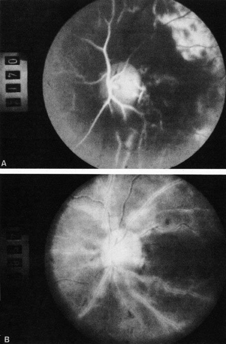
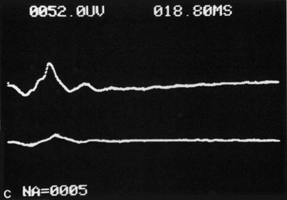
| Fig. 9. Intravenous fluorescein angiogram of a patient with hypotensive or hypoperfusion retinopathy. A: There is a marked delay in the choroidal and retinal filling. B: In the recirculation of the angiogram, there is a characteristic staining of both arteries and veins. C: Electroretinogram shows normal a- and b-waves in the normal right eye (upper tracing) and marked redirection of the a- and b-waves in the affected left eye (lower tracing). |