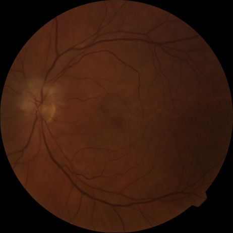
Fig. 5.
A:
Ischemic optic neuropathy in a patient with giant cell arteritis.
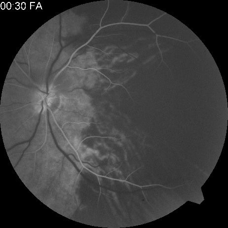
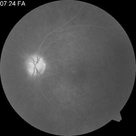
B
and
C:
Intravenous fluorescein angiography demonstrates delayed filling of the lateral posterior ciliary artery.