|
|
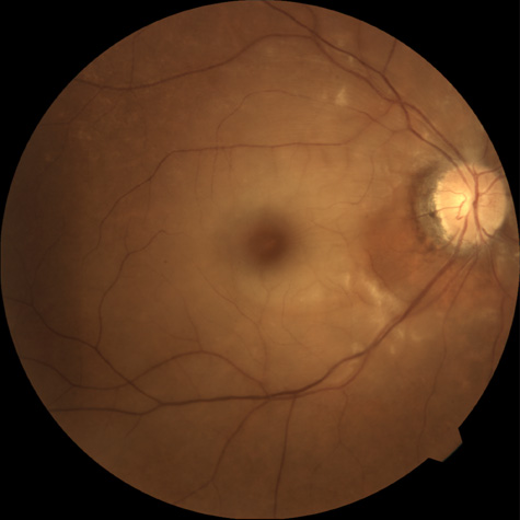
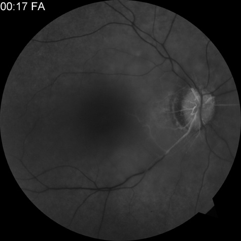
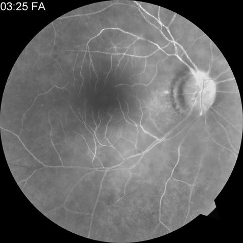
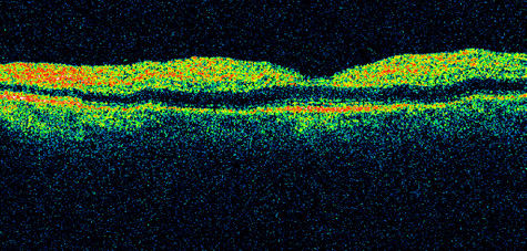
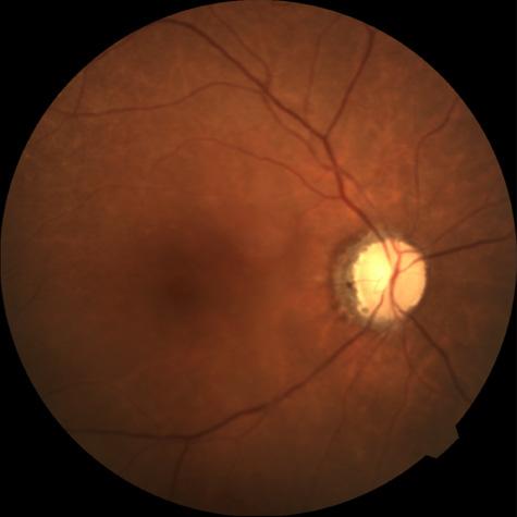
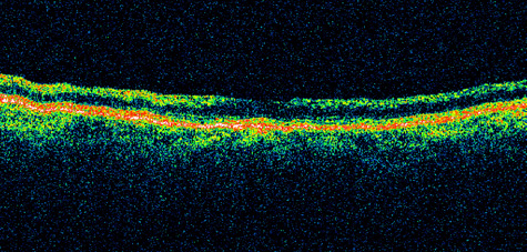
| Fig. 1. A: Acute central retinal artery obstruction with a cherry-red spot. B and C: Intravenous fluorescein angiography. There is a delay of dye appearance in the central retinal artery, and when it does appear, it does not fill the arteries completely. D: Ocular coherence tomography (OCT) at the time of occlusion showing the increase in retinal thickness and reflectivity of the inner layers of the retina. E: Seven months later there is significant optic atrophy(G), and the OCT (F) shown now has marked thinning of the retina. |