|
|
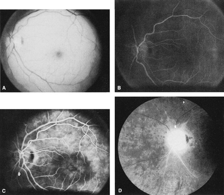
| Fig. 4. A: Ophthalmic artery obstruction showing a cherry-red spot: the visual acuity was no light perception. B: At 26.2 seconds, the retinal vessels are filled, but no dye is seen in the choroidal circulation. C: By 37.7 seconds, the choroidal circulation is more visible, and a doughnut-shaped area of hypofluorescence is noted. D: Six months after the initial photographs, optic atrophy is present, as is atrophy of the retinal vessels. Diffuse pigment disturbance is evident. |