|
|
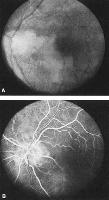
| Fig. 3. A: Cilioretinal artery obstruction showing opacity of the retina in the area of obstruction. B: The cilioretinal artery has begun to fill after the branches of the central retinal artery but is still not completely filled at this point in the angiogram. |