Chapter 13
Systemic Hypertension and the Eye
JOSEPH B. WALSH, RICHARD B. ROSEN and DANIEL M. BERINSTEIN
Main Menu   Table Of Contents

Search

HYPERTENSIVE RETINOPATHY
ARTERIOSCLEROTIC CHANGES
HYPERTENSIVE CHOROIDOPATHY
HYPERTENSIVE NEUROPATHY
CLASSIFICATION OF HYPERTENSIVE RETINOPATHY
REFERENCES

More than 50 million adults in the United States have arterial hypertension. The third National Health and Nutrition Examination Survey (NHANES III) estimates the current prevalence to be 31% of the adult American population.1 The strong correlation linking arterial hypertension with heart disease, stroke, and renal failure makes it a leading cause of morbidity and mortality among adults in the United States. The most recent classification categorizes blood pressure as normal, high-normal, or hypertension2 (Table 1). A systolic blood pressure greater than 130 mmHg or diastolic blood pressure greater than 85 mmHg on two or more readings taken on two or more visits is considered elevated, putting the patient at increased risk for developing cardiovascular disease. Identified causes of hypertension include renal-vascular diseases, primary aldosteronism, pheochromocytoma, Cushing's disease, and coarctation of the aorta. In over 90% of patients with hypertension, however, the cause remains unknown.


TABLE 13-1. Fifth Joint National Committee on Detection, Evaluation, and Treatment of High Blood Pressure Classification of Blood Pressure for Adults Aged 18 Years and Older


Category*Systolic Blood Pressure (mmHg)Diastolic Blood Pressure (mmHg)
Normal<130<85
High-normal130–13985–89
Hypertension  
 Stage 1140–15990–99
 Stage 2160–179100–109
 Stage 3180–209110–119
 Stage 4210120

*When systolic and diastolic pressure fall into different categories, the higher category should be selected to classify the individual's blood pressure status. For instance, 160/92 mmHg should be classified as stage 2, and 180/120 mmHg should be classified as stage 4. Isolated systolic hypertension is defined as systolic blood pressure > 140 mmHg and diastolic blood pressure < 90 mmHg and staged appropriately (e.g., 170/85 mmHg is defined as stage 2 isolated systolic hypertension based on the average of two or more readings taken at each of two or more visits after an initial screening).
†Joint National Committee on Detection, Evaluation and Treatment of High Blood Pressure: The fifth report of the Joint National Committee on detection, evaluation and treatment of high blood pressure. Arch Intern Med 153:154, 1993


The retinal vasculature is unique in its accessibility to the clinician for viewing noninvasively the arteries and veins of a patient. In addition, ancillary tests, such as fluorescein angiography and indocyanine green angiography, can assess capillary perfusion and integrity of the blood-retinal barrier and study the dynamics of the retinal, choroidal, and optic nerve head circulations. These techniques, along with the development of animal models,3–6 have led to the understanding that the retinal, choroidal, and optic nerve head circulations each possess distinct anatomic and physiologic properties that respond differently to blood pressure changes. The result is three distinct types of hypertensive disease visible in the fundus: hypertensive retinopathy, hypertensive choroidopathy, and hypertensive optic neuropathy.

Back to Top
HYPERTENSIVE RETINOPATHY
Retinal vascular response and other retinal changes seen in hypertension are variable and depend on several factors. The most important factors are the rate and degree of hypertension and the baseline condition of the retinal vasculature.7 Concomitant diseases, such as diabetes or renal or connective tissue disorders also play a role in the severity of findings.

Clinical findings seen in the retinal vasculature in hypertensive retinopathy include the following:

  • Arteriolar narrowing (focal and diffuse)
  • Arteriovenous crossing changes
  • Arterial sclerosis
  • Arterial tortuosity
  • Increased branching angles

One of the earliest and classic signs of hypertensive retinopathy is arteriolar narrowing (Fig. 1). An increase in vascular wall tone initiated acutely by autoregulatory mechanisms causes a decrease in caliber of the vessel, in what has been described as the vasoconstrictive phase.8,9 A more focal or diffuse vasoconstrictive state may be observed, depending on the initial condition of the retinal vasculature (Fig. 2). Vessels with areas of sclerosis lack muscle tone and tend to dilate secondary to elevated intraluminal pressure. Nonsclerotic vessels exhibit narrowing because of intact muscular walls and preserved autoregulatory response. Hayreh and colleagues,10,11 in comparing prehypertensive fluorescein fundus angiograms to those with hypertensive retinopathy, found no narrowing of arterioles.They concluded that the apparent narrowing was caused by an ophthalmoscopic artifact produced by retinal edema masking the arteriole walls from the sides along with the contrast of dilated venules, which made the arterioles appear more tapered.

Fig. 1. Fundus photograph demonstrating vasoconstrictive and early sclerotic changes seen in hypertensive retinopathy, including areas of diffuse arteriolar narrowing, sinusoidal tortuosity, and copper wire appearance. There also are arteriovenous crossing changes, with tapering of the veins and increased arteriolar branching angles.

Fig. 2. The changes seen in the fundus secondary to hypertension are representative of changes taking place in the arterioles throughout the body. Schema of hypertensive grading: grade I, generalized arteriolar narrowing; grade II, generalized narrowing and focal constrictions; grade III, more narrowing, focal constriction, hemorrhage, and exudation; grade IV, marked narrowing and focal constrictions with hemorrhages, exudates, and edema of the disc. (Scheie HG: Evaluation of ophthalmoscopic changes of hypertension and arteriolar sclerosis. Arch Ophthalmol 49:117, 1953)

Clinical findings of the retina in hypertensive retinopathy are as follows12:

  • Hemorrhages
  • Retinal and macular edema
  • Edema residues (hard exudates)
  • Inner retinal ischemic spots (cotton-wool spots)
  • Nerve fiber layer loss
  • Focal intraretinal periarteriolar transudates (FIPTs)

One common feature is hemorrhage (Fig. 3). Hemorrhages within the more superficial layers of the inner retina have a flame-shaped appearance because they track along axons in the nerve fiber layer. Deeper hemorrhages in the retina have a dot or blot appearance, which varies with the configuration of the neural elements of layer to which they are confined. Hayreh and associates11,13 concluded that retinal hemorrhages were only a minor feature of hypertensive retinopathy. They found that nerve fiber layer hemorrhages in the distribution of the radial peripapillary capillaries were more common than dot or blot hemorrhages or subhyaloid hemorrhages. This probably reflects the arteriolar character of the radial peripapillary capillary bed.14 Hard exudates, cotton-wool spots, and retinal edema are additional manifestations of the exudate phase of hypertensive retinopathy (Fig. 4) and indicate a more serious stage of the disease.

Fig. 3. The fundus changes in hypertension, in order of appearance, are generalized narrowing of the arterioles, focal constriction, hemorrhages, exudates, and finally, edema of the disc. They reflect the severity of the hypertension. A. Earliest sign of hypertension showing only generalized narrowing of the arterioles with no change in the vessel wall thickness or reflex. B. Grade II hypertension showing generalized narrowing plus focal constriction and grade I arteriolar sclerosis with widening of the reflex stripe. C. Grade III hypertension showing generalized narrowing, focal constriction, hemorrhages, and exudate, and grade I arteriolar sclerosis with widening of the light reflex. D. Grade IV hypertension showing generalized narrowing, focal constriction, hemorrhages, and exudates and edema of the disc with grade I arteriolar sclerosis. (Scheie HG, Ashley BJ, Yanoff M: Medical ophthalmology: Hypertension and arteriosclerosis. In Scheie HG [ed]: Medical Ophthalmology: Ophthalmologic Manifestations of Systemic Diseases. Medicolor Illustration and Editorial Service, 1966)

Fig. 4. Fundus photographs. A. Exudative phase findings seen in hypertensive retinopathy. Note areas of flame hemorrhages extending off the superior temporal and nasal arcade. Cotton-wool spots are seen in the peripapillary area, and there is a small serous detachment inferior to the disc. Macular edema is present, and there are multiple scattered pale spots due to choroidal ischemia. B. Fluorescein angiogram revealing early areas of hypofluorescence corresponding to cotton-wool spots and nonperfusion. There also are early hyperfluorescent spots corresponding to the pale spots seen clinically. C. Later phase shows hyperfluorescence caused by extensive leakage in areas of retinal edema.

Hard exudates—or, more appropriately, edema residues—are formed from extravasated plasma during the exudative phase. The residues are composed of lipids and cholesterol, giving them a characteristic waxy yellow or glistening appearance, and seem to settle in a bathtub ring-like configuration. They generally are found in the posterior pole and assume patterns that reflect the source of the leakage (i.e., circinate rings) and the neural elements of the layer in which they are found (i.e., macular star).

The development of clinical edema of the retina and macula may stem from a variety of events at a cellular level and may have multiple causes. Loss of autoregulation, as in episodes of acute hypertension, may result in an increase in transmural pressure in the capillaries leading to transudation of plasma into surrounding retina-producing extracellular edema.3,15 Intracellular edema is the direct result of retinal ischemia.3,15 Breakdown of the retinal pigment epithelium (RPE) blood-retinal barrier secondary to hypertensive choroidopathy produces serous retinal detachments overlying regions of choroidal ischemia. Diffusion of subretinal fluid into the retina eventually may create tissue edema.11,16,17

Cotton-wool spots, or so-called soft exudates,18 are areas of acute inner retinal ischemia caused by occlusion of terminal arterioles. They have a fluffy white appearance, irregular borders, and most commonly are found in the posterior pole and along the distribution of the radial peripapillary capillary bed. They are localized within the nerve fiber layer, often involving the underlying ganglion cells and inner nuclear while sparing the deeper retinal layers.19 Fluorescein angiography demonstrates nonperfusion of the cotton-wool spots and adjacent capillaries. Typically, they resolve ophthalmoscopically in 4 to 6 weeks, leaving a corresponding nerve fiber layer defect.

Cases of accelerated hypertension often are manifest by FIPTs.12 These appear as dull-white round, focal areas surrounding arterioles. The proposed mechanism of FIPT formation is the focal breakdown of the blood-retinal barrier, with the accumulation of plasmatic deposits in the retina following an accelerated hypertension event.12

Back to Top
ARTERIOSCLEROTIC CHANGES
Sclerotic changes are observed within the vessel walls as constriction of the retinal vasculature persists (Fig. 5). The arteriole wall normally is invisible, appearing only as an erythrocyte column with central light reflex by ophthalmoscopy (Fig. 6). As the wall thickens from continuous vasoconstriction, the light reflex becomes more diffuse and partially obscures the blood column, giving the once transparent arteriole a yellowed or copper wire appearance. Progression of the thickening and sclerotic changes eventually obscures the blood column completely, producing a silver wire appearance. Along with reflective changes, the thickening produces arteriovenous crossing changes. Retinal arterioles and venules share a common adventitial sheath at their crossing points. As the arteriolar wall thickens, the venule appears tapered if posterior to the arteriole or elevated if over the arteriole (Fig. 7). Other characteristic changes common but not unique to arteriosclerosis are increased tortuosity secondary to fibrous replacement of the vessel wall and increased arteriolar branching angles, known as perpendicularization. Progression of arteriosclerosis leads to endothelial damage and necrosis of the muscular component of the vessel walls.3 This leads to a breakdown of the blood-retinal barrier, causing exudative leakage into the retina. The formation of fibrin and thrombosis within the vessel may cause closure of the lumen, resulting in ischemic changes within the retina. This has been described as the exudative phase.8,9 Concomitant choroidopathy may contribute to further retinal changes seen in this phase of hypertensive retinopathy.

Fig. 5. Schema of ophthalmoscopic grading of arteriolar sclerosis. (Scheie HG: Evaluation of ophthalmoscopic changes of hypertension and arteriolar sclerosis. Arch Ophthalmol 49:117, 1953)

Fig. 6. Arteriolar sclerosis develops in prolonged hypertension and is manifest by increased light reflex, arteriovenous nicking copper or silver wire arterioles. A. Grade I hypertension and grade I arteriolar sclerosis showing the earliest generalized narrowing with increase light reflex. B. Grade I hypertension showing generalized narrowing and grade II arteriolar sclerosis showing increase in light reflex plus arteriovenous compression. C. Grade I hypertension showing generalized narrowing and grade III arteriolar sclerosis showing broad light reflex, arteriovenous compression plus copper wire arterioles. D. Grade I hypertension plus grade IV arteriolar sclerosis showing increase of light reflex, arteriovenous compression, copper and silver wire arterioles. (Scheie HG, Ashley BJ, Yanoff M: Medical ophthalmology: Hypertension and arteriosclerosis. In Scheie HG [ed]: Medical Ophthalmology: Ophthalmologic Manifestations of Systemic Diseases, p 17. Elgin, IL: Medicolor Illustration and Editorial Service, 1966)

Fig. 7. Scanning electron micrograph (× 226) demonstrating venous compression at arteriovenous crossing. (Fryczkowski AW, Smith DR: Contemp Ophthalmol 5:184, 1987)

Complications related to hypertensive retinopathy include the following:

  • Central or branch artery occlusion
  • Central or branch vein occlusion
  • Macroaneurysms
  • Epiretinal membrane
  • Retinal neovascularization
  • Vitreous hemorrhage
  • Cystoid macular edema

Artery and vein occlusions and macroaneurysms are the most common results of arteriosclerotic changes found in this condition (Fig. 8). The development of neovascularization with or without vitreous hemorrhage is the result of subsequent retinal ischemia. Cystoid macular edema also may result from ischemia, as well as from the breakdown of the retinal vascular and RPE blood barrier. Epiretinal membranes may form in the cicatricial phase of any of these events. Consequent development of a full macular hole after severe hypertensive retinopathy with macular edema and vitreous hemorrhage also has been described.20 Other recent findings have shown abnormal electroretinogram and visually evoked potential studies 2 to 4 years after an accelerated hypertensive event likely related to retinal infarction and ischemic optic neuropathy, respectively.21

Fig. 8. Complications seen in hypertensive retinopathy. A. Branch retinal vein occlusion (chronic with vascular remodeling at the site of occlusion) B. Fluorescein angiogram demonstrates collateral across the median raphe and multiple microaneurysms. C. Macroaneurysms straddling arteriovenous crossing with cuff of edema and circinate edema residue. D. Fluorescein angiogram demonstrating halo of nonperfusion surrounding macroaneurysm pair. E. Central retinal arterial occlusion with partial macular sparing cilioretinal artery. F. Fluorescein angiogram demonstrating absence of central retinal arterial filling with cilioretinal arterial filling and early retrograde perfusion. G. Retrograde flow continues to diffuse into the retinal arteries from cilioretinal source.

The overall rates of hypertensive retinopathy in the nondiabetic population ranges from 0.8% to 7.8%.22–24 The study of populations is difficult and highly variable because of different evaluation methods, grading classifications of retinopathy, selection bias groups, and the association of other systemic diseases.

Back to Top
HYPERTENSIVE CHOROIDOPATHY
The choroid is very sensitive to blood pressure changes that only indirectly affect the overlying RPE and neurosensory retina. Its pathophysiologic response to arterial blood pressure changes also is very different than that of the retinal vasculature. The choroid receives sympathetic innervation and is sensitive to circulating vasoconstrictive factors such as angiotensin II, adrenaline, and vasopressin. These factors and neural stimulation can initiate vasoconstriction of the choroid and choriocapillaris, leading to focal ischemia. The overlying RPE and the outer blood-retinal barrier may be compromised as a result.5,17

The clinical features of hypertensive choroidopathy include the following:

  • Elschnig's spots and Siegrist's spots (RPE and choroidal infarcts)
  • Subretinal exudates
  • Serous retinal detachments
  • RPE depigmentation (chronic)
  • Choroidal sclerosis

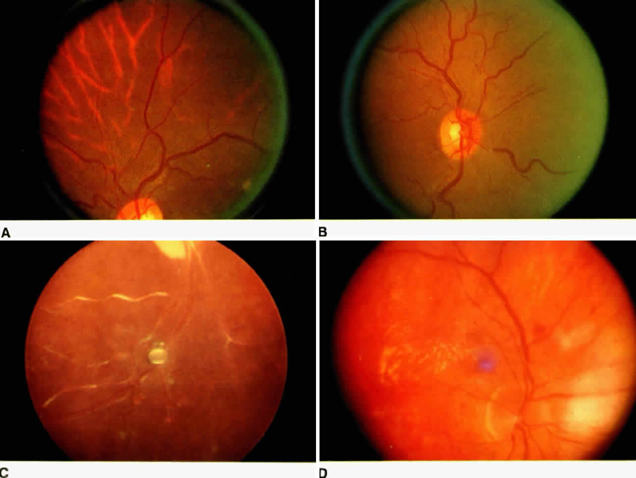
Clinically, direct changes to the choroidal vasculature are difficult to detect by ophthalmoscopy. Many findings seen as retinal changes are a result of choroidal vasculature response to blood pressure change. Elschnig's spots25 are ischemic infarcts of the RPE that coincide to hypoperfusion of the underlying choroid (Fig. 9). They appear as focal subretinal lesions with yellowish halo. In an experimental model,26 Elschnig's spots appeared within 24 hours of accelerated hypertension. Ischemic infarcts at the equator have a more linear appearance and are referred to as Siegrist's streaks. The presence of Siegrist's streaks may indicate a more advanced vascular sclerosis.27 As ischemic RPE becomes edematous, the blood-retinal barrier becomes disrupted, allowing leakage of fluid from the choroid into the subretinal space and forming serous detachments. Resolution of such detachments can follow rapidly the restoration of blood pressure control.28

Fig. 9. Fundus photograph showing retinal changes secondary to hypertensive choroidopathy. A. Elschnig's spots are ischemic infarcts of the RPE that appear in the posterior pole. B. Siegrist's spots are ischemic infarcts of the RPE that appear at the equator. They become hyperpigmented as the RPE repairs the injury. C. Fluorescein angiogram reveals early hyperfluorescence caused by disruption of the RPE blood-retinal barrier in areas of the Elschnig's spots.

Over time, the Elschnig's spots and areas of serous detachment develop central areas of pigmentation with surrounding atrophy. Sclerotic choroidal vessels become visible through areas of atrophic RPE.17,29 Pathologic correlation confirms the presence of hyalinization and necrosis of the vessels.5,30 Fluorescein angiographic studies demonstrate an irregular choroidal filling pattern with areas of hypofluorescence caused by hypoperfusion. Elschnig's spots typically display hyperfluorescence because of leakage at sites of blood-retinal barrier disruption. Hyperfluorescence may extend into the subretinal space if a serous detachment is present. Fluorescein angiography in chronic cases reveals a hyperfluorescent pattern corresponding to the RPE window defects. Indocyanine green videoangiography provides an enhanced view of the choroidal circulation, which in these cases appears sluggish.31 Kishi and colleagues5 provided the most comprehensive demonstration of the sequential progression of experimental hypertensive choroidopathy. Pathologic features were classified into three phases of development: acute ischemic phase, chronic occlusive phase, and chronic reparative phase. In the acute ischemic phase, choroidal arterioles constrict, narrowing the lumina. The adjacent choriocapillaris displays necrotic endothelial cells, hydropic degeneration of pericytes, and fibrinous deposits in Bruch's membrane. Overlying RPE exhibits intercellular edema and subretinal exudates. The acute ischemic phase lasts for the first 2 months. After 2 to 4 months, a chronic occlusive phase characterized by retinal detachments and subretinal exudates develops. Choroidal arteries remain narrowed or occluded with proliferation of intimal cells and fibrin deposition in the arterial walls. The choriocapillaris remains occluded by fibrin with its endothelium and pericyte layer denuded. Vessel walls undergo hyalinization, and the overlying RPE becomes necrotic. A chronic reparative phase was observed after 4 to 21 months. RPE depigmentation developed in areas of previous serous detachment and occluded choroidal arteries recanalized with smaller lumina lined by a new elastic laminae. Ghost choriocapillaris was seen in areas in which recanalization failed to occur, and new vessels appeared to be composed of nonfenestrated endothelial cells.

Back to Top
HYPERTENSIVE NEUROPATHY
In malignant hypertension, one of the earliest changes seen is optic nerve head edema6,32:

Clinical Findings in Neuropathy

  • Optic disc edema
  • Optic disc pallor
  • Optic disc ischemia

The clinical appearance is indistinguishable from other causes of optic nerve head swelling, such as elevated intracranial pressure (Fig. 10). The mechanism of disc swelling remains controversial. In Tso and Jampol's study of baboons,8 the edema was believed to be the result of an accumulation of axoplasmic components. However, they could not determine whether this accumulation was related to ischemia or to some mechanical phenomena. Kishi and associates6 demonstrated axonal hydropic swelling that was secondary to ischemia. They later concluded that the ischemia produced was related to vasoconstriction of the peripapillary choroidal and optic nerve head vessels.11,32 The direct vasoconstrictive and occlusive properties seen in hypertensive choroidopathy lead to ischemic changes in the optic nerve head because it receives most of its blood supply from the peripapillary choroidal vessels.11,32,33 Further, vascular endothelial substances can diffuse easily into the optic nerve head from the surrounding choroidal bed and cause vasoconstriction of the optic nerve head vessels.11,32,33

Fig. 10. A. Disc photos showing optic nerve head edema with cotton-wool spots and splinter hemorrhages seen in hypertensive optic neuropathy. B. Fluorescein angiogram shows hyperfluorescence caused by late leakage.

Late findings seen in hypertensive neuropathy are optic nerve head pallor and atrophy resulting from chronic ischemia. The clinician must be careful to rule out other possible causes of these findings, including anterior ischemic optic neuropathy and temporal arteritis when evaluating patients for hypertensive optic neuropathy.

Back to Top
CLASSIFICATION OF HYPERTENSIVE RETINOPATHY
Since Marcus Gunn's description in 1898 of the changes in retinal vessels in patients with arterial hypertension,34 various classification systems have been attempted to explain observed changes and correlate them with the systemic disease. The first major classification scheme, by Keith and colleagues,35 was designed to relate survival to retinal vascular changes in the hypertensive population. Patients are classified according to the severity of their fundus changes into four groups and morbidity is looked at over a 5-year period (Table 2). They found that changes correlated directly with the degree of systemic hypertension and, inversely, with the prognosis for survival. Later, Wagener and coworkers36 developed more quantitative criteria for classifying hypertensive retinal vascular changes. This system was based on the narrowing of arterioles and graded focal arterial constriction (Table 3). In addition, they grouped vascular hypertension with associated retinal changes within each group. Although more precise in design, reproducibility of the system was poor, and it was soon replaced by a more complete classification scheme described by Harold Scheie in 1953.37 He graded changes of hypertension and arteriolar sclerosis separately in five stages (Table 4). He defined hypertensive changes as those related to arteriolar constriction and vascular changes from long-standing hypertension as arteriolar sclerosis.


TABLE 13-2. Keith-Wagener-Barker Classification


GroupDescription
1Mild to moderate narrowing and sclerosis of the arteries
2Moderate to marked sclerosis of the retinal arterioles; exaggeration of the light reflex; arteriovenous compression changes or generalized or localized narrowing of the arterioles
3Retinal arteriolar narrowing and focal constriction; retinal edema; cotton-wool spots; hemorrhage
4Group 3 plus papilledema
(Walsh JB: Hypertensive retinopathy: Description, classification, and prognosis. Ophthalmology 89:1127, 1982)


TABLE 13-3. Wagener-Clay-Gipner Modification of Generalized Arteriolar Narrowing


GroupDescription
1Reduction of caliber of arterioles to three fourths of average caliber
2Reduction of caliber of arterioles to half of average caliber
3Reduction of caliber of arterioles to one third of average caliber
4Arterioles thread-like or invisible
*(Walsh JB: Hypertensive retinopathy: Description, classification, and prognosis. Ophthalmology 89:1127, 1982)


TABLE 13-4. Scheie Classification


GroupHypertensionArteriolar sclerosis
0No changesNormal
1Barely detectable arteriolar narrowingBarely detectable light reflex changes
2Obvious arteriolar narrowing with focal irregularitiesObvious increased light reflex changes
3Grade 2 plus retinal hemorrhages or exudatesCopper wire arterioles
4Grade 3 plus papilledemaSilver wire arterioles
(Walsh JB: Hypertensive retinopathy: Description, classification, and prognosis. Ophthalmology 89:1127, 1982)


One of the problems that complicates all classification systems is the ophthalmoscopic variation in the extent of the acute hypertensive changes and those of the duration-related sclerotic changes observed in the same patient (Fig. 11). In 1957, Leishmann7 presented a seven-part classification that took into account the development of arteriolar sclerosis as part of the natural aging process and emphasized the modified ability of retinal arterioles with involutional sclerosis to respond to hypertension. Subsequent to these discussions, clinicians have become sensitive to the need to account for the presence or absence of arteriolar sclerosis in interpreting the fundi of hypertensive patients. In 1966, the original study by Keith and associates35 was updated and modified to include the grading of generalized and focal arteriolar narrowing and arteriolar sclerosis38 (Table 5). This modified system served as a helpful prognosticator of hypertensive disease. Arteriolar narrowing and focal constrictions in the absence of retinal hemorrhages or disc edema seem to be the most sensitive indicators employed by the system. It currently remains the most commonly used classification for grouping hypertensive patients ophthalmoscopically and enjoys worldwide acceptance.

Fig. 11. Any combination of the hypertensive changes and arteriolar sclerosis may be seen in the fundus of a particular case, depending on the severity and duration of the hypertension. A. Grade IV hypertension with generalized and focal constriction, hemorrhages, exudates, and papilledema, and grade IV sclerosis with silver-wire arterioles. B. Grade IV hypertension and grade II arteriolar sclerosis compatible with a marked exacerbation of hypertension in a long-standing hypertensive patient. C. Grade II hypertension and grade IV arteriolar sclerosis compatible with controlled hypertension in a long-standing hypertensive patient. D. Grade IV hypertension and arteriolar sclerosis. (Scheie HG, Ashley BJ, Yanoff M: Medical ophthalmology: Hypertension and arteriosclerosis. In Scheie HG [ed]: Medical Ophthalmology: Ophthalmologic Manifestations of Systemic Diseases, pp 5, 8. Elgin, IL: Medicolor Illustration and Editorial Service, 1966)


TABLE 13-5. Modified Keith-Wagener-Barker Classification


 Group
 1234
Sclerosis< 11 0–40–4
General arteriolar narrowing0–40–40–40–4
Focal arteriolar narrowing0–40–40–40–4
Hemorrhage-±±±
Exudate--+±
Papilledema---+

0–4 depends on amount or extent of vascular change.
+ Present.
- Absent.
(Walsh JB: Hypertensive retinopathy: Description, classification, and prognosis. Ophthalmology 89:1127, 1982)


The manifestations of hypertension in the eye have been studied for more than 150 years. There have been many attempts at correlating ocular findings with the systemic disease, its prognosis, and success of blood pressure control. With the development of fluorescein angiography and animal models, the understanding of hypertensive changes has improved. Further studies looking at the function and role of vascular endothelial products may enhance further our understanding at a more molecular level. Currently, the retina remains the most accessible organ for the clinician to detect undiagnosed hypertension and to monitor blood pressure control.

Back to Top
REFERENCES

1. Burt VL, Whelton R, Roccella EJ: Prevalence of hypertension in the U.S. adult population: Results from the Third National Health and Nutrition Examination Survey, 1988-1991. Hypertension 2S:305, 1995

2. The Joint National Committee on Detection, Evaluation and Treatment of High Blood Pressure: The fifth report of the Joint National Committee on detection, evaluation and treatment of high blood pressure. Arch Intern Med 153:154, 1993

3. Garner A, Ashton N, Tripathi R et al: Pathogenesis of hypertensive retinopathy: An experimental study in the monkey. Br J Ophthalmol 59:3, 1975

4. Hayreh SS, Servais G, Virdi PS: Fundus lesions in malignant hypertension: III. Arterial blood pressure, biochemical, and fundus changes. Ophthalmology 93:45, 1986

5. Kishi S, Tso MOM, Hayreh SS: Fundus lesions in malignant hypertension: I. A pathologic study of experimental hypertensive choroidopathy. Arch Ophthalmol 103:1189, 1985

6. Kishi S, Tso MOM, Hayreh SS: Fundus lesions in malignant hypertension: II. A pathologic study of experimental hypertensive optic neuropathy. Arch Ophthalmol 103:1198, 1985

7. Leishman R: The eye in general vascular disease: Hypertension and arteriosclerosis. Br J Ophthalmol 41:641, 1957

8. Tso MOM, Jampol LM: Pathophysiology of hypertensive retinopathy. Ophthalmology 89:1132, 1982

9. Tso MOM, Abrams GW, Jampol LM: Hypertensive retinopathy, choroidopathy, and optic neuropathy: A clinical and pathophysiological approach to classification. In Singerman LJ, Jampol LM (eds): Retinal and Choroidal Manifestations of Systemic Disease, pp 79–127. Baltimore: Williams & Wilkins, 1991

10. Hayreh SS, Servais G, Virdi PS: Retinal arteriolar changes in malignant arterial hypertension. Ophthalmologica 198:178, 1989

11. Hayreh SS: Duke-Elder Lecture: Systemic arterial blood pressure and the eye. Eye 10:5, 1996

12. Hayreh SS, Servais G, Virdi PS: Fundus lesions in malignant hypertension. IV. Focal intraretinal periarteriolar transudates. Ophthalmology 93:60, 1986

13. Hayreh SS, Servais G: Retinal hemorrhages in malignant arterial hypertension. Int Ophthalmol 12:137, 1988

14. Wise GN, Dollery CT, Henkind P: The Retinal Circulation. New York: Harper and Row, 1971

15. Ashton N: The eye in malignant hypertension. Trans Am Acad Ophthalmol Otolaryngol 76:17, 1972

16. Hayreh SS, Servais G, Virdi PS: Macular lesions in malignant arterial hypertension. Ophthalmologica 198:230, 1989

17. Hayreh SS, Servais G, Virdi PS: Fundus lesions in malignant hypertension: VI. Hypertensive choroidopathy. Ophthalmology 93:1383, 1986

18. Hayreh SS, Servais G, Virdi PS. Cotton-wool spots (inner retinal ischemic spots) in malignant arterial hypertension. Ophthalmologica 198:197, 1989

19. Garner A: Vascular diseases. In: Garner A, Klintworth GK (eds). Pathobiology of Ocular Disease: A Dynamic Approach, pp 1625–1710. New York: Marcel Dekker Inc, 1994

20. Cohen SM, Gass JDM: Macular hole following severe hypertensive retinopathy. Arch Ophthalmol 112:878, 1994

21. Talks SJ, Good P, Clough CG et al: The acute and long-term ocular effects of accelerated hypertension: A clinical and electrophysiological study. Eye 10:321, 1996

22. Klein R, Klein BEK, Moss SE et al: Hypertension and retinopathy, arteriolar narrowing, and arteriovenous nicking in a population. Arch Ophthalmol 112:92, 1994

23. Leibowitz HM, Krueger DE, Maunder LR et al: The Framingham Eye Study monograph. Surv Ophthalmol 24:335, 1980

24. McDonough JR, Garrison GE, Harnes CG: Blood pressure and hypertensive disease among Negroes and whites: A study in Evans County, Georgia. Ann Intern Med 61:208, 1964

25. Elschnig A: Die diagnostische und prognostische Bedeutung der Netzhauterkrankungen bei Nephritis. Wien Med Wochenschrift 54:446, 1904

26. deVenecia G, Wallow I, Houser D et al: The eye in accelerated hypertension: I. Elschnig's spots in non human primates. Arch Ophthalmol 98:913, 1980

27. Klien BA: Ischemic infarcts of the choroid (Elschnig spots): A cause of retinal separation in hypertensive disease with renal insufficiency: A clinical and histopathologic study. Am J Ophthalmol 66:1069, 1968

28. deVenecia G, Jampol LM: The eye in accelerated hypertension: II. Localized serous detachments of the retina in patients. Arch Ophthalmol 102:68, 1984

29. Morse PH: Elschnig's spots and hypertensive choroidopathy. Am J Ophthalmol 66:844, 1968

30. Friedman E, Smith TR, Kuwabara T et al: Choroidal vascular patterns in hypertension. Arch Ophthalmol 71:842, 1964

31. MacCumber MW, Flower RW, Langham ME: Ischemic hypertensive choroidopathy: Fluorescein angiography, indocyanine green videoangiography, and measurement of pulsatile blood flow. Arch Ophthalmol 111:704, 1993

32. Hayreh SS, Servais G, Virdi PS: Fundus lesions in malignant hypertension: V. Hypertensive optic neuropathy. Ophthalmology 93:74, 1986

33. Sossi N, Anderson DR: Blockage of axonal transport in optic nerve induced by elevation of intraocular pressure; Effect of arterial hypertension induced by angiotensin I. Arch Ophthalmol 101:94, 1983

34. Gunn M: On ophthalmoscopic evidence of general arterial disease. Trans Ophthalmol Soc UK 18:356, 1898

35. Keith NM, Wagener HP, Barker NW: Some different types of essential hypertension: Their course and prognosis. Am J Med Sci 197:332, 1939

36. Wagener HP, Clay GE, Gipner JF: Classification of retinal lesions in the presence of vascular hypertension. Trans Am Ophthalmol Soc 45:57, 1947

37. Scheie HG: Evaluation of ophthalmoscopic changes of hypertension and arteriolar sclerosis. Arch Ophthalmol 49:117, 1953

38. Hayreh SS: Structure and blood supply of the optic nerve. In Heilmann K, Richardson KT (eds): Glaucoma: Conceptions of a Disease, Pathogenesis, Diagnosis, Therapy, pp 78–96. Philadelphia: WB Saunders, 1978

Back to Top