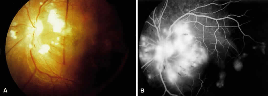
Fig. 10.
A.
Disc photos showing optic nerve head edema with cotton-wool spots and splinter hemorrhages seen in hypertensive optic neuropathy.
B.
Fluorescein angiogram shows hyperfluorescence caused by late leakage.