|
|
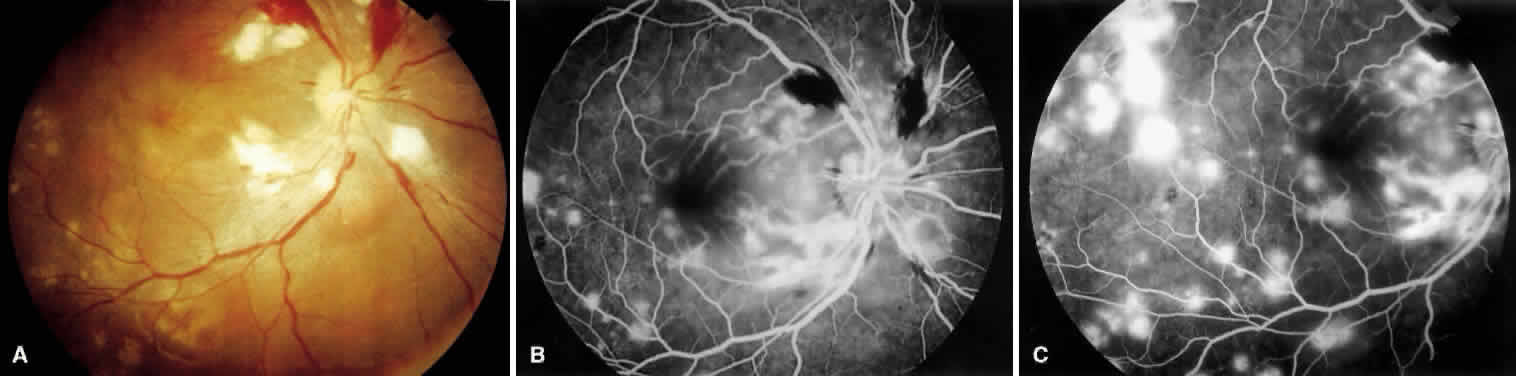
| Fig. 4. Fundus photographs. A. Exudative phase findings seen in hypertensive retinopathy. Note areas of flame hemorrhages extending off the superior temporal and nasal arcade. Cotton-wool spots are seen in the peripapillary area, and there is a small serous detachment inferior to the disc. Macular edema is present, and there are multiple scattered pale spots due to choroidal ischemia. B. Fluorescein angiogram revealing early areas of hypofluorescence corresponding to cotton-wool spots and nonperfusion. There also are early hyperfluorescent spots corresponding to the pale spots seen clinically. C. Later phase shows hyperfluorescence caused by extensive leakage in areas of retinal edema. |