Chapter 24
Computed Tomography of the Orbit
JAMES A. GARRITY and JULIE R. GILBERTSON
Main Menu   Table Of Contents

Search

VASCULAR LESIONS OF THE ORBIT
INFLAMMATION OF THE ORBIT
INFECTIONS
TUMORS OF THE OPTIC NERVE
SECONDARY ORBITAL TUMORS
METASTATIC TUMORS TO THE ORBIT
ORBITAL TRAUMA
LESIONS OF THE GLOBE
SUMMARY
REFERENCES

This chapter initially was written 10 years ago. With the opportunity for an update, we assert that many of the stated beliefs about computed tomography (CT) scanning being an effective, cost-efficient clinical tool remain true and many of the potential applications for CT have become routine. In the interim, helical CT scanning with slip-ring technology and rapid contrast injection followed by CT imaging has evolved and is readily available on most state-of-the-art CT scanners. This technology has permitted shorter scan times, facilitating rapid scanning of unstable trauma patients and pediatric patients with reduced need for anesthesia. Additionally, the contrast dose is reduced and radiation dose is also decreased. There has been considerable improvement in multiplanar reformations with this technology as well as three-dimensional (3D) reconstruction capabilities providing exquisite anatomic detail. These factors permit CT to remain a screening examination of choice for the orbit for many disease processes, particularly for trauma, proptosis, and childhood leukocoria.1

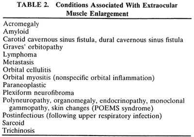
CT revolutionized the fields of neuro-ophthalmology and orbital diseases by its ability to show many of the lesions responsible for various clinical symptoms and signs. Magnetic resonance imaging (MRI) has surpassed CT in terms of clinical utility in most neuro-ophthalmic applications such as the evaluation of cranial nerve palsies and optic nerve lesions, especially if intracranial extension is suspected. CT remains a quick, clinically effective, and cost-effective method for evaluating most orbital problems. Most orbital lesions are adequately visualized, which is helpful with differential diagnosis or planning a surgical approach to minimize morbidity. The effects on adjacent bone are better appreciated with CT. The strength of being able to evaluate bone also is one of the weaknesses in that visualization of the orbital apex is compromised by the dense bone. Although one may be able to imply certain characteristics of tissue type with CT, MRI is better. Finally, there are certain instances in which CT and MRI are complementary such as with a patient with an ethmoid sinus tumor who also has an opacified frontal sinus. Bony changes around the ethmoid sinus are seen with the CT, but MRI is valuable in differentiating tumor from benign inflammatory sinus disease.

CT traces its beginnings to research initiated by Hounsfield in Great Britain in 1969. Scanning was accomplished by inserting a patient's head intoa latex cap that projected into a plastic water-containing box. The head was scanned by a slitx-ray beam, and the emergent photons were detected by two crystal photomultiplier detectors that moved parallel to and in accurate alignment with the x-ray tube across the patient's head. Data were displayed on an 80 × 80-matrix cathode ray tube in which the brightness of each point was proportional to its absorption coefficient. Initial slices (axial only) were 13-mm thick, although 8-mm slices could be produced. Scanning times ranged from 5 to 10 minutes per slice. Iodinated contrast material was not used.

CT of the head was introduced into clinical practice in the United States in June 1973. Two reports detailing early experience quickly followed.2,3 Orbital applications of this new technique were subsequently developed in 1974.4,5

The early CT scanners (EMI scanners) were specifically designed for scanning the head, and technical difficulties were encountered in obtaining tomographic slices low enough to visualize the entire orbit adequately. Evolutionary changes in technology now permit a 512 × 512 matrix, overlapping slices 1.5 to 3 mm thick, computer software that can reformat images in three dimensions and in other planes, a gantry that angulates, faster data acquisition, and intravenous contrast material. In addition, dynamic scans every 2 to 4 seconds can be taken while intravenous contrast material is being given to assess vascularity of a lesion while ultrafast scanning (five scans per second) permits detailed views of the globe and optic nerve in motion.

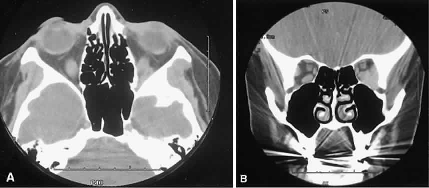
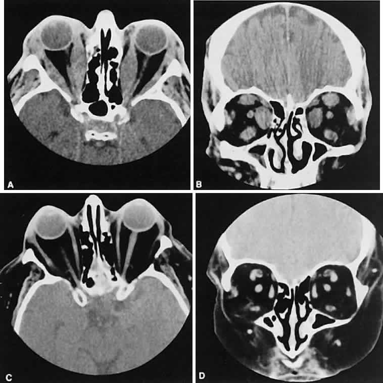
Optimal visualization of the orbit requires imaging from at least two planes. Axial slices should be oriented parallel to the optic nerve (-10 degreesto the orbitomeatal line) and no thicker than 3 mm. Axial views, because of volume averaging, may miss lesions located along the floor or roof. Additional views, typically coronal, can be obtained by reformatting data obtained during axial imaging or by direct coronal scanning.

Direct coronal views usually are preferable because of better resolution.6,7 They can be obtained by having patients lie either prone or supine, extending their neck, and angulating the gantry sufficiently to provide coronal imaging while avoiding artifacts from the teeth. Direct coronal scans also should be no thicker than 3 mm.

Coronal views may need to be reformatted from axial scans if a patient has extensive dental fillings, is anesthetized, or cannot extend the neck sufficiently for direct coronal scanning. The resolution on these images can be improved if data are collected from 1.5-mm contiguous axial slides.6 Spiral CT has resulted in improved multiplanar reformation with thin section (1 to 1.5 mm) axial images. High-resolution images necessary for leukocoria or foreign body imaging are obtained with 1-mm axial slices at 1:1 pitch at 1-mm intervals. Ideally, most screening orbit studies are performed at 3-mm direct axial and direct coronal images.

After the orbit has been visualized adequately, it also has been our practice to obtain 10-mm axial slices through the remaining portion of the head to complete the study. Intravenous contrast material usually is given, although the low-density orbital fat produces an inherent high level of contrast for most orbital CT studies. Intravenous administration of iodinated contrast medium is most helpful in detecting intracranial extension of an orbital process or identifying a pathologic process involving the optic nerve/sheath, most notably optic nerve sheath meningioma. Specific contraindications for contrast material include allergy or renal failure.

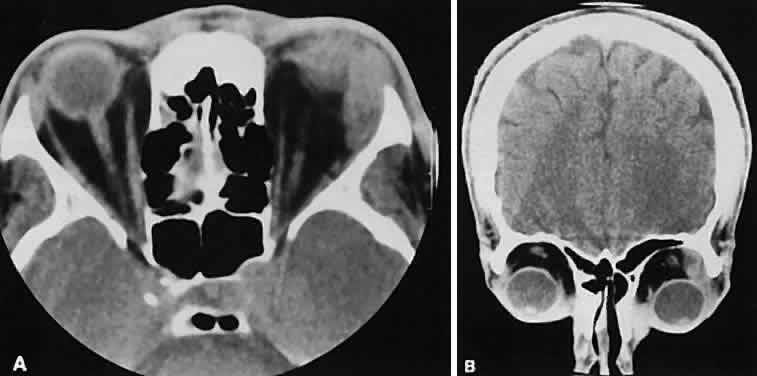
If a patient with severe Graves' orbitopathy, in whom the diagnosis is not in question, requires orbital CT scanning, it generally is performed without intravenous contrast.8 The administered iodine load would interfere with therapy directed at the thyroid gland and generally is not necessary to evaluate muscle enlargement or other features of Graves' orbitopathy.

For proper interpretation, it is not necessary to understand all of the technical aspects of CT scanning, just as it is not necessary to completely understand the inner workings of a personal computer to benefit from it. Familiarity with certain terms and concepts, however, facilitates a more fundamental appreciation for some of the limits of CT scanning.

The CT image consists of a matrix (or grid) of small squares known as pixels. Resolution is increased by increasing the numbers of pixels that are used to create the image. Total pixel number is limited by image contrast necessary to differentiate the various tissue densities in the image. A higher numbered matrix is composed of smaller pixels. A voxel is the 3D counterpart that includes the added dimension of slice thickness.

By altering window setting and level of attenuation on the console, subtle differences in tissuecomposition or bone changes can be appreciated.Window setting refers to the amount of the pixel sampling used by the computer to determine image contrast. Level refers to the scale of density used by the computer to create the image. Both the window setting and level of attenuation are extremely important, for example, when evaluating lesions in the fossa of the lacrimal gland, when the surrounding bone must be examined carefully for either pressure thinning or tumor erosion.

The display console also has the ability to move a cursor to any area on the screen. The computer can then examine this area to determine its den-sity. By convention, these density units are calledHounsfield units. Air and fat have the lowest values, whereas bone has the highest.

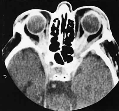
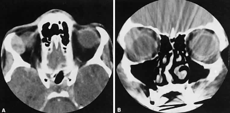
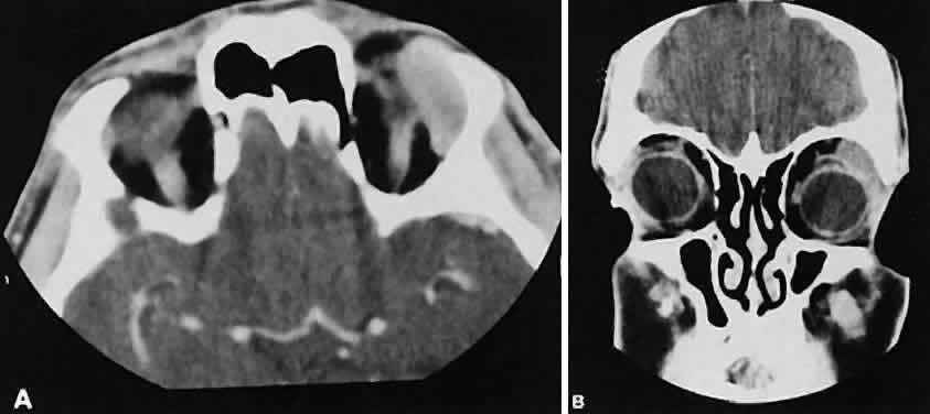
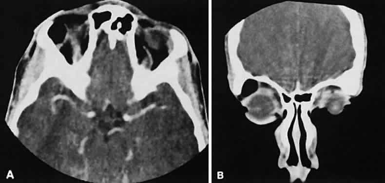
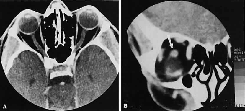
An appreciation for the various pathologic processes that affect the orbit is facilitated by an understanding of the normal orbital anatomy (Fig. 1). The orbit is a pyramid-shaped bony structure bounded inferiorly by the maxillary sinus, medially by the ethmoidal sinus, and superiorly by the frontal sinus. The sphenoidal sinus is situated posteriorly along the medial orbital wall and has a common wall with the optic canal. The lacrimal gland lies within its fossa located in the superior temporal aspect of the orbit and can be seen on both axial and coronal views.

Fig. 1. Normal anatomy. A. Axial scan through the plane of the optic nerve. Note the normal size of the horizontal rectus muscles and the undulating course of the optic nerve. B. Coronal view of the right orbit. The plane of section is slightly oblique to avoid dental artifacts and is immediately behind the right globe, which is partially volume averaged inferiorly. The arrow points to the optic nerve.

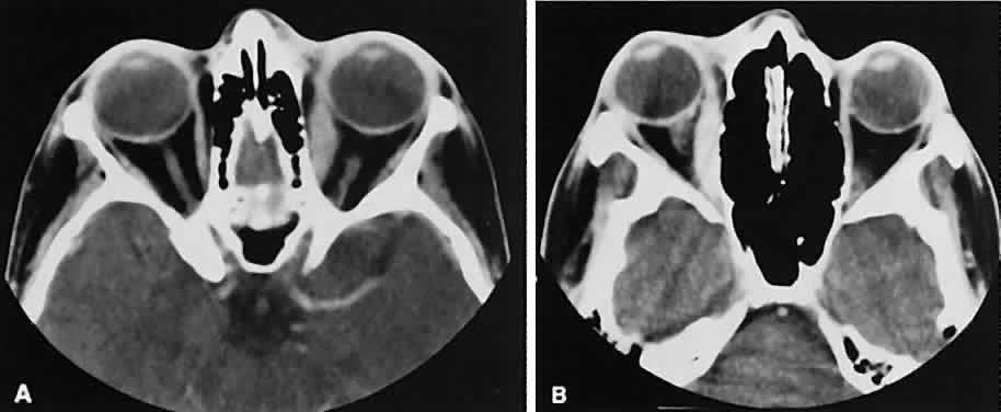
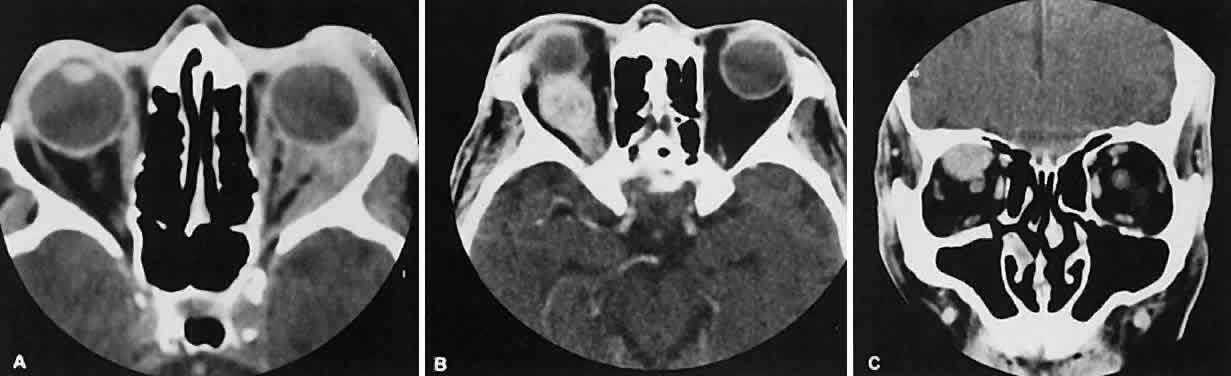
The extraocular muscles (EOMs), with the exception of the inferior oblique, originate from the anulus of Zinn in the orbital apex. The inferior oblique takes its origin from the frontal process of the maxilla and is seen occasionally on CT imaging. The superior oblique, after originating from the anulus, courses along the superior nasal orbital wall just above the medial rectus muscle before passing through the trochlea. The rectus muscles conveniently form a muscle cone, which is sometimes helpful in terms of differential diagnosis. Before thinner axial slices and multiplanar imaging were available, an enlarged inferior rectus muscle often was imaged as an apical mass, especially if dysthy-roid optic neuropathy was present. The importance of imaging from two different planes cannot be overemphasized in this situation.

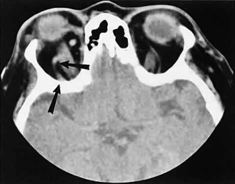
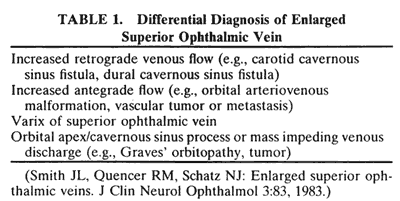
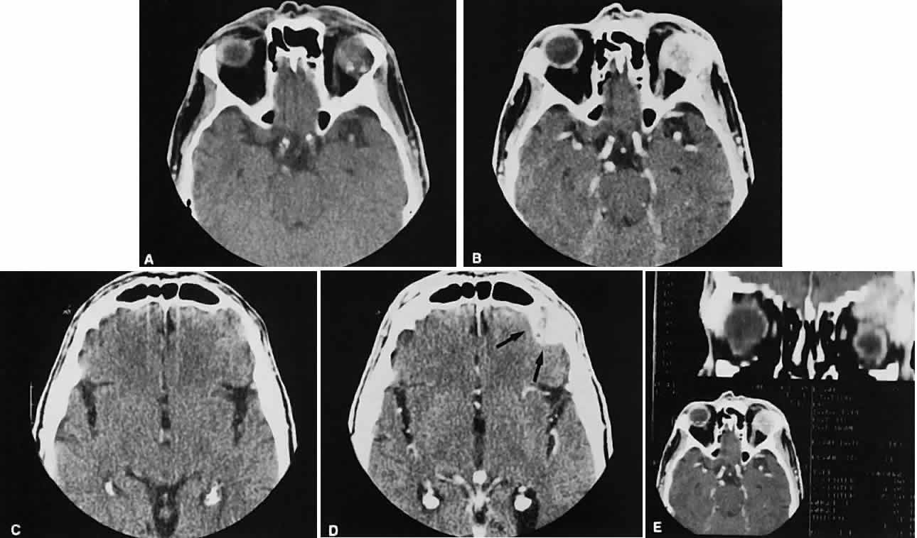
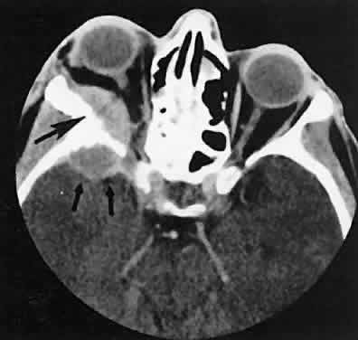
The superior ophthalmic vein (SOV) is an important vascular structure to recognize. It begins in the superior nasal quadrant near the trochlea before coursing posteriorly and laterally beneath the superior rectus muscle, exiting the orbit through the superior orbital fissure. Drainage is into the cavernous sinus. Asymmetric enlargement, especially in the presence of an ipsilateral cavernous sinus enlargement, suggests a vascular anomaly, which may require selective carotid angiography for further definition. Enlargement of one or multiple EOMs in this setting is likely. The SOV also may be enlarged as a result of any process impeding drainage from the orbital apex, such as dysthyroid orbitopathy or metastatic disease.

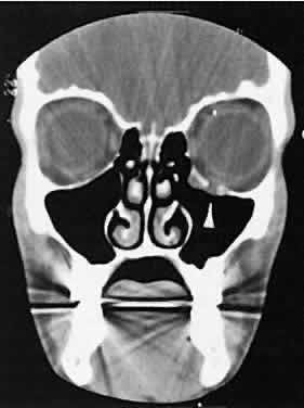
The optic nerve occupies the central intraconal space. By necessity, the nerve has a certain amount of slack, which is necessary to permit movement of the globe. In the axial plane, the optic nerve has an undulating course and thus may appear thicker or thinner as a result of partial volume averaging as it passes in and out of the axial plane. It is imperative to recognize this normal pattern for proper interpretation of axial images.

The optic nerve itself is invested by the same meningeal layers that cover the brain, and the intracranial space may extend along the course of the optic nerve to the back of the globe. Enlargement of this space may be recognized as pseudomeningoceles of the optic nerve sheath. It is sometimes possible to tell whether the nerve, the sheath, or both are enlarged by CT scanning, although MRI affords the better view.

Back to Top
VASCULAR LESIONS OF THE ORBIT
The orbital manifestations of a vascular process are dependent on whether the lesion is on the venous or the arterial circulation. Patients with venous lesions (varix) characteristically report a history of intermittent proptosis with either a Valsalva's-type maneuver or having the head in a dependent position. Suspicion of a venous varix should alert the radiologist to perform the scan before and after a Valsalva's maneuver.9,10 Color flow doppler imaging showing reversal of flow during a Valsalva maneuver also is a helpful finding. Phleboliths may or may not be present. Another clue to the diagnosis of a varix is the location, many times in the posterior, inferior lateral orbit, seemingly coming out of the inferior orbital fissure near the apex. Lesions that appear to change size or shape from the axial view to the coronal view also have turned out to represent a varix based on our experience (Fig. 2).

Fig. 2. Orbital varix. A. Axial view shows a small soft tissue mass within the inferolateral left orbital apex. B. Coronal view with patient repositioned prone with neck extension. Increased venous pressure distends the varix, accounting for increased size.

Arterial lesions may either have high or low flow. The high-flow lesions are carotid cavernous sinus fistulas and result from a direct communication between the carotid artery and the cavernous sinus. These arise spontaneously or as a result of trauma. The orbit is involved as an innocent bystander, because all signs and symptoms of orbital involvement follow from the retrograde transmission of increased venous pressure from the cavernous sinus. EOMs generally are enlarged, as is the SOV. The ipsilateral cavernous sinus also is enlarged. Intercommunication between the cavernous sinuses also may account for the occasional bilateral findings.

Low-flow lesions result from increased blood flow through the cavernous sinus, but the intracavernous carotid artery itself is intact (Fig. 3). These typically are dural-cavernous sinus fistulas. A high index of suspicion may be required to diagnose these, but a fairly stereotyped presentation is a unilateral red-eyed glaucoma with proptosis. Abduction weakness also may be present. Enlargement of one or more EOMs along with an enlarged SOV are noted with CT scanning. A small lesion may escape detection with CT scanning and require high-resolution MRI or even selective internal and external carotid angiography for diagnosis if a high degree of clinical suspicion exists. In a patient with a known dural fistula, a sudden and dramatic deterioration in the clinical picture may be seen with a thrombosis of the SOV.11 The radiographic picture, at least regarding the CT appearance, probably will not change. MRI can nicely show the thrombosis in the SOV.

Fig. 3. Axial view showing a dilated superior ophthalmic vein (SOV; arrows) secondary to dural-cavernous sinus fistula. Note the normal-sized SOV in the contralateral orbit.

The finding of an enlarged SOV is most often associated with vascular lesions involving the orbit. The differential diagnosis of this finding is listed in Table 1.12


Back to Top
INFLAMMATION OF THE ORBIT
In a qualitative way, CT scanning has redefined some of the concepts regarding nonspecific orbital inflammations (orbital pseudotumor). We now appreciate that any or all structures within the orbit or orbits may be affected. Inflammation can be localized to an EOM (myositis), lacrimal gland (dacryoadenitis), optic nerve sheath (perioptic neuritis), orbital soft tissues (nonspecific inflammation), posterior scleritis, or trochlea (trochleitis). There is no satisfactory explanation about why or how this inflammation occurs and remains so localized, although some types may be associated with various systemic diseases such as sarcoidosis, Wegener's granulomatosis, or other vasculitic conditions.

The CT characteristics of each subgroup are different and are considered separately. The nonspecific inflammation, if diffuse and acute, can affect the entire orbit with an amorphous infiltrate, which enhances with contrast (Fig. 4).13,14 The inflammation has been reported on occasion to spill over into the nasal15–18 and intracranial compartments.19–29 Chronic inflammation also may appear dense and relatively homogeneous. The margins may or may not be regular, and the lesions usually enhance with contrast. Changes in the adjacent bone, either sclerosis or hyperostosis or both, were seen on the CT in 30 (17%) of 176 patients with biopsy-proven pseudotumor.30

Fig. 4. Axial view of a chronic “sclerosing” orbital pseudotumor with enhancing amorphous infiltration involving each orbital apex and enveloping the optic nerves and rectus muscles.

The CT evaluation of orbital myositis may show diffuse irregular enlargement of one or more EOMs; enlargement can be bilateral (Fig. 5). Incontradistinction to Graves' orbitopathy, involvement of the tendinous insertion and edema ofthe adjacent fat may be noted. Other conditions associated with EOM enlargement are listed in Table 2.31–33

Fig. 5. Axial views of orbital myositis. A. Note increased attenuation with enhancement of the thickened medial rectus and thickened scleral insertion. B. Thickening of the medial rectus muscle along the entire length of muscle is more evident.


Nonspecific inflammatory dacryoadenitis shows enlargement of the lacrimal gland, which typically molds to the shape of the globe (Fig. 6). The adjacent bone is normal, and contrast enhancement of the gland usually is fairly homogeneous. Bilateral involvement may be apparent with certain conditions such as Sjogren's syndrome, sarcoidosis, lymphoma, or even Graves' orbitopathy, although enlarged EOM in the latter condition might make this a more obvious diagnosis.

Fig. 6. Nonspecific inflammatory dacryoadenitis. An isodense enlargement of the lacrimal gland is noted on axial (A) and coronal (B) views. Note the similarity to Figure 18.

On most occasions, the inflammatory categories listed earlier are idiopathic and nonspecific. In-volvement of the adjacent paranasal sinuses should alert the clinician to the possibility of a systemic process such as Wegener's granulomatosis.34–38

Perioptic neuritis and trochleitis are the least common of the orbital inflammations. Clinically, perioptic neuritis shows signs of optic nerve dysfunction with pain and proptosis. The optic nerve may enhance, resembling acute optic neuritis39 or an optic nerve sheath meningioma.40

Inflammation of the trochlea presents with pain localized to the trochlea and can be associated with limitation of superior oblique muscle movement similar to a Brown's syndrome. Most patients are treated clinically without the benefit of CT scanning. One would expect an enhancing lesion centered on the trochlea. Calcification of the trochlea has been associated with advancing age; however, its presence, especially in those younger than 40 years of age, is strongly associated with diabetes.41

Graves' orbitopathy probably represents the most frequent cause of proptosis and EOM enlargement. The CT findings are fairly stereotyped and typically display various degrees of EOM enlargement (Fig. 7). The inferior rectus muscle usually is affected earliest, followed by the medial rectus, superior rectus, and finally the lateral rectus muscle. Rootman and colleagues13 noted more frequent involvement of the superior rectus/levator and medial rectus muscles than what had been reported previously with Graves' orbitopathy. These muscles can be affected in isolation, with the exception of the lateral rectus. To the best of our knowledge, isolated lateral rectus enlargement has not been reported in Graves' orbitopathy and in our experience usually is associated with a sphenoid wing meningioma.

Fig. 7. Graves' orbitopathy with two variations. Axial (A) and coronal (B) views show symmetric fusiform enlargement of the extraocular muscles with tapered muscle insertions. Note the predominant enlargement of the inferior, medial, and superior rectus muscles with lesser involvement of the lateral rectus muscle, a frequent pattern of enlargement in Graves' orbitopathy. Axial (C) and coronal (D) views of Graves' orbitopathy with expansion of retrobulbar ground substance and relative sparing of the extraocular muscles.

CT evidence of Graves' orbitopathy tends to be bilateral. Approximately 86% of patients with unilateral clinical findings have bilateral CT findings in our experience, which is consistent with the experience of others.42

Morphologically, the EOM belly is enlarged, with a gradual tapering toward and sparing of the tendinous portion of the muscle. Tendon involvement is a typical feature of orbital myositis. Tendon involvement helps to differentiate this lesion from Graves' orbitopathy, although Rootman and Nugent43 have noted a rare patient with Graves' orbitopathy with this finding.

The muscle belly has a smooth contour with no edema of the adjacent orbital fat. We recently evaluated a patient with a referral diagnosis of Graves' orbitopathy. The patient was euthyroid but had severe orbital congestion typical of advanced Graves' orbitopathy. Imaging showed “dirty” orbital fat and lateral rectus muscle morphology that seemed atypical (Fig. 8). Biopsy specimen results showed a low-grade lymphoma. Hypertrophy of the medial rectus muscle can cause the medial wall to bow in toward the ethmoidal sinus from the chronic effects of pressure on the bone. The hypertrophied muscles also can give rise to a compressive optic neuropathy in the orbital apex as the enlarged muscles take their origin from the anulus of Zinn. Axial views of the apex show an apparent mass if the inferior rectus muscle is enlarged. It is imperative that additional views, sagittal or preferably coronal, be obtained to show the true nature of this apparent mass. Intracranial fat prolapse, seen by CT, may be another sign of optic neuropathy.44 An optic neuropathy also can be seen with relatively normal-sized EOM. An expanded fat compartment with optic nerve stretch has been associated with an optic neuropathy.45,46

Fig. 8. Low-grade lymphoma confined to orbit mistaken for Graves' ophthalmopathy in a 65-year-old man. A. On the axial view, orbital fat appears “dirty” with marked increase in soft tissue stranding. Muscles do not have a smooth appearance, lateral rectus muscles have lumpy appearance (arrow), and both lateral rectus muscles are disproportionately large for what typically is seen in Graves' orbitopathy. B. Coronal view also shows dirty orbital fat. Note left inferior rectus, which is small (arrowhead), and also is atypical in Graves' orbitopathy when there is enlargement of the other extraocular muscles.

Apical congestion/compression also may give rise to an enlarged SOV (see Table 1). If the veins appear asymmetric, the consideration of a carotid cavernous or dural sinus fistula should be entertained. Expansion of the orbital fat compartment also causes the orbital septum to bulge, an ancillary finding helpful in support of the radiographic picture of Graves' orbitopathy.

We prefer CT scanning over MRI in the evaluation of patients with Graves' orbitopathy. Although there is no need to scan every patient, we do use imaging to corroborate the diagnosis of Graves' orbitopathy in instances of markedly asymmetric orbital involvement or any instance in which the diagnosis is in question. We also obtain a CT scan before orbital decompression to survey the paranasal sinus anatomy, to note the location of the infraorbital nerves giving an idea of how much floor is available for decompression, and, most important, to use the direct coronal views for inspection of the cribriform plate, which identifies patients at risk of having cerebrospinal fluid leaks.8

Back to Top
INFECTIONS
The edema and inflammatory infiltrate that accompany infections are indistinguishable from that associated with orbital inflammations. The presence of sinus disease, however, is a more constant feature with infections, because the sinus represents the infectious nidus in most cases (Fig. 9). In addition to the frequent appearance of sinus disease, the adjacent EOM is invariably enlarged. Less often, orbital infections may be transmitted through the blood, related to a retained foreign body, or spread from the lids. Chandler and colleagues47 classified orbital infections into five groups. Group 1 (preseptal cellulitis) represents inflammatory edema and reflects congestion of venous outflow. Group 2 has actual orbital infiltration/edema with mass effects and functional deficits. Group 3 includes patients with subperiosteal abscesses. Most of these dome-shaped subperiosteal abscesses are located along the medial wall. Group 4 includes patients with orbital abscesses who may display a ring-enhancing lesion with mass effect. Group 5 represents intracranial extension of the inflammation into the cavernous sinus or sinuses, which appear engorged and opacified.

Fig. 9. Orbital cellulitis. A. Axial view shows a subperiosteal soft-tissue mass displacing the medial rectus muscle. The ethmoidal sinus is opacified secondary to inflammatory disease, which invariably precedes the orbital inflammation. B. Coronal view further shows the subperiosteal and ethmoidal sinus inflammation. Inflammation changes also are seen in each maxillary sinus.

Harris48 related the clinical course of patients with subperiosteal abscesses to their CT scans. The sub-periosteal material could not be predicted from the size or the relative radiodensity of the collections on the scan. Serial scans also showed enlargement of the abscess during the first few days of intravenous antibiotic therapy, regardless of the ultimate response to therapy.

Back to Top
TUMORS OF THE OPTIC NERVE
Abnormal size of the optic nerve and sheath on the CT scan usually is pathologic. Rothfus and colleagues49 suggested a differential approach based on high-resolution CT morphology.

An enlarged optic nerve also is associated with an age-related differential diagnosis. Most gliomas are found in children, whereas optic nerve sheath meningiomas tend to afflict adults, primarily women. Nevertheless, there are exceptions for each.

Optic nerve gliomas are associated with neurofibromatosis. A prospective study determined that a child with known neurofibromatosis has a 15% chance of having a glioma of the anterior visual pathway. Conversely, a child with an optic nerve glioma has a 25% chance of having neurofibromatosis.50

Although an optic nerve meningioma can occur in association with neurofibromatosis, particu-larly NF-2,51 the association is not as firmly established as with gliomas. It is uncommon but well-established that optic nerve meningiomas can occur during childhood. Not only can they create confusion regarding differentiation from optic nerve glioma, but, most important, they behave in a more aggressive fashion.52

The CT evaluation of optic nerve lesions is facilitated by the use of 1.5-mm axial slices and intravenous contrast.49 Gliomas usually appear as a fusiform enlargement with sharp delineation from the surrounding tissue due to circumscription by an intact dura (Fig. 10). Kinking and buckling of the optic nerve along with infarctive cysts are typical findings in an optic nerve glioma. Bilaterality and intracranial involvement of the anterior visual pathway also may be noted.

Fig. 10. Optic nerve glioma. Axial section shows lobulated iso-dense enlargement of the optic nerve glioma compared with the intense contrast enhancement and peripheral calcification characteristic of an optic nerve sheath meningioma.

The radiographic signature of an optic nerve meningioma is more variable (Fig. 11). The optic nerve shadow tends to be diffusely enlarged, with irregular expansion along the optic nerve. Jakobiec and colleagues53 found diffuse enlargement of the optic nerve with apical expansion to be the most frequent pattern. Calcification within the optic nerve shadow, which is unusual in optic nerve gliomas, is common with optic nerve meningioma. The irregular excrescent margins most likely signify extradural invasion into the adjacent orbital soft tissues. A central linear lucency extending the length of the optic nerve shadow also is a typical finding of an optic nerve meningioma.49

Fig. 11. Axial view of optic nerve sheath meningioma. There is subtle thickening and contrast enhancement of the apical portion of the left optic nerve sheath in a train-track pattern (arrow) characteristic of an optic nerve sheath meningioma.

These two lesions represent the most frequent tumors affecting the optic nerve. Other reported tumors include hemangioblastoma, leukemic infiltration, and involvement by metastatic disease.

Enlargement of the dural sheath around the optic nerve, an optic nerve meningocele, can be confused with a tumor.54 The radiographic procedure of choice for evaluating suspected optic nerve lesions is MRI with gadolinium and high-resolution, fat-suppression techniques.

ORBITAL TUMORS

In a patient with an orbital mass, CT can show not only the mass but also other valuable information such as shape, location, consistency, intralesional calcium, and effect on surrounding structures.55 Contrast enhancement offers some ideas about vascularity.

VASCULAR TUMORS

The vascular tumors under consideration are capillary hemangioma, cavernous hemangioma, lymphangioma, and hemangiopericytoma. Capillary hem-angiomas and lymphangiomas are benign infiltrativetumors seen in a pediatric population. Capillary hemangiomas have a homogeneous consistency, display intense uniform contrast enhancement, and do not usually displace any orbital structures (Fig. 12). Phleboliths are a characteristic finding in hemangiomas and, when present, are virtually pathognomonic of this diagnosis.56

Fig. 12. A. Axial view shows an exuberant homogenous soft-tissue mass infiltrating the retrobulbar space and periorbital tissue, typical of large infantile capillary hemangioma. B. Extension (arrow) into the adjacent periorbital tissues can be seen on the coronal view.

Lymphangiomas have a cystic component, which, on occasion, may hemorrhage into itself, accounting for rapid onset of symptoms and clinical/radiographic evidence of mass effect (Fig. 13). In all likelihood, scans require sedation for an adequate study. Thin (1.5-mm) axial views with computer reformatting for coronal views along with contrast enhancement should provide an adequate study.

Fig. 13. Axial view of a lymphangioma that shows a large mixed-density mass with a cystic component.

Both cavernous hemangiomas (Fig. 14) and hemangiopericytomas typically are seen in adults. Their radiographic appearance is similar and shows a well-demarcated, contrast-enhancing mass, although more intense enhancement may be seen with the hemangiopericytoma. The differential diagnosis of a well-demarcated, contrast-enhancing lesion is listed in Table 3.

Fig. 14. Axial (A) and coronal (B) views of a cavernous hemangioma depicted as a hyperdense-enhancing, well-circumscribed mass.


NEUROGENIC TUMORS

In addition to the schwannoma and the isolated neurofibroma mentioned in Table 3, the remaining neurogenic tumor of importance is the plexiform neurofibroma. It is the most common peripheral nerve sheath tumor and is a feature of neurofibromatosis. It diffusely infiltrates all the orbital structures and can be associated with an ipsilateral absence of the greater wing of the sphenoid, glaucoma, and enlarged EOMs. Although CT cannot allow unequivocal presurgical diagnosis, it is certainly suggestive and can facilitate biopsy.57

MESENCHYMAL TUMORS

The prototypical mesenchymal tumor is the rhabdomyosarcoma. Clinical evaluation of a child with acute or subacute proptosis includes a CT scan, which shows a homogeneous isodense infiltrative process with little or no contrast enhancement (Fig. 15). Bone destruction and invasion of extraorbital compartments also may be seen.58,59 Other disease processes also must be considered, such as nonspecific inflammation, retained foreign bodies, cellulitis, inflammation associated with a ruptured der-moid, neuroblastoma, chloroma, or lymphangioma.60Most entities on this list can be ruled out using radiography, but a tissue diagnosis is ultimately required.

Fig. 15. Axial views of rhabdomyosarcoma. Note the large iso-dense homogeneous mass extending into the adjacent periorbital tissues and infratemporal fossa. Bone destruction is present in both views.

LACRIMAL FOSSA LESIONS

Lacrimal fossa lesions deserve separate consideration and do not fit any other convenient classification scheme. The lacrimal gland is a substrate for various tumors both benign and malignant. Benign lesions are typified by the pleomorphic adenoma (benign mixed tumor). The clinical profile of painless progressive proptosis is matched with a CT picture that shows an enlarged, sometimes nodular, well-delineated lacrimal gland (Fig. 16). Bone windows may show pressure expansion and thinning of the bone in the lacrimal fossa. Intralesional calcium61 or intralesional hemorrhage62 also may be present.

Fig. 16. Benign mixed tumor of the lacrimal gland. Axial (A) and coronal (B) views show a well-demarcated hyperdense mass that expands the fossa of the lacrimal gland by pressure effects rather than erosion. Suggestion of mixed density is apparent on the axial view, and globe displacement is evident on the coronal view.

Malignant lesions of the lacrimal gland, conversely, often appear invasive, with bone destruction evident on the CT scan (Fig. 17). Intralesional calcium may be present.63,64 The use of contrast material facilitates detection of lesions that extend beyond the confines of the orbit.

Fig. 17. Adenocarcinoma of the lacrimal gland. A. Unenhanced axial view shows the speckled calcification of a lacrimal gland tumor. B. Contrast-enhanced view at about the same level shows intense enhancement and vascularity. In axial views at a higher level, an unenhanced scan (C) is unremarkable, although a contrast-enhanced view (D) at the same level highlights intracranial extension (arrows). E. Reconstructed coronal view has bone destruction with extension into the intracranial and temporalis fossae.

Lymphomatous involvement of the lacrimal gland resembles inflammatory dacryoadenitis radiographically. The enlarged lacrimal gland conforms to the shape of the globe, and the adjacent bone is normal (Fig. 18). We are aware of three high-grade lymphomas that affected the orbit and lacrimal gland and indented the globe, but this is distinctly unusual. Non-Hodgkin's B-cell lymphomas represent the most common type of lymphoma to affect the orbit. Literally any structure in the orbit can be subject to lymphomatous involvement; therefore, a lymphoma should be suspected in the presence of any amorphous isointense infiltrative process.

Fig. 18. Axial (A) and coronal (B) views of lacrimal gland lymphoma. There is homogenous enhancement of the enlarged lacrimal gland. Compared with Fig. 6, there is no difference.

Other lesions in the lacrimal fossa include eosinophilic granuloma, seen especially in children with complaints of local pain and swelling. Radiographically, a soft-tissue mass with adjacent bone destruction may be evident.65

Hematic cysts (cholesterol granuloma), a residual of remote trauma, also can be associated with bone destruction in the lacrimal fossa. Evidence of mass effect also is invariably present. MRI may prove more efficacious in detecting old blood and evaluating these lesions.66

Dermoid cysts are located most frequently in the superior temporal quadrant of the orbit. They may be located anywhere within the orbital confines. Lesions in the lacrimal fossa or along the posterior lateral wall may communicate intracranially or into the temporalis fossa. Radiographically, the cysts are well-delineated and have lucent interiors with CT attenuation densities in the fat range (Fig. 19). Calcification along the rim of the cyst may be present. The bony changes have irregular, notched borders rather than the moth-eaten appearance associated with malignancy. The range of CT appearances can be quite wide.67

Fig. 19. Axial (A) and coronal (B) scans of a dermoid cyst. Note the low attenuation (lower than retrobulbar fat) within the well-demarcated cyst located in the right lacrimal gland fossa.

Back to Top
SECONDARY ORBITAL TUMORS
The orbit, by virtue of its location, is subject to invasion by extension from the lid, the globe, the paranasal sinus, or intracranial tumors. The radiographic appearance of each is dependent on the primary tumor.

Orbital extension of basal cell, squamous cell, or sebaceous cell carcinoma accounts for most cases of secondary orbital tumors. A soft-tissue mass often is apparent in the lids, with an infiltrating mass present behind the orbital septum.

The paranasal sinuses play host to various processes that can extend into the orbit. For purposes of this discussion, mucoceles have been included in this classification. An expanded sinus cavity with loss of normal internal septa generally can be discerned. The expanded sinuses are associated with thinning, sometimes to the point of absence rather than destruction of the sinus wall. The orbital portion usually is well-delineated and smooth. The sinus portion usually is homogeneous, and predisposing conditions such as old trauma may be apparent.

Neoplastic disease originating in a sinus cavity must transgress a bony barrier to enter the orbit. A universal feature in this setting is the presence of actual bone destruction, although inflammation (pseudotumor) affecting the sinus also can be associated with bone destruction and cause diagnostic confusion.17 A squamous cell carcinoma of either the maxillary or the ethmoidal sinus is the most frequent offender.

Perhaps the most common intracranial tumor to extend into the orbit is the meningioma. Those located along the spheroid wing tend to be associated with hyperostosis and tumor infiltration of the posterior lateral wall and roof of the orbit (Fig. 20). In our experience, isolated enlargement of the lateral rectus muscle is never caused by Graves' orbitopathy but rather is secondary to infiltration by spheroid wing meningiomas.

Fig. 20. Axial scan of a sphenoid wing meningioma showing an enhancing intracranial component (small arrows) with intraorbital extension. Note reactive hyperostosis of the sphenoid bone (large arrows).

Another secondary orbital tumor that is much less frequent than meningioma is esthesioneuro-blastoma. One may find a mass with bony destruction in the region of the cribriform plate. Extension into the superior nasal orbit and the ethmoidal sinus is likely.68

Back to Top
METASTATIC TUMORS TO THE ORBIT
Metastatic disease accounts for 2% to 10% of all orbital tumors.69,70 As a generalization, childhood metastases are from undifferentiated sarcomas, whereas adult metastases are from postembryonal carcinomas.

Radiographic evaluation can show either infiltrative or circumscribed masses. Some tumors display a predilection for metastasizing to certain structures, such as prostate carcinoma to bone and cutaneous melanoma to EOMs (Fig. 21). Evidence of bilateral disease at presentation ranges from 7% to 9%, with the prime example being neuroblastoma.71

Fig. 21. A. Axial view shows bilateral involvement from metastatic breast carcinoma. An amorphous infiltrative soft-tissue mass is more apparent in the right orbit, which encases the globe, producing clinical and radiographic enophthalmos. Cutaneous melanoma metastatic to the superior rectus muscle is depicted on axial (B) and coronal (C) views. A locally enhancing and asymmetric enlargement is restricted to the right superior rectus muscle. These features distinguish this lesion from Graves' orbitopathy.

Back to Top
ORBITAL TRAUMA
CT scanning is a valuable tool in the management of orbital trauma. Appropriate windows allow optimal visualization of bone (fractures) and soft tissue (foreign bodies, emphysema, hematomas, entrapment). There is no “standard set” of films to obtain for trauma, and communication with a radiologist beforehand is essential for the best study. The recent introduction of spiral (helical) CT has facilitated the evaluation of trauma patients.72–74 Scanning times with this new scanning technique have markedly decreased, an important consideration with a combative or uncooperative patient. There also is less radiation exposure.

Direct coronal views (3-mm) by themselves, without contrast, probably give the most information about the orbit in a trauma evaluation. Subtle nondisplaced fractures can be seen, and soft-tissueentrapment also may be appreciated (Fig. 22).Blowout fractures of the floor and medial wall are straightforward in their radiologic assessment. Rounding of the inferior rectus muscle can be a helpful radiologic clue to the presence of a floor fracture if the fracture itself is not seen on the scan.75 “Blow-in” fractures of the roof need to be scrutinized carefully for frontal sinus fractures and pneumocephalus, which, if untreated, could lead to a cerebral abscess.76,77

Fig. 22. Coronal view depicting a blowout fracture with prolapse of the orbital contents into the maxillary antrum (arrow). There was clinical evidence of muscle entrapment.

The last trauma consideration here is traumatic optic neuropathy associated with an optic canal fracture. Trauma directed at the brow is a common precursor to this type of lesion. Detailed evaluation of the optic canal is not routine, and a high index of suspicion is required on the part of the clinician and the radiologist to adequately evaluate this area radiographically.

Back to Top
LESIONS OF THE GLOBE
As the sensitivity and resolution of CT scanning has increased, its utility in the evaluation of the globe itself has increased.1 Children with leukocoria and in whom the diagnosis of retinoblastoma is suspected can be scanned, looking for the characteristic intraocular focal appearance of calcification.78 Incidental asymptomatic calcification was evaluated in a series of 100 patients.79 Bilateral drusen of the optic nerve were seen in 2%, calcified scleral plaques located anterior to the lateral and medial rectus insertions were seen in 3%, and bilateral ossification of the trochlea were seen in 3%. Trauma evaluations, specifically looking for evidence of a ruptured globe or foreign bodies, are now quick and reliable.72,74
Back to Top
SUMMARY
It must be recalled and emphasized that an orbital CT study differs technically from a head CT study. Head CT simply is inadequate for thorough evaluation of suspected orbital pathology. If the reader is not familiar with CT scanning and how to order the best study, then discussion with an interested neuroradiologist before ordering the scan is the most efficient approach.

Orbital CT represents the current imaging standard for most conditions that affect the orbit. The ability to visualize most normal and abnormal structures is facilitated by the orbital fat, which serves as inherent contrast material. Supplemental intravenous contrast is a helpful addition, particularly in trying to assess vascularity of a lesion or evaluating extraorbital extension. The use of thin sections (minimum 3 mm) and views from two planes (axial and coronal) permits the most thorough evaluation. Shorter data acquisition times make CT more acceptable, although neck extension for direct coronal views may not be well-tolerated. CT techniques are safe, with an estimated radiation exposure of 2 to 4 cGy to the head but little or no measurable radiation to the rest of the body.

CT probably is less effective than MRI for evaluating optic nerve lesions because MRI affords superior spatial resolution. Intracranial optic nerve lesions and orbital apex lesions also are seen more clearly with MRI because of less interference from surrounding bone. Orbital CT scanning remains the most effective and cost-efficient tool for noninvasive orbital examination.

Back to Top
REFERENCES

1. Maya MM, Heier LA: Orbital CT: Current use in the MR era. Neuroimag Clin North Am 8:651–683, 1998

2. Baker HL, Campbell JK, Houser OW et al: Computer assisted tomography of the head: An early evaluation. Mayo Clin Proc 49:17–27, 1974

3. New PF, Scott WR, Schnur JA et al: Computerized axial tomography with the new EMI scanner. Radiology 110:109–123, 1974

4. Baker HL, Kearns TP, Campbell JK et al: Computerized transaxial tomography in neuro-ophthalmology. Trans Am Ophthalmol Soc 72:49–64, 1974

5. Gawler J, Sanders MD, Bull JW et al: Computer assisted tomography in orbital disease. Br J Ophthalmol 58:571–587, 1974

6. Forbes GS, Earnest F, Waller RR: Computed tomography of orbital tumors, including late-generation scanning techniques. Radiology 142:387–394, 1982

7. Leib ML: Computed tomography of the orbit. Int Ophthalmol Clin 26:103–121, 1986

8. Garrity JA, Gorman CA: Pitfalls associated with orbital decompression for thyroid associated orbitopathy. Exp Clin Endocrinol 97:338–343, 1991

9. Rubin PA, Remulla HD: Orbital venous anomalies demonstrated by spiral computed tomography. Ophthalmology 104:1463–1470, 1997

10. Yazici B, Yazici Z, Gelisken O: An unusual case: Bilateral orbital varices. Acta Ophthalmol Scand 77:453–455, 1999

11. Sergott RC, Grossman RI, Savino PJ et al: The syndrome of paradoxical worsening of dural-cavernous sinus arteriovenous malformations. Ophthalmology 94:205–212, 1987

12. Smith JL, Quencer RM, Schatz NJ: Enlarged superior ophthalmic veins. J Clin Neuroophthalmol 3:83, 1983

13. Rootman J, Robertson W, Lapointe JS: Inflammatory diseases. In Rootman J (ed): Diseases of the Orbit. Philadelphia: JB Lippincott, 1988:143–204

14. Weber AL, Romo LV, Sabates NR: Pseudotumor of the orbit. Clinical, pathologic, and radiologic evaluation. Radiol Clin North Am 37:151–168, 1999

15. Eshaghian J, Anderson RL: Sinus involvement in inflammatory orbital pseudotumor. Arch Ophthalmol 99:627–630, 1981

16. Pillai P, Saini JS: Bilateral sino-orbital pseudotumor. Can J Ophthalmol 23:177–180, 1988

17. Som PM, Brandwein MS, Maldjian C et al: Inflammatory pseudotumor of the maxillary sinus: CT and MR findings in six cases. Am J Roentgenol 163:689–692, 1994

18. Weissler MC, Miller E, Fortune MA: Sclerosing orbital pseudotumor: A unique clinicopathologic entity. Ann Otol Rhinol Laryngol 98:496–501, 1989

19. Bencherif B, Zouaoui A, Chedid G et al: Intracranial extension of an idiopathic orbital inflammatory pseudotumor. AJNR 14:181–184, 1993

20. Berger JR, Snodgrass S, Glaser J et al: Multifocal fibrosclerosis with hypertrophic intracranial pachymeningitis. Neurology 39:1345–1349, 1989

21. Clifton AG, Borgstein RL, Moseley IF et al: Intracranial extension of orbital pseudotumour. Clin Radiol 45:23–26, 1992

22. DeJesus O, Inserni JA, Gonzalez A et al: Idiopathic orbital inflammation with intracranial extension: Case report. J Neurosurg 85:510–513, 1996

23. Edwards MK, Zauel DW, Gilmore RL et al: Invasive orbital pseudotumor: CT demonstration of extension beyond the orbit. Neuroradiology 23:215–217, 1982

24. Frohman LP, Kupersmith MJ, Lang J et al: Intracranial extension and bone destruction in orbital pseudotumor. Arch Ophthalmol 104:380–384, 1986

25. Gartman JJ, Powers SK, Fortune M: Pseudotumor of the sellar and parasellar areas. Neurosurgery 24:896–901, 1989

26. Kaye AH, Hahn JF, Craciun A et al: Intracranial extension of inflammatory pseudotumor of the orbit. J Neurosurg 60:625–629, 1984

27. Kitano A, Shimomura T, Okada A et al: Multifocal fibrosclerosis with intracranial pachymeningitis. Int Med 34:267–271, 1995

28. Noble SC, Chandler WF, Lloyd RV: Intracranial extension of orbital pseudotumor: A case report. Neurosurgery 18:798–801, 1986

29. Wild T, Strotzer M, Volk M et al: Idiopathic hypertrophic cranial pachymeningitis associated with an orbital pseudotumor. Eur Radiol 9:1401–1403, 1999

30. Quaghebeur G, Taylor J, Moseley I et al: The incidence and relevance of bone sclerosis in orbital pseudotumor. Clin Radiol 51:123–126, 1996

31. Casteels I, De Bleecker C, Demaerel P et al: Orbital myositis following an upper respiratory tract infection: Contribution of high resolution CT and MRI. J Belge Radiol 74:45–47, 1991

32. Harris GJ, Murphy ML, Schmidt EW et al: Orbital myositis as a paraneoplastic syndrome. Arch Ophthalmol 112:380–386, 1994

33. Patrinely JR, Osborn AG, Anderson RL et al: Computed tomographic features of nonthyroid extraocular muscle enlargement. Ophthalmology 96:1038–1047, 1989

34. Kalina PH, Garrity JA, Herman DC et al: Role of testing for anticytoplasmic autoantibodies in the differential diagnosis of scleritis and orbital pseudotumor. Mayo Clin Proc 65:1110–1117, 1990

35. Nishino H, Rubino FA, DeRemee RA et al: Neurological involvement in Wegener's granulomatosis: An analysis of 324 consecutive patients at the Mayo Clinic. Ann Neurol 33:4–9, 1999

36. Perry SR, Rootman J, White VA: The clinical and pathologic constellation of Wegener granulomatosis of the orbit. Ophthalmology 104:683–694, 1997

37. Provenzale JM, Mukherji S, Allen NB et al: Orbital involvement by Wegener's granulomatosis: Imaging findings. Am J Roentgenol 166:929–934, 1996

38. Weinberger LM, Cohen ML, Remler BF et al: Intracranial Wegener's granulomatosis. Neurology 43:1831–1834, 1993

39. Howard CW, Osher RH, Tomsak RL: Computed tomographic features in optic neuritis. Am J Ophthalmol 89:699–702, 1980

40. Dutton JJ, Anderson RL: Idiopathic inflammatory perioptic neuritis simulating optic nerve meningioma. Am J Ophthalmol 100:424–430, 1985

41. Hart BL, Spar JA, Orrison WW Jr: Calcification of the trochlear apparatus of the orbit: CT appearance and association with diabetes and age. Am J Roentgenol 159:1291–1294, 1992

42. Forbes GS, Gorman CA, Brennan MD et al: Ophthalmopathy of Graves' disease: Computerized volume measurements of the orbital fat and muscle. AJNR 7:651–656, 1986

43. Rootman J, Nugent R: Graves' orbitopathy. In Rootman J (ed): Diseases of the Orbit. Philadelphia: JB Lippincott, 1988:241–280

44. Birchall D, Goodall KL, Noble JL et al: Graves ophthalmopathy: intracranial fat prolapse on CT images as an indicator of optic nerve compression. Radiology 200:123–127, 1996

45. Anderson RL, Tweeten JP, Patrinely JR et al: Dysthyroid optic neuropathy without extraocular muscle involvement. Ophthalmic Surg 20:568–574, 1989

46. Barrett L, Glatt HJ, Burde RM et al: Optic nerve dysfunction in thyroid eye disease. CT Radiol 167:503–507, 1988

47. Chandler JR, Langenbrunner DJ, Stevens ER: The pathogenesis of orbital complications in acute sinusitis. Laryngoscope 80:1414–1428, 1970

48. Harris GJ: Subperiosteal abscess of the orbit: Computed tomography and the clinical course. Ophthal Plast Reconstr Surg 12:1–8, 1996

49. Rothfus WE, Curtin HD, Slamovits TL et al: Optic nerve/sheath enlargement: A differential approach based on high resolution CT morphology. Radiology 150:409–415, 1984

50. Lewis RA, Gerson LP, Axelson KA et al: von Reckling-hausen neurofibromatosis. II. Incidence of optic gliomata. Ophthalmology 91:929–935, 1984

51. Walsh FB: Meningiomas, primary within the orbit and optic canal. In Smith JL (ed): Neuro-Ophthalmology Symposium of the University of Miami and the Bascom Palmer Eye Institute. St Louis: Mosby, 1970:240–266

52. Jakobiec FA, Font RL: Orbit. In Spencer WH (ed): Ophthalmic Pathology: An Atlas and Textbook. Philadelphia: WB Saunders, 1986:2632–2646

53. Jakobiec FA, Depot MJ, Kennerdell JS et al: Combined clinical and computed tomographic diagnosis of orbital glioma and meningioma. Ophthalmology 91:137–155, 1984

54. Garrity JA, Trautmann JC, Bartley GB et al: Optic nerve sheath meningocele: Clinical and radiographic features of 13 cases with a review of the literature. Ophthalmology 97:1519–1531, 1990

55. Warner MA, Weber AL, Jakobiec FA: Benign and malignant tumors of the orbital cavity including the lacrimal gland. Neuroimag Clin North Am 6:123–142, 1996

56. Forbes GS: Vascular lesions in the orbit. Neuroimag Clin North Am 6:113–122, 1996

57. Dervin JE, Beaconsfield M, Wright JE et al: CT findings in orbital tumours of nerve sheath origin. Clin Radiol 40:475–479, 1989

58. Mafee MF, Pai E, Philip B: Rhabdomyosarcoma of the orbit. Evaluation with MR imaging and CT. Radiol Clin N Am 36:1215–1227, 1998

59. Sohaib SA, Moseley I, Wright JE: Orbital rhabdomyosarcoma: The radiological characteristics. Clin Radiol 53:357–362, 1998

60. Harris GJ, Beatty RL: Acute proptosis in childhood. InLinberg JV (ed): Oculoplastic and Orbital Emergencies. Norwalk, CT: Appleton & Lange, 1990:87–103

61. Primary orbital neoplasms. In Peyster RG, Hoover ED (eds): Computed Tomography in Orbital Disease and Neuro-Ophthalmology. Chicago: Year Book Medical Publishers, 1984:21–56

62. Wharton JA, O'Donnell BA: Unusual presentations of pleomorphic adenoma and adenoid cystic carcinoma of the lacrimal gland. Aust N Z J Ophthalmol 27:145–148, 1999

63. Rootman J, Robertson WD: Tumors. In Rootman J (ed): Diseases of the Orbit. Philadelphia: JB Lippincott, 1988:391–405

64. Froula PD, Bartley GB, Garrity JA et al: The differential diagnosis of orbital calcification as detected on computed tomographic scans. Mayo Clin Proc 68:256–261, 1993

65. Hidayat AA, Mafee MF, Laver NV et al: Langerhans' cell histiocytosis and juvenile xanthogranuloma of the orbit. Clinicopathologic, CT and MR imaging features. Radiol Clin North Am 36:1229–1240, 1998

66. Kersten RC, Kersten JL, Bloom HR et al: Chronic hematic cyst of the orbit: Role of magnetic resonance imaging in diagnosis. Ophthalmology 95:1549–1553, 1988

67. Chawda SJ, Moseley IF: Computed tomography of orbital dermoids: A 20-year review. Clin Radiol 54:821–825, 1999

68. Rakes SM, Yeatts RP, Campbell RJ: Ophthalmic manifes-tations of esthesioneuroblastoma. Ophthalmology 1749–1753, 1985

69. Goldberg RA, Rootman J, Cline RA: Tumors metastaticto the orbit: a changing picture. Surv Ophthalmol 35:1–24,1990

70. Metastatic carcinoma. In Henderson JW, Campbell RJ,Farrow GM, Garrity JA (eds): Orbital Tumors. New York: Raven, 1994:361–375

71. Rootman J: Metastatic and secondary tumors of the orbit. In Rootman J (ed): Diseases of the Orbit. Philadelphia: JB Lippincott, 1988:405–480

72. Lakits A, Steiner E, Scholda C et al: Evaluation of intraocular foreign bodies by spiral computed tomography and multiplanar reconstruction. Ophthalmology 105:307–312, 1998

73. Rhea JT, Rao PM, Novelline RA: Helical CT and three-dimensional CT of facial and orbital injury. Radiol Clin North Am 37:489–513, 1999

74. Lakits A, Prokesch R, Scholda C et al: Orbital helical computed tomography in the diagnosis and management of eye trauma. Ophthalmology 106:2330–2335, 1999

75. Levine LM, Sires BS, Gentry LR et al: Rounding of the inferior rectus muscle: A helpful radiologic finding in the management of orbital floor fractures. Ophthal Plast Reconstr Surg 14:141–143, 1998

76. Flanagan JC, McLachlan DL, Shannon GM: Orbital roof fractures: Neurologic and neurosurgical considerations. Ophthalmology 87:325–329, 1980

77. Rothman MI, Simon EM, Zoarski GH et al: Superior blowout fracture of the orbit: The blowup fracture. AJNR 19:1448–1449, 1998

78. Hopper KD, Sherman JL, Boal DK et al: CT and MR imaging of the pediatric orbit. Radiographics 12:485–503, 1992

79. Murray JL, Hayman LA, Tang RA et al: Incidental asymptomatic orbital calcifications. J Neuroophthalmol 15:203–208, 1995

Back to Top