|
|
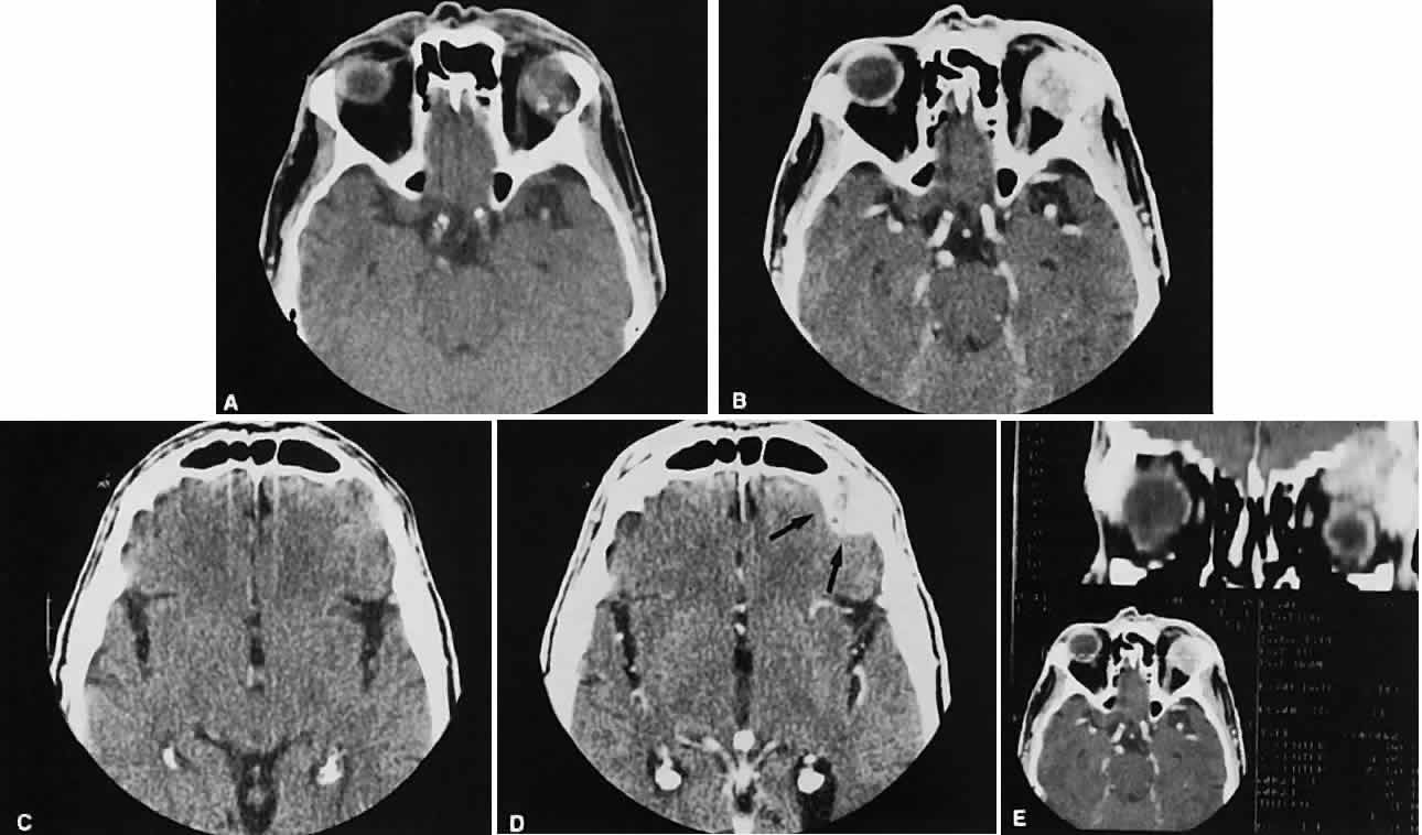
| Fig. 17. Adenocarcinoma of the lacrimal gland. A. Unenhanced axial view shows the speckled calcification of a lacrimal gland tumor. B. Contrast-enhanced view at about the same level shows intense enhancement and vascularity. In axial views at a higher level, an unenhanced scan (C) is unremarkable, although a contrast-enhanced view (D) at the same level highlights intracranial extension (arrows). E. Reconstructed coronal view has bone destruction with extension into the intracranial and temporalis fossae. |