|
|
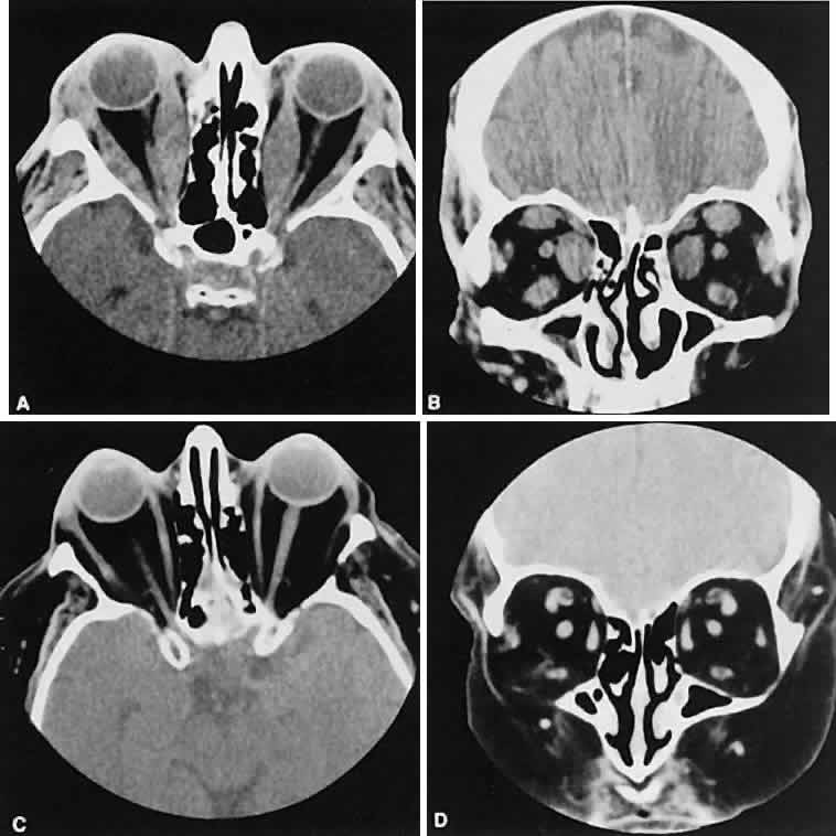
| Fig. 7. Graves' orbitopathy with two variations. Axial (A) and coronal (B) views show symmetric fusiform enlargement of the extraocular muscles with tapered muscle insertions. Note the predominant enlargement of the inferior, medial, and superior rectus muscles with lesser involvement of the lateral rectus muscle, a frequent pattern of enlargement in Graves' orbitopathy. Axial (C) and coronal (D) views of Graves' orbitopathy with expansion of retrobulbar ground substance and relative sparing of the extraocular muscles. |