|
|
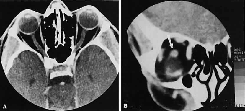
| Fig. 1. Normal anatomy. A. Axial scan through the plane of the optic nerve. Note the normal size of the horizontal rectus muscles and the undulating course of the optic nerve. B. Coronal view of the right orbit. The plane of section is slightly oblique to avoid dental artifacts and is immediately behind the right globe, which is partially volume averaged inferiorly. The arrow points to the optic nerve. |