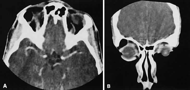
Fig. 19.
Axial (
A
) and coronal (
B
) scans of a dermoid cyst. Note the low attenuation (lower than retrobulbar fat) within the well-demarcated cyst located in the right lacrimal gland fossa.