|
|
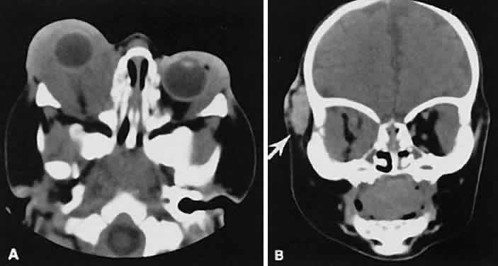
| Fig. 12. A. Axial view shows an exuberant homogenous soft-tissue mass infiltrating the retrobulbar space and periorbital tissue, typical of large infantile capillary hemangioma. B. Extension (arrow) into the adjacent periorbital tissues can be seen on the coronal view. |