Chapter 18
Pathology of Diabetes Mellitus
MARILYN C. KINCAID
Main Menu   Table Of Contents

Search

CONJUNCTIVA AND CORNEA
LENS
IRIS
UVEAL TRACT
RETINA
OPTIC NERVE
VITREOUS
REFERENCES

Diabetes mellitus is the leading cause of blindness in adults 20 to 74 years of age and can affect virtually every ocular structure. It has been estimated that patients with diabetes are 25 to 30 times more likely to become blind than persons of similar age who are not diabetic.1 Diabetes is an enormous public health problem, not only because of the ophthalmic complications, but also because of the neurologic and vascular sequelae, and the problem will only increase in magnitude as the population ages.2 Moreover, it is estimated that up to one half of all diabetic persons are unaware that they have the disease.3 For many patients, however, timely intervention can substantially reduce the likelihood of blindness.4

Although a number of factors have been implicated in the development of diabetic retinopathy and subsequent blindness, the overwhelming predictor is the length of time the patient has had diabetes.5 Moreover, since proliferative retinopathy does tend to occur along with diabetic nephropathy,6 life expectancy after development of severe retinopathy and blindness is often limited.

When insulin was first discovered, it was believed that diabetes would become a readily controlled disease. However, although it is still understood that the underlying problem in diabetes mellitus is glucose intolerance, many of the mechanisms of the disease process and complications are far from clear. Even the concept that longstanding hyperglycemia is responsible, in one way or another, for the complications of diabetes is somewhat controversial.7

The influence of the sorbitol metabolic pathway has only recently been elucidated. Both glucose and galactose are converted to fructose by this pathway, which is composed of two enzymes, aldose reductase and sorbitol dehydrogenase.7 Many ocular complications may be due to activation of aldose reductase, an enzyme that reduces glucose and galactose to sorbitol, the corresponding sugar alcohol. The effects of increased levels of aldose reductase include alterations in cell metabolism and increased production of basement membrane.8 However, a clinical trial of sorbinil, an aldose reductase inhibitor, failed to reduce the incidence of diabetic retinopathy. This may be because the trial lasted only 3 years, and the dosage of sorbinil may have been inadequate to alter aldose reductase in the retina.9

Other investigators7 have proposed that glycosylation of certain intracellular and extracellular proteins may also promote complications of diabetes. This attachment of a glucose molecule to the lysine residues within proteins is dependent on glucose concentration and occurs nonenzymatically but irreversibly. Subsequent crosslinking and deactivation of the proteins may then occur. What specific clinical effects this crosslinking causes are not known.7 Hemoglobin glycosylation, however, is a useful clinical marker: it is a more precise indicator of blood glucose levels in the recent past than is a direct measurement of the blood glucose level. Elevated levels of glycosylated hemoglobin do correlate with increased risk of retinopathy.10

Back to Top
CONJUNCTIVA AND CORNEA
Diabetes affects the cornea by interfering with the hemidesmosomes that anchor the epithelium to its basement membrane. The number of hemidesmosomes in diabetic persons is markedly reduced, a phenomenon that may be due to altered extracellular matrix. As a consequence of this reduction, the epithelium is relatively easily removed, and relatively minimal trauma can lead to corneal abrasions. Moreover, re-epithelialization takes much longer in diabetics than in nondiabetics,8 and these patients may develop recurrent erosion syndrome.11 This is a particular problem in patients undergoing vitrectomy, since the entire epithelium may be removed at surgery for visualization, resulting in delayed healing.8

Neurotrophic ulcers are a common manifestation of diabetic keratopathy. Corneal sensation is markedly decreased in diabetic patients, and they are prone to developing spontaneous erosions and corneal ulcers. The keratopathy can be associated with generalized peripheral neuropathy.12 The reason for epithelial instability in patients with corneal neuropathy is unknown, although nondiabetic causes of impaired sensation, such as sectioning of the trigeminal nerve, also lead to decreased transparency, thickening, and defects in the corneal epithelium. Patients with retinopathy, especially proliferative retinopathy, are more likely to have difficulties with corneal erosions.13

Histologically, studies in animals have disclosed that there is irregularity and thickening in the basement membranes of Schwann cells, the myelin-producing cells of the peripheral nervous system.14 These changes may indicate impaired function.

The corneas of diabetic patients have increased levels of aldose reductase activity in both the epithelium and endothelium. Corneal edema may result in part from an abnormally high level of sorbitol in the endothelium, which interferes with the sodium-potassium adenosine triphosphatase (ATPase) pump.7 Topical aldose reductase inhibitors have been used successfully on individual patients to treat recurrent erosion and neurotrophic ulcers.8 The epithelial mosaic, as disclosed by specular microscopy, reverted to normal in a 7-month period in one treated patient.15

Another finding in diabetic patients is pigment on the posterior surface of the cornea. Histologically, the pigment is located within the corneal endothelium, having been released and phagocytosed from the iris pigment epithelium. This finding is not specific, since there are many causes of endothelial pigment phagocytosis, including prior intraocular surgery.

Wrinkles in Descemet's membrane have been described as being more frequent in diabetic patients than in age-matched controls. These wrinkles clinically are fine, gray, branching linear streaks on the posterior surface of the cornea. They are distinct from the coarser folds seen in conditions of hypotony, although they are not specific for diabetes. No histology has been described.16

Back to Top
LENS
Patients with uncontrolled diabetes often present with unstable refractive error because the lens is subjected to changes in osmotic pressure induced by hyperglycemia. This can result in either a hyperopic or a myopic shift, depending on how the size and curvature of the lens is affected.17 Control of the blood sugar levels helps to stabilize the refractive error.

Cataracts are more frequent in patients with diabetes mellitus than in otherwise comparable nondiabetic persons.1 Lens opacities that occur in diabetic patients are indistinguishable from those occurring in nondiabetic patients, but they may develop in diabetics at an earlier age. Some of the more common lens opacities include anterior cortical spokes and posterior subcapsular plaques. Sometimes these lens opacities, especially the juvenile “snowflake” variety, are completely reversible when the disease is brought under better control.

A contributing factor is the aldose reductase pathway. Sorbitol, the metabolic product of aldose reductase, accumulates in lens fibers, but it cannot escape because the cell membrane is poorly permeable to it. The resulting hypertonicity causes water to enter, swelling the lens and damaging the lens fibers, although early changes may be reversible.8 Clinically and histologically, the cortical, nuclear, and posterior subcapsular cataracts in diabetic patients appear to be no different from those of nondiabetic patients (Fig. 1).18

Fig. 1. Partially liquefied lens fibers in a patient with diabetes mellitus. The lens changes are nonspecific. (H&E, original magnification × 19.5)

Back to Top
IRIS
Patients with diabetes may have tiny pinpoint iris transillumination defects. This phenomenon is particularly well seen in blue-eyed patients. The corresponding change seen histologically is lacy vacuolization of the iris pigment epithelium (Fig. 2; Color Plate 1,* Color Fig. A).18 These vacuoles contain glycogen. Some authors have found that the vacuolization is transient, and thus related to blood glucose levels prior to death,19 whereas others have been less convinced of such a relationship.20

Fig. 2. Lacy vacuolization of the iris pigment epithelium. Note the abrupt cessation of vacuolization at the iris root; also, there is great accentuation of the circumferential ridges (cut here meridionally). Although the pupillary portion is not involved here, it may become so and has been noted clinically in the region of the pupil. (H&E, × 38)

Color Plate 1 A. Lacy vacuolization of the iris pigment epithelium (H&E × 77.5). B. Neovascularization of the iris with peripheral anterior synechiae. The peripheral iris is adherent to the cornea at about the level of the termination of Descemet's membrane. The fibrovascular membrane is anterior to the anterior border of the iris (PAS, × 77.5). C. Ectropion uveae. The iris pigment epithelium is pulled anteriorly by the fibrovascular membrane. Note the folding of the sphincter muscle (H&E, × 77.5). D. Fundus appearance of microaneurysms, dot hemorrhages, and exudates, some in a typical circinate pattern found in background diabetic retinopathy. E. Exudates have collected in the outer plexiform layer, and appear eosinophilic. Two microaneurysms are present to the right in the figure, within the inner nuclear layer (H&E, × 77.5). F. New vessels originating at the disc partially obscure the underlying disc and retinal vessels. G. Dense, fibrous membrane on the retinal surface distorts and obscures the retina. To the lower left is a small retroretinal membrane, identified as such because the retinal vessels overlie it.


See Color Plate 1 for Color Figs. A through G

These glycogen vacuoles can rupture when the intraocular pressure is suddenly reduced, resulting in a cloud of pigment granules carried anteriorly in the aqueous fluid. This pigment may then be phagocytosed by the corneal endothelium. The pupils of patients with diabetes often dilate poorly, and the amount of glycogen in the pigment epithelium may correlate to damage to the dilator muscle.

Rubeosis iridis is a clinical term for the reddish discoloration imparted by neovascularization of the iris surface. It is associated most frequently with retinal ischemia and neovascularization, and indicates the presence of a diffusible vascular proliferative factor secreted by the retina.21 Initially, the vessels can be quite difficult to see. With more advanced rubeosis, there may be ectropion uveae, an eversion of the posterior iris surface at the pupil margin. The earliest location of the new vessels has been a matter of longstanding debate. They appear at the pupil margin and in the angle virtually simultaneously, subsequently spreading across the iris surface, but gonioscopic examination is required for the vessels to be seen in the angle. In some patients, the vessels do form in the angle before being perceptible at the pupil margin.22

Peripheral anterior synechiae form rapidly after neovascularization of the iris, closing off the trabecular meshwork and giving rise to so-called neovascular glaucoma, which is difficult to treat. In addition, the vessels and associated fibrous tissue can proliferate across the pupil, sequestering it. Particularly in aphakic patients, the vessels can also extend through the pupil and onto the posterior iris surface.

Histologically the new vessels are located anterior to the anterior border layer of the iris (Fig. 3, Color Fig. B). They are thin-walled, unlike the normal thick-walled iris stromal vessels. New vessels may be located only at the pupillary margin and in the angle at the iris root, corroborating the clinical finding.18

Fig. 3. Neovascularization glaucoma. Rubeosis iridis tends to bleed and lead to hyphema; the condition has incorrectly been called hemorrhagic glaucoma. The hemorrhage is not responsible for the glaucoma, but the rubeosis iridis and peripheral anterior synechias (arrow) are. Note the lacy vacuolization of the iris pigment epithelium. (H&E, × 75)

Scanning electron microscopy has disclosed a thin, transparent layer of myofibroblastic proliferation anterior to the new vessels. This explains the clinical observation that the anterior iris surface appears smoothed out, with loss of the collarette and crypts typically seen on the normal iris surface. Myofibroblasts have contractile properties and thus may be responsible for the angle closure of neovascular glaucoma and also for the eversion of the posterior iris surface at the pupillary margin (Color Fig. C).23

Back to Top
UVEAL TRACT
For reasons that remain obscure, the basement membrane of the pigment epithelium of the pars plicata of the ciliary body is thickened in diabetes. The basement membrane of the nonpigmented epithelium is also thickened, but this is much more subtle (Fig. 4).18

Fig. 4. Marked thickening of the basement membrane of the ciliary pigmented epithelium. (PAS, × 125)

Arteriosclerotic changes in the choroidal vessels are more common in diabetic patients than in matched nondiabetics. These changes include fibrosis and hyalinization of the media with plaque formation in larger vessels.18

The choriocapillaris shows diffuse thickening of the basement membrane, sometimes to the point of total vascular occlusion. Both pericytes and endothelial cells appear to participate in this process of excess basement membrane synthesis. The obliterated capillaries form eosinophilic nodules that superficially resemble drusen, although they are on the choroidal side of Bruch's membrane. They also resemble the nodules seen in kidneys of patients with diabetic glomerulosclerosis.24

Two patients in Hidayat and Fine's series,24 both less than 30 years old and who had not been treated with photocoagulation, showed peripheral subretinal neovascularization, one near the equator and one near the ora. The significance of this finding is uncertain.

Back to Top
RETINA
Diabetic retinopathy can be divided into two types: nonproliferative retinopathy (sometimes called “background” retinopathy) and proliferative retinopathy. “Retinitis proliferans” is an obsolete term for proliferative retinopathy and is inaccurate because the neovascularization is not caused by an inflammatory process. Nonproliferative changes take place within the sensory retina. Proliferative retinopathy includes neovascularization and its sequelae; these changes occur internal to the retinal surface.

In fact, it is not known what prompts the development of diabetic retinopathy, although ischemia clearly plays a role.25 Certain alterations have been observed in blood from diabetic patients, including increased rigidity and aggregation of the erythrocytes, altered platelet activity, changes in plasma proteins, and increased affinity of hemoglobin for oxygen. Both the increased aggregates of platelets and the abnormally rigid erythrocytes can occlude small blood vessels, contributing to retinal ischemia. The increased affinity of hemoglobin for oxygen means that less oxygen is released to the tissues.26

Whether tightly controlling blood sugar levels improves retinopathy or even halts its progression is controversial. In one investigation,27 dogs were made diabetic and put into three groups: good control for 5 years, poor control for 5 years, and poor control for 2½ years followed by good control for the remaining time. The third group had developed almost as many microaneurysms at 5 years as did the group under poor control for the entire time. This study indicates that tight control is maximally beneficial when undertaken early in the course of the disease.

Human studies to corroborate or refute this have been understandably difficult to carry out. Transplantation of the pancreas or pancreatic islet cells is one way to normalize blood glucose by restoring endogenous insulin. In one study, eight patients who received pancreatic transplants exhibited no difference in retinopathy between the four whose transplants functioned and the four whose transplants failed.28 In another small study, the proliferative retinopathy of patients who received pancreatic transplants remained stable or worsened, despite their being euglycemic.29

Recently, results from a large prospective series, the Diabetes Control and Complications Trial, have shown that tight control, including three or more daily insulin injections or continuous infusion, based on at least four glucose determinations daily, does prevent development or progression of retinopathy to a significant degree. All patients in the primary prevention group had diabetes for 5 years or less and did not have retinopathy at the outset of the study. Thus, tight control begun early may indeed help prevent or delay the onset of retinopathy.30

As noted previously, duration of disease is a strong predictor for development of retinopathy.31,32 Prepubertal children may develop minimal retinopathy,33 but the blood-retinal barrier is altered during puberty, apparently through hormonal influence, leading to retinopathy.34

Unless the macula is edematous, nonproliferative retinopathy is asymptomatic and may be found only on routine ophthalmoscopy. In patients who develop diabetes after puberty, retinopathy may be a presenting sign of the disease.

The earliest manifestation of diabetic retinopathy is the appearance of microaneurysms (Fig. 5, Color Fig. D). They appear as small red dots clinically and often are seen more readily on fluorescein angiography, where they fill and leak, with staining of the vessel wall.35 Histologically they are small “outpouchings” of the capillary vessel wall and are usually saccular but occasionally fusiform in shape. They can occur anywhere along the capillary network between the arteriole and venule.18 Some microaneurysms, however, do not fill with fluorescein and have been shown to be thin-walled capillary outpouchings filled with erythrocytes. They may represent either one stage of microaneurysm formation or a specific type of it.36

Fig. 5. Retinal capillary microaneurysm (arrow) is characterized by its thin wall and location in the capillary area of the retina (middle retinal layers) rather than the major vessel area (inner retinal layers). Inset. Fundus appearance of microaneurysms and hard or waxy exudates. (Main figure, H&E, × 176)

As shown by trypsin digestion, capillary pericytes are lost first,27 followed by endothelial cells (Figs. 6 and 7). Aldose reductase, one of the enzymes in the sorbitol metabolic pathway, is present in significant quantities in retinal capillary pericytes, but not in endothelial cells, and may thus be implicated in the formation of microaneurysms.37 The final result is an acellular segment of capillary that is nonperfused on fluorescein angiography.36

Fig. 6. Diabetic retinal capillary. A. Basement membrane shell (arrows) is the only remaining indication of where the pericytes had been. B. Nondiabetic normal capillary shows the basement membrane shell (arrow) around the pericyte. C. Diabetic capillary has only a basement membrane shell (arrow), with the nucleus absent. (A, PAS, × 630; B, PAS, × 850; C, PAS, × 630)

Fig. 7. Retinal capillary microaneurysm (RCM). A. RCMs occur in random distribution between the arteriole (a) and venule (v). “Young” RCMs are seen as saccular capillary outpouchings with proliferated endothelial cells (arrows). “Old” RCMs appear as solid black balls with their lumens obliterated by PAS-positive material. Note the darker color of the capillaries with thickened basement membranes and arteriolar-venular connections. B. Very large RCM (arrow) or the tiny hemorrhages associated with abnormal vessels are probably responsible for the RCMs seen clinically. (A, PAS, × 40; B, PAS, × 115)

The normal retinal vasculature, including the capillaries, forms a barrier to the free exchange of metabolites.38 This blood-retinal barrier is damaged in diabetes, allowing leakage of larger molecules, which are clinically manifested as hard exudates and retinal edema. Ultrastructurally, the normal junctions between capillary endothelial cells are disrupted.39

Hard exudates clinically appear as shiny yellow deposits (see Fig. 5; Color Fig. D). When they surround a microaneurysm in a circular pattern, the condition is called circinate retinopathy. Histologically, the deposits accumulate at the level of the outer plexiform layer (Fig. 8, Color Fig. E) and consist of lipids, complex carbohydrates, and proteins, as demonstrated histochemically. Hard exudates are normally asymptomatic unless they involve the macula, where they cause a drop in visual acuity. If a leaking microaneurysm can be identified on fluorescein angiography, laser photocoagulation can obliterate it. Over time, the fluid is resorbed, and sometimes visual acuity can improve.40

Fig. 8. Hard or waxy exudates. Retinal exudates accumulate in the outer plexiform (Henle's fiber) or in inner nuclear (bipolar cell) layers. Note that in this case the middle limiting membrane separates the layers. Inset. Fundus appearance of a moderate background diabetic retinopathy. (Main figure, H&E, × 220)

Cystoid macular edema can be caused by many different diseases as well as diabetes. It is characterized by diffuse leakage of the capillaries in the posterior pole. In advanced cases, the cystoid spaces are ophthalmoscopically visible, resembling the petals of a flower. Cystoid edema is seen more easily on fluorescein angiography, since the spaces fill with fluorescein.

Histologically, cystoid fluid is seen at the outer plexiform layer, but may also accumulate in the inner nuclear layer. The synapses of the outer plexiform layer are slanted in the fovea because the inner retinal layers are pushed away from the foveal pit. Because of these slanted connections, this area can be distended by fluid more readily. Ultrastructurally, the fluid appears to be located within Müller cells, and there is evidence of capillary endothelial damage.41

In some patients, the cystoid spaces can coalesce, forming lamellar or full-thickness macular holes.42 Other nonproliferative changes include intraretinal hemorrhages. Flame-shaped hemorrhages occur in the nerve fiber layer, and dot-blot hemorrhages occur in deeper retina, in the outer plexiform and inner nuclear layers (Fig. 9).

Fig. 9. Dot and blot hemorrhages consist of small collections of blood in the inner nuclear and outer plexiform layers of the retina. Insets. Fundus appearance of dot (1) and blot (2) hemorrhages, respectively. (Main figure, H&E, × 260)

Nonproliferative retinal changes that have been associated statistically with an increased likelihood of development of proliferative retinopathy have sometimes been termed proproliferative retinopathy. These changes indicate an increased level of retinal ischemia.

Cotton-wool spots are areas of capillary closure. The ischemic axons swell, with resultant loss of retinal transparency. They occur frequently in diabetics with and without hypertension.43 Clinically, they appear as white, feathery spots, commonly at the bifurcation of retinal vessels. On fluorescein angiography, these areas appear dark, both because of focal capillary closure and nonperfusion, and because the focal retinal swelling obscures the subjacent choroidal fluorescence.

Histologically, the axons of the nerve fiber layer are focally swollen in a fusiform configuration and have been called cytoid bodies because the central density superficially resembles a cell with its nucleus.44 Depending on the severity of the ischemia, individual ganglion cells may recover or be lost.20

Capillary dropout in the fovea is an important cause of untreatable visual loss, even if there is no macular edema.43 The foveal avascular zone is significantly larger and more irregular in contour in patients with diabetic retinopathy than in control patients,45 even when the patients have no clinical visual loss.43

The retinal arterioles may become narrowed and irregular. Patients with diabetes are likely to have arteriosclerotic changes in the retina as well as systemically. The arterial wall is thickened, with resultant narrowing of the blood column. Clinically, this narrowing is manifest as a “copper-wire” change and with greater severity, as a “silver-wire” change. These descriptions reflect the narrowed and relatively less visible column of erythrocytes. Even severely narrowed, silver-wire arterioles, however, may still allow some passage of fluorescein.

The arterioles may ultimately become occluded,20 and this phenomenon can proceed rapidly, with accompanying capillary dropout.43 Extensive capillary and arteriolar nonperfusion can be even more prominent in the midperipheral retina than posteriorly.46

The retinal veins can also become irregular and tortuous with sausage-shaped dilatations. The vascular walls become diffusely permeable so that they both leak and stain with fluorescein. These changes also seem to be associated with ischemia. As shown by trypsin digestion, the beaded areas are hypercellular (Fig. 10).43 Diabetes is also a risk factor for branch retinal vein occlusion47 and central vein occlusion.43

Fig. 10. A and B. Sausage-shaped venules (arrows) result from irregularities in the venular walls. Note the arteriolar-venular connections and thickened capillary basement membranes (dark-colored capillaries). Retinal capillary microaneurysms tend to arise from cellular (viable) capillaries and cluster around acellular (nonviable) capillaries. The venular walls not only are irregular, but they also have an unusual presence of saccular microaneurysms (Inset 1, and Inset 3 from the area of the double arrows). Inset 2. Fundus appearance of microaneurysms, a cotton-wool spot, irregular venules, and intraretinal neovascularization in the form of a rete mirabile. (a = arteriole; v = venule) (A and B, PAS, × 16; Inset 1, PAS, × 40; Inset 2, fundus; Inset 3, PAS × 54)

Other capillary changes, more severe and extensive than microaneurysms, are collectively termed intraretinal microvascular abnormalities (IRMAs). Clinically, these manifest as dilated channels that leak fluorescein and show vascular wall staining (Fig. 11). De Venecia and co-workers36 were able to distinguish between IRMAs arising from arteriolar capillaries and IRMAs arising from venular capillaries. The IRMAs associated with arterioles exhibited early filling and late leakage on angiography. Histologically, they appeared as solid proliferations of endothelial cells, adjacent to acellular capillaries. In some instances, they may represent true intraretinal neovascularization. The IRMAs adjacent to venules filled in the early venous phase and leaked late. Histologically, they were irregularly dilated, had thin walls, and may have represented shunts.

Fig. 11. Arteriolar-venular connection (collateral) consists of a dilated cellular capillary (arrow) with an increased number of anuclear (apparently nonviable) capillaries. (PAS, × 16)

All of these changes are forerunners of proliferative retinopathy. Patients are unlikely to develop proliferative retinopathy before 10 years' duration of diabetes, but thereafter the incidence rises steadily.31,32 Rarely, proliferative retinopathy with vitreal hemorrhage can be the presenting sign in younger patients.48

As in other diseases associated with retinal neovascularization, the stimulus for new vessel formation in diabetes appears to be ischemia. The retina is uniquely dependent on oxygen: its demand on a unit-to-weight basis exceeds that of all other tissues, including the brain.7 A number of growth factors may act on the retina, including somatomedin C (insulin-like growth factor I), platelet-derived endothelial growth factor, fibroblast growth factors, angiogenin, and others.49 According to one recent study of samples of neovascular membranes obtained at vitrectomy from diabetic patients, only vascular endothelial growth factor was present in all specimens.50 This same factor is also present at significantly higher levels in the vitreous in eyes with proliferative retinopathy than in those without the disease.51 The precise role of the different factors in the promotion of neovascularization remains to be elucidated.

New vessels that develop on or within a disc diameter of the optic nerve head are termed new vessels at the disc (NVD) (Color Fig. F). Those that develop anywhere else on the retina are called new vessels elsewhere (NVE). NVD indicate a diffuse retinal ischemia, whereas with NVE, the ischemia may be more localized, occurring at a site of previous arteriolar closure or capillary dropout. NVE can occur peripherally as well as in the posterior pole.52

Histologically, new vessels develop between the retinal surface and the posterior surface of the formed vitreous (Fig. 12). With NVE, there is a demonstrable break through the internal limiting membrane (Fig. 13).20 Early-developing new vessels have lumina even at the proliferating tips.53 Active new vessels have endothelial cells and pericytes, with fenestrations. Morphologically, fenestrations are areas ranging from 40 to 80 nm in diameter where the cell membranes are focally fused, and are normal findings in capillaries outside of the retina and brain. They allow increased permeability and are responsible for the diffuse leakage of fluorescein seen from new vessels on angiography. Inactive or “ghost” new vessels are acellular, having lost both endothelium and pericytes.39 With time, fibrous tissue develops and accompanies the new vessels. The vessels themselves may leak and bleed into the vitreous, leading to visual loss.

Fig. 12. Neovascularization at the disc with fibrous proliferation. The fibrovascular membrane overlies the internal limiting membrane, which is wrinkled (arrows). The retinal detachment is artifactual. (PAS, × 19.5)

Fig. 13. Neovascularization away from the disc. The fibrous tissue has contracted, imparting a triangular shape to the membrane. Active new vessels are present throughout. The dark folds are artifactual. (H&E, × 31.25)

The fibrous tissue is contractile, and has been shown to contain actin filaments.54 Clinically, fibrous proliferation appears as whitish bands on the retinal surface, sometimes obscuring the neovascularization. With retinal elevation, retroretinal membranes also can form.

Traction can cause cystoid changes in the retina20 and venous loops,55 as well as retinal detachment (Color Fig. G). Extensive traction can cause macular heterotopia reminiscent of retinopathy of prematurity.56

Histologically, the cells in the membranes appear to be fibrous astrocytes. At least in some cases, however, there is immunohistochemical evidence that cells of Müller can also migrate and form membranes. Intraretinal migration and replacement of outer retinal layers by cells of Müller can appear clinically as retroretinal proliferation.57 Other cells found in preretinal membranes include inflammatory cells (i.e., lymphocytes, plasma cells, and macrophages) and retinal pigment epithelial cells.58

Panretinal photocoagulation, usually accomplished with an argon laser, is used to treat neovascularization of the retina and iris. The Diabetic Retinopathy Study Research Group59 has established the efficacy of this procedure in preventing severe visual loss, a measure that appears to last for at least 15 years.60

Radiant energy in the visible spectrum passes through the transparent retina and is absorbed by melanin pigment, hemoglobin, or both, depending on the wavelength chosen. The result is a focal scar of the outer retina, or if intense enough, full-thickness retina (Fig. 14).61 Full-thickness scars can cause extensive visual field loss because of obliteration of the nerve fiber layer.20

Fig. 14. Low-power view of panretinal photocoagulation. Normal retina is to the extreme right. Centrally there are areas of outer retinal loss (center arrows), and to the extreme left (single arrow) is an area of full-thickness retinal loss with migrated pigment epithelium. (H&E, × 19.5)

The choriocapillaris is effectively closed by photocoagulation, as demonstrated by studies of vascular casts.62 After panretinal photocoagulation, the oxygen tension on the inner retinal surface is greater over areas of scarring compared with adjacent areas of intact retina, presumably due in part to greater oxygen diffusion from the larger vessels of the choroid. The increased oxygen levels appear to alleviate retinal ischemia and help explain how photocoagulation causes retinal neovascular regression.63 This finding has been substantiated by direct measurements of oxygen tension in human patients undergoing vitrectomy.64

In the juxtafoveal area, krypton red was associated with less inner retinal damage than argon green.65,66 This is of clinical importance in the treatment of macular edema. The wavelength used, however, appears to be unimportant in extrafoveal areas of the retina.66

Macular grid photocoagulation has been proposed as an effective way to treat macular edema when specific leaking points cannot be identified. In animal studies, this technique appeared to work by occluding some capillaries and reducing the lumen in others, thereby decreasing the ratio of the retinal capillary area to the retinal area. The photoreceptors and retinal pigment epithelium were initially damaged but later recovered.67

Back to Top
OPTIC NERVE
Diabetic optic nerve swelling is also called diabetic papillopathy. The pathogenesis is unknown, but it is usually bilateral and usually occurs in young diabetic patients. The clinical course is generally benign, with spontaneous recovery of vision and peripheral fields. There is no specific test, and other causes of disc swelling, including papilledema from increased intracranial pressure, must be ruled out. It may also occur in older patients. Occasionally it is more serious, producing optic nerve head neovascularization and permanent visual loss.68 Diabetic papillopathy may thus be a result of microangiopathy and ischemia in some patients, but the histology is unknown.

Diabetes is a risk factor for anterior ischemic optic neuropathy, a unilateral cause of optic atrophy that is more frequent in older patients. Typically, the patient experiences a sudden, profound visual loss and has a poor prognosis for recovery. An inferior altitudinal field defect is typical clinically, corresponding histologically to loss of superior fibers in the optic nerve. Also lost are the fibers serving the peripheral visual fields.69

Diabetic patients may also develop an orbital apex syndrome consisting of retro-orbital ischemic optic neuropathy, ophthalmoplegia, and pain. In one study,70 two patients who had mild diabetes lost all vision but did recover extraocular motility. Histologic study of the optic nerve from one of the patients revealed ischemic necrosis, and the authors concluded that this is another example of diabetic microangiopathy.

Back to Top
VITREOUS
The incidence of posterior vitreous detachment increases with age, and this phenomenon is more frequent and occurs earlier in diabetic patients, even in those without retinopathy.71

Neovascular fronds grow along the surface of the retina and along the posterior formed vitreous. Shifts in the position of the formed vitreous may thus lead to preretinal hemorrhage, although this most frequently occurs during sleep rather than activity.

Removal of the vitreous, or vitrectomy, is performed on diabetic eyes for several reasons: one indication is the presence of nonresorbing preretinal and vitreal blood; another is membrane peeling or division, often along with repair of traction retinal detachment. Postoperatively, foci of new vessels appear to regress, although in the case studied by Foos and associates,72 the tissue sites appeared as cellular and vascular as those of the opposite, unoperated eye.

One complication that may occur after cataract extraction73 and vitrectomy74 in diabetic eyes is anterior hyaloidal fibrovascular proliferation. Neovascularization accompanied by fibrous tissue extends from the retina close to the ora serrata, along the anterior hyaloid face inward along the posterior surface of the lens or lens remnant, and may lead to recurrent traction retinal detachment. This condition differs from fibrovascular ingrowth from the sclerotomy site or sites, and can be present in any meridian. It may be possible, however, to differentiate the two only on a histologic basis. The patient may have a good prognosis, having limited fibrovascular growth,73 but the eye may be lost as a result of secondary rubeosis iridis and traction retinal detachment.74

Asteroid hyalosis occurs as an aging change and in a variety of ocular diseases, including malignant melanoma. There is no specific association between diabetes and asteroid hyalosis.75 Patients with asteroid hyalosis who had diabetes were generally younger than those with asteroid who did not have diabetes.76


Supported in part by an unrestricted grant from Research to Prevent Blindness, Inc.
Back to Top
REFERENCES

1. Sprafka JM, Fritsche TL, Baker R et al: Prevalence of undiagnosed eye disease in high-risk diabetic individuals. Arch Intern Med 150:857, 1990

2. Javitt JC, Canner JK, Frank RG et al: Detecting and treating retinopathy in patients with type I diabetes mellitus: a health policy model. Ophthalmology 97:483, 1990

3. Harris MI, Hadden WC, Knowler WC et al: Prevalence of diabetes and impaired glucose tolerance and plasma glucose levels in U.S. population aged 20–74 yr. Diabetes 36:523, 1987

4. Diabetic Retinopathy Study Research Group: Photocoagulation treatment of proliferative diabetic retinopathy: Clinical application of Diabetic Retinopathy Study (DRS) findings, DRS report number 8. Ophthalmology 88:583, 1981

5. Marshall G, Garg SK, Jackson WE et al: Factors influencing the onset and progression of diabetic retinopathy in subjects with insulin-dependent diabetes mellitus. Ophthalmology 100:1133, 1993

6. Kostraba JN, Klein R, Dorman JS et al: The epidemiology of diabetes complications study: IV. Correlates of diabetic background and proliferative retinopathy. Am J Epidemiol 133:381, 1991

7. Frank RN: On the pathogenesis of diabetic retinopathy: a 1990 update. Ophthalmology 98:586, 1991

8. Kinoshita JH: Aldose reductase in the diabetic eye: XLIII Edward Jackson Memorial Lecture. Am J Ophthalmol 102:685, 1986

9. Sorbinil Retinopathy Trial Research Group: A randomized trial of sorbinil, an aldose reductase inhibitor, in diabetic retinopathy. Arch Ophthalmol 108:1234, 1990

10. Goldstein DE, Blinder KJ, Ide CH et al: Glycemic control and development of retinopathy in youth-onset insulin-dependent diabetes mellitus: results of a 12-year longitudinal study. Ophthalmology 100:1125, 1993

11. Azar DT, Spurr-Michaud SJ, Tisdale AS et al: Altered epithelial-basement membrane interactions in diabetic corneas. Arch Ophthalmol 110:537, 1992

12. Schultz RO, Peters MA, Sobocinski K et al: Diabetic keratopathy as a manifestation of peripheral neuropathy. Am J Ophthalmol 96:368, 1983

13. Hyndiuk RA, Kazarian EL, Schultz RO et al: Neurotrophic corneal ulcers in diabetes mellitus. Arch Ophthalmol 95:2193, 1977

14. Ishida N, Rao GN, del Cerro M et al: Corneal nerve alterations in diabetes mellitus. Arch Ophthalmol 102:1380, 1984

15. Tsubota K, Yamada M: The effect of aldose reductase inhibitor on the corneal epithelium. Cornea 12:161, 1993

16. Henkind P, Wise GN: Descemet's wrinkles in diabetes. Am J Ophthalmol 51:371, 1961

17. Eva PR, Pascoe PT, Vaughn DG: Refractive change in hyperglycemia: hyperopia, not myopia. Br J Ophthalmol 66:500, 1982

18. Yanoff M: Ocular pathology of diabetes mellitus. Am J Ophthalmol 67:21, 1969

19. Smith ME, Glickman P: Diabetic vacuolation of the iris pigment epithelium. Am J Ophthalmol 79:875, 1975

20. Kincaid MC, Green WR, Fine SL et al: An ocular clinicopathologic correlative study of six patients from the Diabetic Retinopathy Study. Retina 3:218, 1983

21. Gartner S, Henkind P: Neovascularization of the iris (rubeosis iridis). Surv Ophthalmol 22:291, 1978

22. Blinder KJ, Friedman SM, Mames RN: Diabetic iris neovascularization. Am J Ophthalmol 120:393, 1995

23. John T, Sassani JW, Eagle RC Jr: The myofibroblastic component of rubeosis iridis. Ophthalmology 90:721, 1983

24. Hidayat AA, Fine BS: Diabetic choroidopathy: light and electron microscopic observations of seven cases. Ophthalmology 92:512, 1985

25. D'Amore PA: Mechanisms of retinal and choroidal neovascularization. Invest Ophthalmol Vis Sci 35:3974, 1994

26. Little HL: Alterations in blood elements in the pathogenesis of diabetic retinopathy. Ophthalmology 88:647, 1981

27. Engerman RL, Kern TS: Progression of incipient diabetic retinopathy during good glycemic control. Diabetes 36:808, 1987

28. Petersen MR, Vine AK, Committee T: Progression of diabetic retinopathy after pancreas transplantation. Ophthalmology 97:496, 1990

29. Friberg TR, Tzakis AG, Carroll PB et al: Visual improvement after long-term success of pancreatic transplantation. Am J Ophthalmol 110:564, 1990

30. The Diabetes Control and Complications Trial Research Group: Progression of retinopathy with intensive versus conventional treatment in the Diabetes Control and Complications Trial. Ophthalmology 102:647, 1995

31. Klein R, Klein BEK, Moss SE et al: The Wisconsin Epidemiologic Study of Diabetic Retinopathy: II. Prevalence and risk of diabetic retinopathy when age at diagnosis is less than 30 years. Arch Ophthalmol 102:520, 1984

32. Klein R, Klein BEK, Moss SE et al: The Wisconsin Epidemiologic Study of Diabetic Retinopathy: III. Prevalence and risk of diabetic retinopathy when age at diagnosis is 30 or more years. Arch Ophthalmol 102:527, 1984

33. Murphy RP, Nanda M, Plotnick L et al: The relationship of puberty to diabetic retinopathy. Arch Ophthalmol 108:215, 1990

34. Faria de Abreu JR, Silva R, Cunha-Vaz JG: The blood-retinal barrier in diabetes during puberty. Arch Ophthalmol 112:1334, 1994

35. Bresnick GH, Davis MD, Myers FL et al: Clinicopathologic correlations in diabetic retinopathy: II. Clinical and 
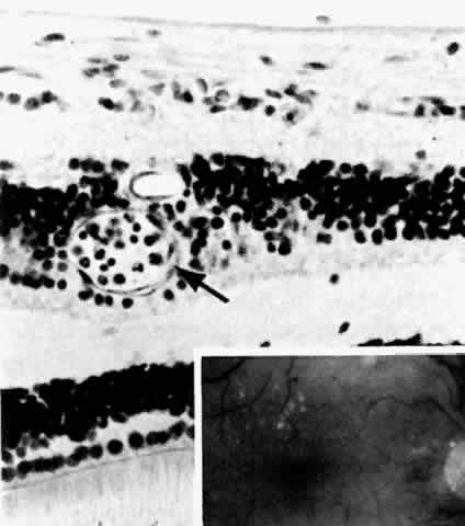
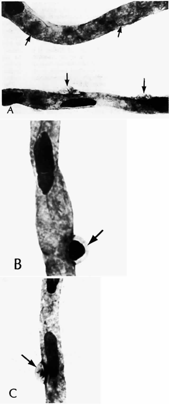
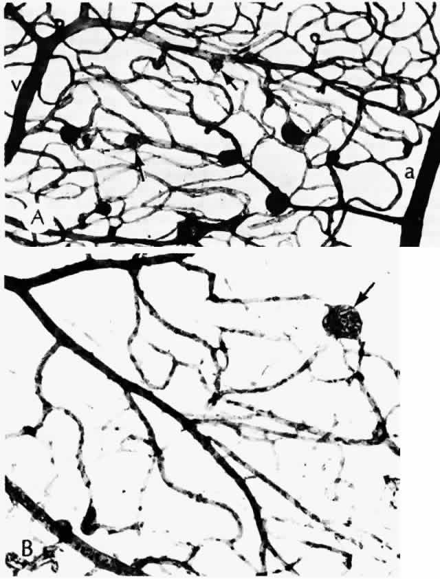
histologic appearances of retinal capillary microaneurysms. Arch Ophthalmol 95:1215, 1977

36. de Venecia G, Davis M, Engerman R: Clinicopathologic correlations in diabetic retinopathy: I. Histology and fluorescein angiography of microaneurysms. Arch Ophthalmol 94:1766, 1976

37. Akagi Y, Kador PF, Kuwabara T et al: Aldose reductase localization in human retinal mural cells. Invest Ophthalmol Vis Sci 24:1516, 1983

38. Cunha-Vaz J: The blood-ocular barriers. Surv Ophthalmol 23:279, 1979

39. Wallow IHL, Geldner PS: Endothelial fenestrae in proliferative diabetic retinopathy. Invest Ophthalmol Vis Sci 19:1176, 1980

40. Reeser F, Fleischman J, Williams GA et al: Efficacy of argon laser photocoagulation in the treatment of circinate 
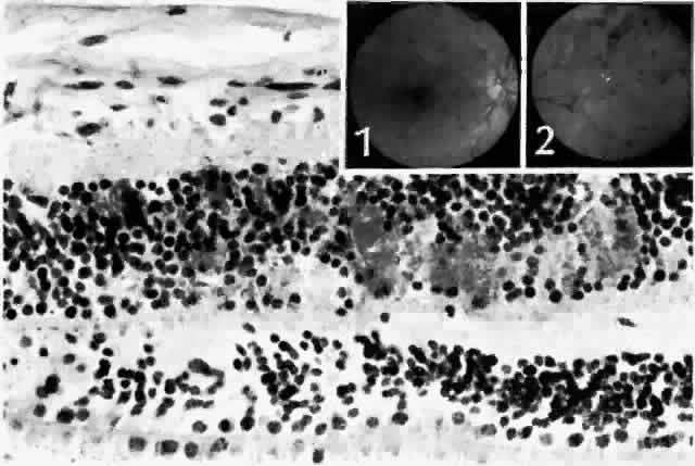
diabetic retinopathy. Am J Ophthalmol 92:762, 1981

41. Fine BS, Brucker AJ: Macular edema and cystoid macular edema. Am J Ophthalmol 92:466, 1981

42. Frangieh GT, Green WR, Engel HM: A histopathologic study of macular cysts and holes. Retina 1:311, 1981

43. Bresnick GH, Engerman R, Davis MD et al: Patterns of ischemia in diabetic retinopathy. Trans Am Acad Ophthalmol Otolaryngol 81:OP-694, 1976

44. Wolter JR: Axonal enlargements in the nerve-fiber layer of the human retina. Am J Ophthalmol 65:1, 1968

45. Mansour AM, Schachat A, Bodiford G et al: Foveal avascular zone in diabetes mellitus. Retina 13:125, 1993

46. Shimizu K, Kobayashi Y, Muraoka K: Midperipheral fundus involvement in diabetic retinopathy. Ophthalmology 88:601, 1981

47. Bowers DK, Finkelstein D, Wolff SM et al: Branch retinal vein occlusion: A clinicopathologic case report. Retina 7:252, 1987

48. O'Hare JA: Proliferative retinopathy and nephropathy at presentation in young insulin dependent diabetics. Br Med J 300:579, 1990

49. Wiedemann P: Growth factors in retinal diseases: Proliferative vitreoretinopathy, proliferative diabetic retinopathy, and retinal degeneration. Surv Ophthalmol 36:373, 1992

50. Malecaze F, Clamens S, Simorre-Pinatel V et al: Detection of vascular endothelial growth factor messenger RNA and vascular endothelial growth factor-like activity in proliferative diabetic retinopathy. Arch Ophthalmol 112:1476, 1994

51. Adamis AP, Miller JW, Bernal M-T et al: Increased vascular endothelial growth factor levels in the vitreous of eyes with proliferative diabetic retinopathy. Am J Ophthalmol 118:445, 1994

52. Jampol LM, Goldbaum MH: Peripheral proliferative retinopathies. Surv Ophthalmol 25:1, 1980

53. Williams JM Sr, de Juan E Jr, Machemer R: Ultrastructural characteristics of new vessels in proliferative diabetic retinopathy. Am J Ophthalmol 105:491, 1988

54. Wallow IHL, Greaser ML, Stevens TS: Actin filaments in diabetic fibrovascular preretinal membrane. Arch Ophthalmol 99:2175, 1981

55. Hersh PS, Green WR, Thomas JV: Tractional venous loops in diabetic retinopathy. Am J Ophthalmol 92:661, 1981

56. Michael JC, de Venecia G, Bresnick GH: Macular heterotopia in proliferative diabetic retinopathy. Arch Ophthalmol 112:1455, 1994

57. Nork TM, Wallow IHL, Sramek SJ et al: Müller's cell involvement in proliferative diabetic retinopathy. Arch Ophthalmol 105:1424, 1987

58. Hamilton CW, Chandler D, Klintworth GK et al: A transmission and scanning electron microscopic study of surgically excised preretinal membrane proliferations in diabetes mellitus. Am J Ophthalmol 94:473, 1982

59. The Diabetic Retinopathy Study Research Group: Photocoagulation treatment of proliferative diabetic retinopathy: the second report of Diabetic Retinopathy Study findings. Ophthalmology 85:82, 1978

60. Blankenship GW: Fifteen-year argon laser and xenon photocoagulation results of Bascom Palmer Eye Institute's patients participating in the Diabetic Retinopathy Study. Ophthalmology 98:125, 1991

61. Swartz M: Histology of macular photocoagulation. Ophthalmology 93:959, 1986

62. Wilson DJ, Green WR: Argon laser panretinal photocoagulation for diabetic retinopathy: scanning electron microscopy of human choroidal vascular casts. Arch Ophthalmol 105:239, 1987

63. Landers MB, III, Stefansson E, Wolbarsht ML: Panretinal photocoagulation and retinal oxygenation. Retina 2:167, 1982

64. Stefánsson E, Machemer R, de Juan E Jr et al: Retinal oxygenation and laser treatment in patients with diabetic retinopathy. Am J Ophthalmol 113:36, 1992

65. Thomas EL, Apple DJ, Swartz M et al: Histopathology and ultrastructure of krypton and argon laser lesions in a human retina-choroid. Retina 4:22, 1984

66. Smiddy WE, Fine SL, Green WR et al: Clinicopathologic correlation of krypton red, argon blue-green, and argon green laser photocoagulation in the human fundus. Retina 4:15, 1984

67. Wilson DJ, Finkelstein D, Quigley HA et al: Macular grid photocoagulation: an experimental study on the primate retina. Arch Ophthalmol 106:100, 1988

68. Ho AC, Maguire AM, Yannuzzi LA et al: Rapidly progressive optic disk neovascularization after diabetic papillopathy. Am J Ophthalmol 120:673, 1995

69. Quigley HA, Miller NR, Green WR: The pattern of optic nerve fiber loss in anterior ischemic optic neuropathy. Am Ophthalmol 100:769, 1985

70. Jabs DA, Miller NR, Green WR: Ischaemic optic neuropathy with painful ophthalmoplegia in diabetes mellitus. Br J Ophthalmol 65:673, 1981

71. Foos RY, Kreiger AE, Forsythe AB et al: Posterior vitreous detachment in diabetic subjects. Ophthalmology 87:122, 1980

72. Foos RY, Kreiger AE, Nofsinger K: Pathologic study following vitrectomy for proliferative diabetic retinopathy. Retina 5:101, 1985

73. Ulbig MRW, Hykin PG, Foss AJE et al: Anterior hyaloidal fibrovascular proliferation after extracapsular cataract extraction in diabetic eyes. Am J Ophthalmol 115:321, 1993

74. Lewis H, Abrams GW, Foos RY: Clinicopathologic findings in anterior hyaloidal fibrovascular proliferation after diabetic vitrectomy. Am J Ophthalmol 104:614, 1987

75. Streeten BW: Vitreous asteroid bodies: ultrastructural characteristics and composition. Arch Ophthalmol 100:969, 1982

76. Wasano T, Hirokawa H, Tagawa H et al: Asteroid hyalosis: posterior vitreous detachment and diabetic retinopathy. Ann Ophthalmol 19:255, 1987

Back to Top