|
|
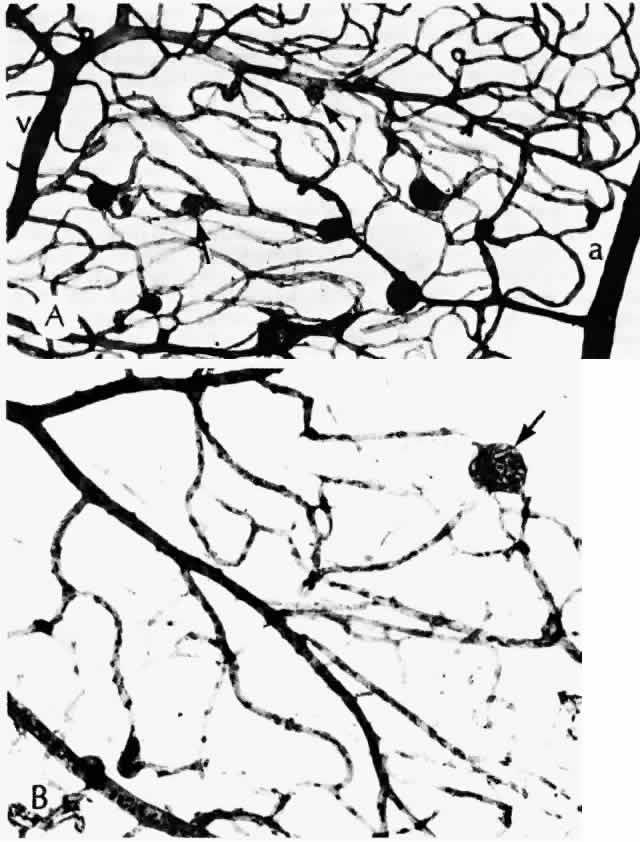
| Fig. 7. Retinal capillary microaneurysm (RCM). A. RCMs occur in random distribution between the arteriole (a) and venule (v). “Young” RCMs are seen as saccular capillary outpouchings with proliferated endothelial cells (arrows). “Old” RCMs appear as solid black balls with their lumens obliterated by PAS-positive material. Note the darker color of the capillaries with thickened basement membranes and arteriolar-venular connections. B. Very large RCM (arrow) or the tiny hemorrhages associated with abnormal vessels are probably responsible for the RCMs seen clinically. (A, PAS, × 40; B, PAS, × 115) |