|
|
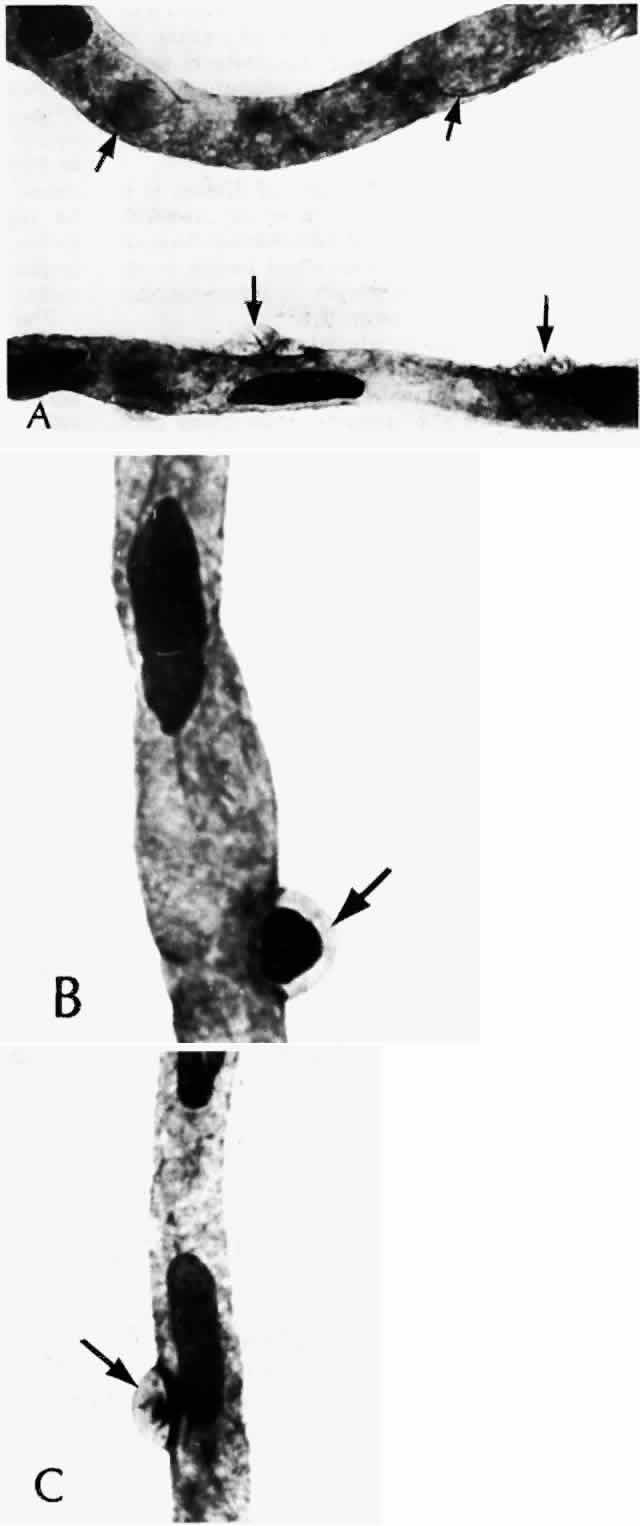
| Fig. 6. Diabetic retinal capillary. A. Basement membrane shell (arrows) is the only remaining indication of where the pericytes had been. B. Nondiabetic normal capillary shows the basement membrane shell (arrow) around the pericyte. C. Diabetic capillary has only a basement membrane shell (arrow), with the nucleus absent. (A, PAS, × 630; B, PAS, × 850; C, PAS, × 630) |