|
|
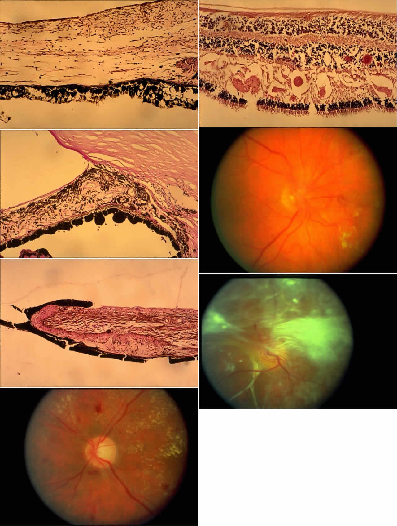
| Color Plate 1 A. Lacy vacuolization of the iris pigment epithelium (H&E × 77.5). B. Neovascularization of the iris with peripheral anterior synechiae. The peripheral iris is adherent to the cornea at about the level of the termination of Descemet's membrane. The fibrovascular membrane is anterior to the anterior border of the iris (PAS, × 77.5). C. Ectropion uveae. The iris pigment epithelium is pulled anteriorly by the fibrovascular membrane. Note the folding of the sphincter muscle (H&E, × 77.5). D. Fundus appearance of microaneurysms, dot hemorrhages, and exudates, some in a typical circinate pattern found in background diabetic retinopathy. E. Exudates have collected in the outer plexiform layer, and appear eosinophilic. Two microaneurysms are present to the right in the figure, within the inner nuclear layer (H&E, × 77.5). F. New vessels originating at the disc partially obscure the underlying disc and retinal vessels. G. Dense, fibrous membrane on the retinal surface distorts and obscures the retina. To the lower left is a small retroretinal membrane, identified as such because the retinal vessels overlie it. |