|
|
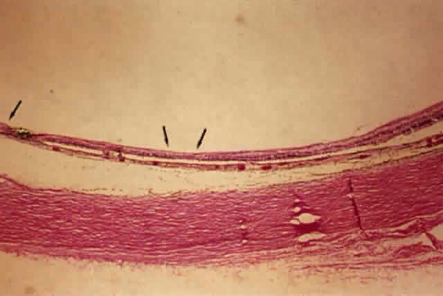
| Fig. 14. Low-power view of panretinal photocoagulation. Normal retina is to the extreme right. Centrally there are areas of outer retinal loss (center arrows), and to the extreme left (single arrow) is an area of full-thickness retinal loss with migrated pigment epithelium. (H&E, × 19.5) |