Chapter 11
Vitreous Humor
ALAN J. RUBY , GEORGE A. WILLIAMS and MARK S. BLUMENKRANZ
Main Menu   Table Of Contents

Search

VITREOUS STRUCTURE
VITREOUS MOLECULAR STRUCTURE
PATHOPHYSIOLOGIC MECHANISMS IN VITREOUS DISEASES
CLINICAL CONDITIONS OF THE VITREOUS
PHARMACOLOGIC VITREOLYSIS
REFERENCES

VITREOUS STRUCTURE

GENERAL FEATURES

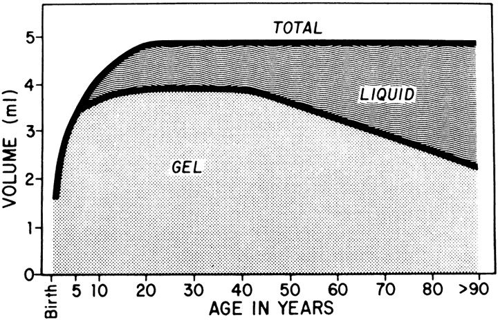
The vitreous of the normal human eye weighs approximately 4 g and occupies a volume of almost 4 ml. The precise weight and volume vary with the age and the size of the eye. The vitreous body is spherical with a depression in the anterior surface, the patellar fossa, corresponding to the posterior surface of the crystalline lens. Larsen1 ultrasonically measured the axial length of the vitreous body in 926 children. He noted that the mean value in newborns was 10.48 mm in boys and 10.22 mm in girls. A sex difference of 0.23 to 0.38 mm greater mean length was noted in males at birth and persisted throughout the growth period. By the age of 13 years, when the axial growth of the vitreous body is essentially completed, the average length was 16.09 mm in males and 15.59 mm in females. Larsen further noted that the relationship between vitreous length and refraction was already established by 1 year of age.

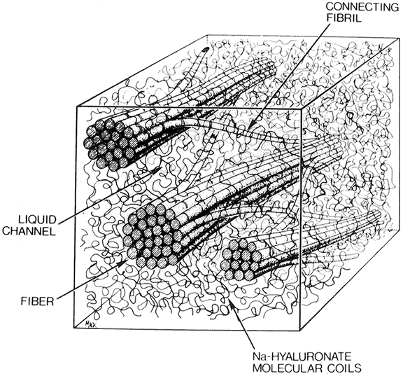
The vitreous body can be divided into two zones or regions. The more peripheral zone, the cortical vitreous, encases the medullary vitreous. The cortical vitreous consists of a relatively more condensed, fibrillar vitreous. Although the cortical vitreous represents only 2% of the total vitreous volume, it is the metabolic center of the vitreous body, because it contains the hyalocytes.2,3 Another connective tissue cell, the fibrocyte, is also found in the cortical vitreous. Most of the vitreous body, the medullary vitreous, is essentially a cell-free mixture of collagens and hyaluronic acid (HA) existing either in a gel or a liquid state depending on the age, refraction, and condition of the eye.

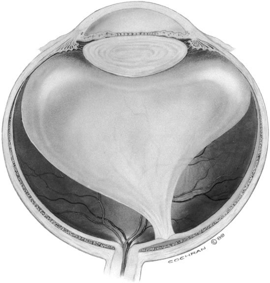
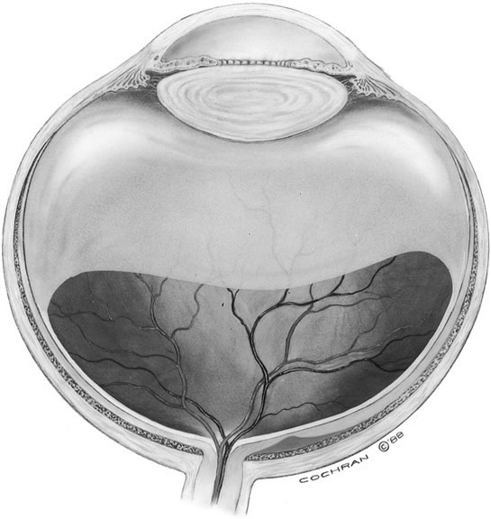
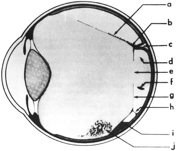
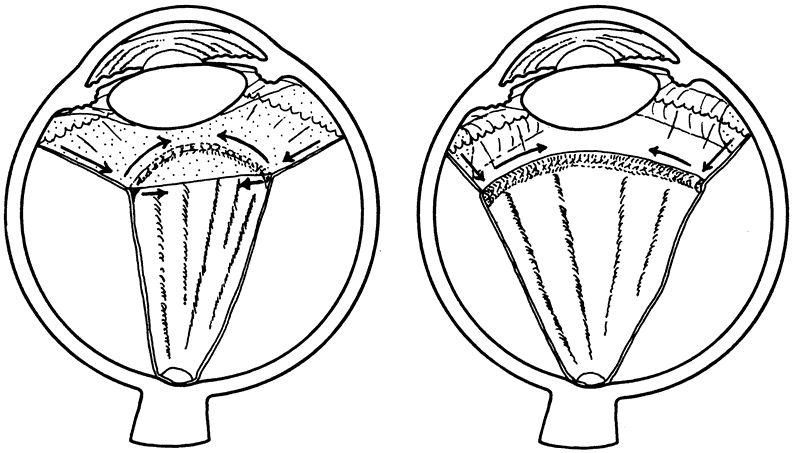
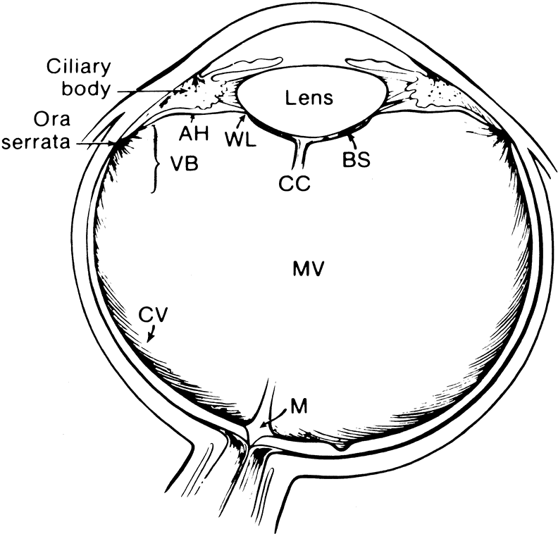
The vitreous body interfaces with a number of ocular structures through the vitreous cortex (Fig. 1). The vitreous cortex extends anteriorly from the vitreous base to form the anterior vitreous cortex and posteriorly to form the posterior vitreous cortex. Recently, the clinical importance of vitreous cortex has become increasingly apparent. The vitreous cortex has been implicated as a primary factor in a variety of vitreoretinal disorders, including retinal breaks, proliferative vitreoretinopathy (PVR), anterior hyaloidal fibrovascular proliferation, macular holes, and epiretinal membranes.

Fig. 1. Diagram of the eye showing the relationship of the vitreous to the other intraocular structures. (AH, anterior hyaloid; VB, vitreous base; MV, medullary vitreous; WL, Wieger's ligament; BS, Berger's space; CC, Cloquet's canal; M, area of Martegiani).

ANTERIOR VITREOUS CORTEX

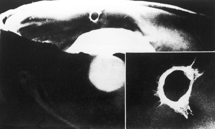
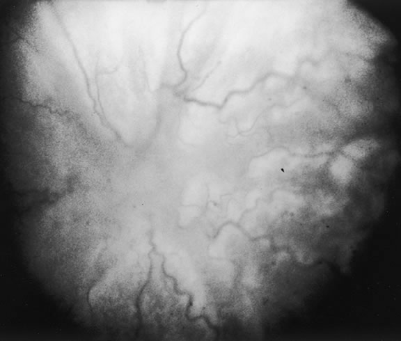
The anterior vitreous cortex or anterior hyaloid is the anterior surface layer or condensation of the vitreous body. There is no specialized membrane that constitutes the anterior vitreous cortex, but instead there is a greater density of collagen fibrils. Microscopically, the framework of collagen fibrils that run throughout the vitreous body ends in an interwoven network to form the anterior vitreous cortex. The superficial fibrils tend to run parallel to the surface in this region. The smooth surface and membrane-like appearance are due to the lamellar distribution of the cortical fibers and the associated highly polymerized mucoproteins.4,5 Anatomically, the anterior hyaloid forms the posterior limits of the posterior chamber. This portion of the vitreous cortex functions in the physiologic communication between the vitreous cavity and the aqueous humor. The anterior surface of the vitreous body separates from the pars plana approximately 1.5 mm anterior to the ora serrata. It extends medially to contact the lens posterior to the lens equator. Thus, the anterior hyaloid is in contact with the ciliary processes and the lens zonules, as well as the posterior lens capsule. The vitreous attaches to the lens capsule in a ring-like manner, forming the hyaloideocapsular ligament of Wieger. This ligament is believed by some to be synonymous with the attachment ring of the posterior zonular fibers.4 The circular area of attachment measures approximately 9 mm in diameter and is especially firm in younger persons or after intraocular inflammation.5 In this circular area the anterior hyaloid is thickened. Central to the attachment of Wieger's ligament (also known as Egger's line), the vitreous lens attachment is less pronounced and appears to be due to surface tension. This central area contains a potential space within the 9-mm ring known as Berger's space, or the patellar fossa. The anterior hyaloid then turns posteriorly to form the anterior portion of Cloquet's canal in the midportion of Berger's space. Cloquet's canal represents the remnants of the primary vitreous and can sometimes be seen with the slit lamp. It arises from the optic disc in a funnel-shaped manner, in the area of Martegiani, and extends forward to the posterior lens surface. The canal is 1 to 2 mm in width and has a down turn in the central vitreous cavity. The area of contact with the posterior lens capsule can at times be identified by a tag of embryonic tissue, known as a Mittendorf dot, located slightly nasal to the posterior pole of the lens. Similarly, a remnant of the posterior primary vitreous can occasionally be identified on the optic disc. This remnant, representing the embryonic point of exit of the hyaloid vascular system from the optic nerve head, is known as Bergmeister's papilla. The walls of Cloquet's canal are formed by a vitreous condensation rather than a true membrane (see Fig. 1).

POSTERIOR VITREOUS CORTEX

The mechanical relationship between the vitreous and the retina is mediated by the posterior vitreous cortex, which is also called the posterior hyaloid. The posterior vitreous cortex consists of relatively densely packed type II collagen fibrils arranged tangentially to the retina. The retinal basal lamina is the basement membrane of the Müller's cells that comprise the internal limiting membrane (ILM) of the retina.6,7 Ultrastructurally, the ILM consists of three layers.8 Adjacent to the end feet of the Müller's cells is the lamina rara interna. The lamina rara externa is contiguous with the vitreous cortex. In between these layers is the lamina densa. Collagen fibers of cortical vitreous are tangential to the lamina rara externa. The ILM is composed of primarily type IV collagen but also contains fibronectin, laminin, and type I collagen.9 The morphology of the ILM varies topographically in the retina. The ILM, and in particular the lamina densa, is thin in the retinal periphery and becomes increasingly thicker and irregular in the posterior retina.6,8

The ILM thickens from 50 nm at the vitreous base to 300 nm at the equator to 1900 nm posteriorly. In the foveal region the ILM thins to 10 to 20 nm.10

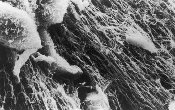
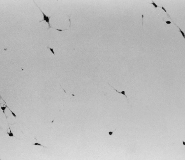
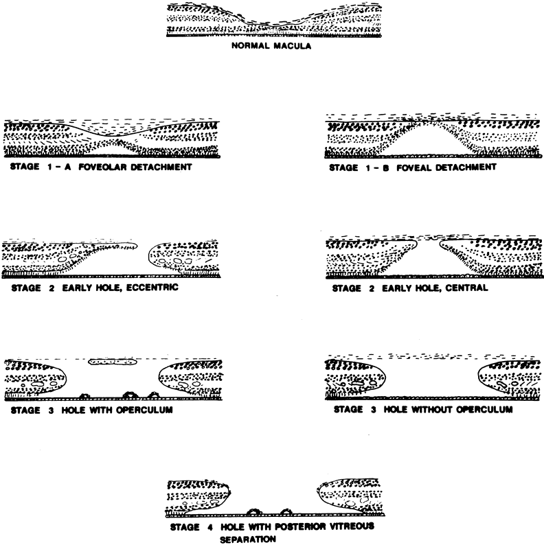
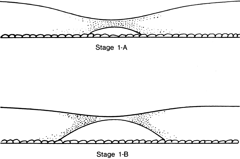
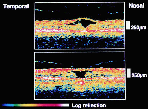
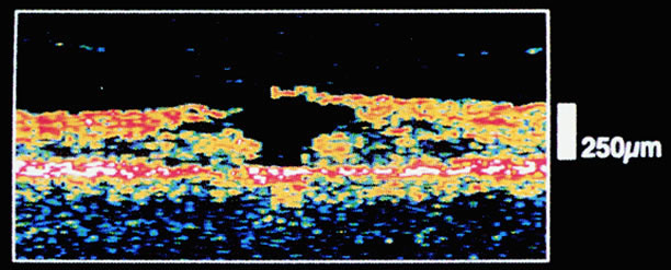
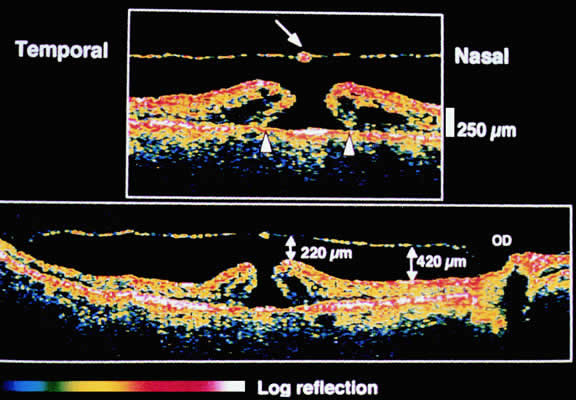
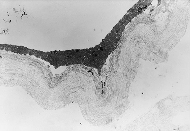
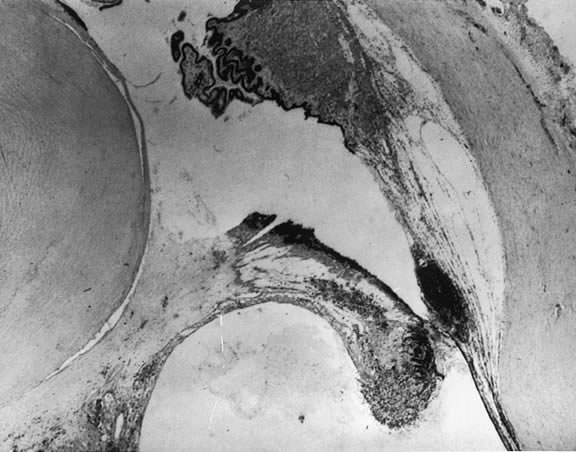
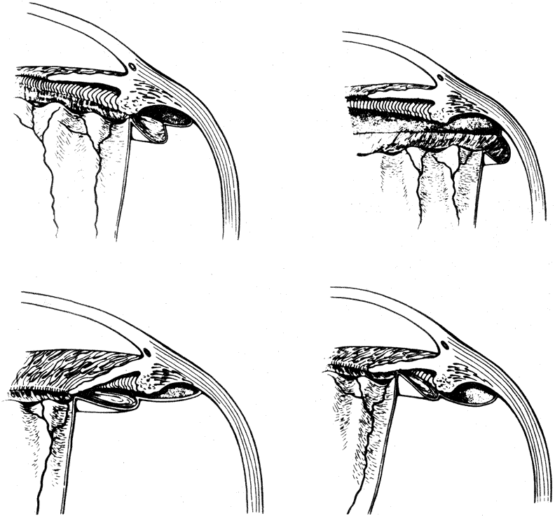
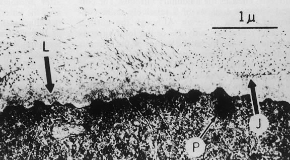
The nature of the adhesion between the vitreous cortex and the ILM is enigmatic. This adhesion is strongest at the optic nerve, the macula, the vitreous base, and retinal vessels. Foos6 demonstrated attachment plaques between Müller's cell cytoplasm and the ILM in the basal and equatorial retina (Fig. 2). Vitreous traction mediated by vitreous fibrils may contribute to these adhesions. These attachment plaques are not present posterior to the equator except where the ILM is thinned in the fovea. This anatomic variation in the fovea region may play a role in the pathogenesis of some of the vitreomacular disorders.

Fig. 2. Electron micrograph of the vitreoretinal junction (J) 1 mm posterior to the equator of the eye. This demonstrates the insertion of the vitreous fibers to the inner limiting lamina (L). Attachment plaques (P), which anchor the inner limiting lamina to the surface glial cells, are illustrated. (Foos RY: Anatomic and pathologic aspects of the vitreous body. Trans Am Acad Ophthalmol Otolaryngol 1973;77:171)

The ILM also thins over major blood vessels. Vitreous strands extend through pores in the ILM to surround the vessels11 and have been termed vitreoretinovascular bands.12 These bands may explain the strong adhesion between the vitreous and retinal vessels.

Despite the presence of these anatomic interfaces between the vitreous cortex and ILM, the adhesion of these structures is still poorly understood. Biochemical investigations suggest that vitreoretinal adhesion involve molecular adhesion mediated by glycoproteins such as fibronectin, laminin, and other glycoconjugates. The ILM consists of type IV collagen, laminin, fibronectin, type I collagen, and as yet uncharacterized glycoproteins. It is believed these substances mediate vitreoretinal adhesion.13

The ILM undergoes age-related changes that are anatomic, biochemical, and functional. Anatomically, the ILM thickens with age.6,8 Biochemically, the distribution of fibronectin and laminin changes with age. In young eyes, anti-fibronectin antibody binding has a homogeneous distribution. In older eyes, the posterior ILM shows a segmented bilaminar distribution of both antifibronectin and antilaminin antibodies.14 An age-dependent distribution of other ILM glycoconjugates has also been demonstrated.13 Variations in the distribution of these and other glycoproteins may play a role in conditions that are characterized by alterations in vitreoretinal adhesion.15

VITREOUS BASE

The vitreous base is a three-dimensional zone centered on the ora serrata where the vitreous is the most adherent to the retina and pars plana epithelium. It extends approximately 1.5 mm anteriorly to the ora serrata. Nasally, it extends 3.0 mm posterior to the ora and 1.8 mm posterior to the ora temporally. The functional base of the vitreous extends several millimeters into the vitreous body in this region. As the eye ages, the firm attachment of the vitreous base may extend posteriorly for several millimeters. This may lead to localized areas of enhanced vitreoretinal traction and result in retinal tears.

In the vitreous base, collagen fibers are relatively coarse, are numerous, and insert perpendicularly to the retina and pars plana. These fibers consist of collagen fibrils with diameters of 10.8 to 12.4 nm.16 The microscopic detail of the vitreous fibers attachment to the vitreous base varies from anterior to posterior. Anterior to the ora the fibers are less dense than posterior to the ora. Electron microscopic studies by Gartner17 demonstrate that the vitreous fibers have complex interdigitations with the reticular fibrillar materials of the basement membrane of the nonpigmented ciliary epithelium but do not pass between the cells. The anteriormost fibers splay out anteriorly to form the anterior loop of the vitreous base, which is important in the pathogenesis of anterior PVR.18 Posterior to the ora, bundles of vitreous fibrils attach to the ILM. Cords of vitreous collagen insert into gaps between the neuroglia. Gloor and Daicker19 likened this arrangement to Velcro and suggested that this may explain the strong vitreoretinal adhesion of the vitreous base. Cellular elements are also present in the vitreous base. Fibroblast-like cells are present anterior to the ora, and macrophage-like cells are posterior to the ora.17 These cells may play a role in the hypocellular gel contraction that characterizes anterior PVR.

VITREOUS CELLS

The vitreous normally contains relatively few cells compared with other connective tissues. Most of these are found within the thin layer of cortical vitreous. These few cells account for the metabolic activity of the vitreous body, which as a result is among the lowest in metabolic activity in the body. The cells are variously called vitreous cells, vitreous cortex cells, vitreocytes, or, most commonly, hyalocytes. In the physiologic state, the vitreous body appears to have a mixed population of cells. Morphologically, most are hyalocytes, whereas approximately 10% are similar to fibroblasts.

Hyalocytes are found throughout the vitreous cortex, but they appear to be concentrated in the basal region.3 These cells are more numerous in the fetal vitreous than in older eyes, and their exact origin and function are open to question. Some investigators believe hyalocytes are remnants of the primary vitreous.20 Based on developmental studies in animals and humans, hyalocytes arise from either monocytes originating from mesenchymal cells in the optic cup or embryonic fissure or from blood elements in the vasa hyaloidea propria. These cells migrate through the vitreous cavity and accumulate in the cortical vitreous as hyalocytes by the fifth month of gestation. Following birth, there is no further migration of cells to the vitreous cortex and existing hyalocytes do not normally proliferate. As a result, ocular growth results in increasing vitreous cortex surface area with a corresponding decrease in hyalocyte density. Gloor21 contests the previously mentioned hypothesis and believes hyalocytes are continuously derived from blood monocytes through life. The hyalocytes remain in the vitreous cortex for about a week and are then replaced by blood borne monocytes to maintain a constant cell population.

The hyalocytes appear as flattened, 10 to 15 μm in diameter, spindle-shaped cells when found over the retina. Whereas, in the region of the vitreous base, the cells are larger, rounder, and at times star-shaped with prominent nuclei.21,22 The hyalocytes are active metabolically with numerous lysosomal granules, phagosomes, and Golgi complexes. Hyalocytes synthesize vitreous glycosaminoglycans (GAGs) including HA and hexosamine.23 The highest density of hyalocytes is in the region of the vitreous base followed by the posterior pole, with the lowest density at the equator.

In addition to the hyalocytes and fibroblasts, there are cells found in the vitreous that appear to be in varying stages of degeneration.24 These cells are believed to represent the involutional remnants of the original cell population of the embryonic vitreous and are seen more often in young eyes. By electron microscopy, in the region of the ora serrata, cells are seen that appear to be damaged retinal cells that have been sloughed into the vitreous base.17

There is a mechanism for the elimination of foreign and degenerated substances from the vitreous that is found partly in the region of the optic disc. When India ink is injected into the anterior portion of the vitreous in a rabbit eye and the enucleated eye examined at varying intervals, it is noted the ink particles are eliminated from the vitreous by way of the optic nerve.25 Later the particles are removed by phagocytic cells that appear to enter the vitreous body from the region of the optic nerve and ciliary body. Surgical treatment may also affect the cellular composition of the vitreous. Cryopexy induces a macrophage influx into the vitreous cavity.26 This may play a role in the clearing of vitreous opacities.

Back to Top
VITREOUS MOLECULAR STRUCTURE
In this section we present a brief overview of the macromolecular structure of the vitreous. For a more complete discussion of this complex area, the reader should consult the excellent works of Sebag, Eisner, and Balazs.27–29

The major macromolecular components of the vitreous are HA, also known as hyaluronan, and collagens. HA is a GAG consisting of repeating disaccharide units of glucuronic acid and N-acetyl glucosamine. It is arranged in a linear helix that undergoes conformational changes into both a compressed or extended conformation. The molecular weight of the sodium salt of HA ranges from 3 to 4.5 × 10⁶. In vivo, HA is highly hydrated with a large specific volume of 2000 to 3000 ml/g. This results in the formation of a highly entangled open coil. HA is produced by the hyalocytes that possess the necessary enzymes for HA synthesis. HA concentration is highest in the regions of greater hyalocyte density. Therefore, it is highest in the cortical gel.

Vitreous collagen consists of nonbranching fibrils 7 to 28 nm in diameter. The collagen molecules forming these fibrils are covalently linked. Transmission electron microscopy reveals that the fibrils have uniform banding density and are associated with a filament consisting of a succession of linked 20-nm beads with a spacing ranging from 35 to 85 nm. The vitreous base has the highest density of collagen fibrils, followed by the posterior and then anterior vitreous cortex.30

The fibrils consist of a mixture of collagen types distributed along each fibril. Semiquantitative analysis of bovine vitreous reveals collagen types II, V, and IX distributed in a ratio of 69:24:7, respectively.30 Although most of the collagen is type II, types IX and V are believed to be important in promoting stability of the vitreous gel. Type IX collagen probably promotes cohesion between the fibrils and HA. Type V collagen coats the type II collagen to produce a continuous filament structure in the fibril. The assembly of these collagens results in a fibril of enhanced stability and strength.

There may be some topographic variation in vitreous fibril composition. Transmission electron microscopy of the vitreous base shows fibrils consistent with type VI collagen.30 It is the interaction between the collagen fibrils and HA that provides the vitreous gel with its unique mechanical and optical properties. The large domains of HA stabilize and spread apart the collagen fibrils, thus, minimizing light scattering. The viscoelastic properties of vitreous are the result of complex molecular interactions between the collagen fibrils and HA27–29 (Fig. 3).

Fig. 3. Schematic diagram of adult vitreous ultrastructure depicting the relationship between hyaluronic acid molecules and collagen fibrils. (Sebag J, Balazx EA: Morphology and ultrastructure of human vitreous fibers. Invest Ophthalmol Vis Sci 1989;30:1871)

Scott31 has suggested that there is some order to the association of the vitreous GAGs and collagens. He proposed a three-component complex that forms an infinite meshwork contributing to the semirigidity of the vitreous. The spaces between collagen fibrils are filled by HA and other GAGs such as chondroitin 6-sulfate. The GAGs link together as aggregates via hydrophobic and hydrogen bonding, resulting in a three-dimensional honeycomb structure. This ordered aggregation of GAGs is fragile and may have clinical implications. HA fragments may destabilize the aggregation of GAGs.32 Therefore, a small amount of HA breakdown may have a marked effect on the integrity of the whole gel. HA is sensitive to damage by free radical mechanisms. Irradiation of the vitreous with 300-nm light or with white light in the presence of photosensitizers results in partial depolymerization of HA. Exposure to neodymium:yttrium-aluminum-garnet (Nd:YAG) laser energy has a similar effect. These photodynamic events generate hydroxyl radicals that are responsible for the HA degradation. Depolymerization alters the hydration of HA resulting in liquefaction. This mechanism may explain the vitreous liquefaction seen with age, inflammation, or after cataract extraction.

The vitreous contains a group of enzymes known as matrix metalloproteinases (MMPs) which are zinc-dependent enzymes that mediate the structure and function of the extracellular matrix (ECM). Because most of the vitreous is ECM, it is not surprising that MMPs are present and that MMPs have been implicated in a variety of vitreoretinal disorders. The enzymatic activity of MMPs is regulated by tissue inhibitors of metalloproteinases (TIMPs). The functional proteolytic activity of MMPs in the vitreous is, thus, dependent on the relative concentrations of regulatory TIMPs and active MMPs. There are at least 18 MMPs and 4 TIMPs that have been reported. The MMPs have varying substrate specificity and patterns and location of expression. In the vitreous, MMP-1, MMP-2, MMP-3, and MMP-9 are present. TIMP-1, TIMP-2, and TIMP-3 have also been reported in the vitreous. The source of vitreous MMPs and TIMPs is not known, but hyalocytes may be responsible.33

MMPs and TIMPs play both a physiologic and a pathologic role in the vitreous. These enzymes are probably responsible for the proteolytic state that clears the vitreous of potential opacification and maintains optical clarity. In disease states, MMPs have been implicated in PVR, retinal neovascularization, retinopathy of prematurity, and proliferative diabetic retinopathy.33,34

Back to Top
PATHOPHYSIOLOGIC MECHANISMS IN VITREOUS DISEASES
There appear to be at least two distinct mechanisms by which alterations in vitreous morphology lead to clinical disease syndromes: (1) mechanical interfacial separation of the vitreous from the retina and (2) cell-mediated contraction of the cortical vitreous gel.

MECHANICAL INTERFACIAL SEPARATION OF THE VITREOUS FROM THE RETINA

Mechanical separation of the vitreous from the retina occurs when the vitreous separates from the ILM of the retina. This separation may be relatively complete, as in posterior vitreous detachment (PVD), or may be limited, as in macular hole formation. PVD occurs when mechanical stress forces acting on the vitreous body overcome the naturally occurring adhesion of the vitreoretinal interface.

PVDs are usually spontaneous events that are age related, but they may also be precipitated by trauma. They occur earlier in association with vitreous inflammation, vitreous hemorrhage, myopia, and diabetes. As the vitreous ages, there is an increase in the water content and subsequent liquefaction of the vitreous, a process that normally begins by the age of 5 years. At about 40 years, the volume of the vitreous gel begins to decrease in parallel with the increase in liquid vitreous until approximately half the vitreous space is occupied by liquid vitreous29 (Fig. 4). This process of liquefaction is called synchysis. Synchysis usually begins in the midvitreous and is recognized with biomicroscopy as optically empty spaces without the membranous appearance of the normal vitreous. This is because liquid vitreous does not contain collagen fibrils.

Fig. 4. Change of the liquid and gel vitreous volume during aging and development of the human eye.

Once vitreous liquefaction occurs, fluid currents develop within the vitreous. These currents, stemming from the rotational and gravitational forces transmitted to the vitreous body, produce stresses at the vitreoretinal interface that lead to separation at points of weakest attachment. Dark-field slit microscopy studies on human autopsy specimens indicate that persons 21 years or younger demonstrate firm adherence between the ILM and the vitreous cortex in the posterior pole. Older persons demonstrate relatively weak adhesive forces in comparable areas.15

After liquid vitreous has formed, vitreous body collapse or syneresis can occur. This event is precipitated by the passage of liquid vitreous through the vitreous cortex at either the prepapillary hole or the premacular cortex and into the potential space between the vitreous cortex and the ILM. The liquid vitreous then dissects and fills the potential space. The subsequent volume displacement of liquid vitreous through holes in the vitreous cortex results in vitreous collapse and a PVD. Eisner28 terms this event a rhegmatogenous vitreous detachment. Liquid vitreous may also pass through an intact vitreous cortex in what Eisner calls an arrhegmatogenous vitreous detachment, which is a slower detachment occurring as a result of a pathologic condition, such as diabetes.

Defects in the premacular vitreous cortex have been identified in autopsy eyes by Sebag and Balazs18 and confirmed by Kishi and Shimizu35 using differing techniques. The premacular or prefoveal hole appears to correspond to the posterior aspect of the posterior precortical vitreous pocket (PPVP). In this region liquefied vitreous is immediately adjacent to the vitreous cortex. It is unknown what allows the passage of liquid vitreous through the vitreous cortex to disrupt the vitreous cortex ILM adhesion, but mechanical, hormonal, and biochemical changes have been implicated.

Although vitreous detachment typically results in cleavage between the posterior vitreous cortex and the ILM of the retina, clinical and electron microscopic observations suggest that this is not always the case, particularly in younger persons and in pathologic states. Microscopic studies have confirmed the multilaminar nature of the vitreoretinal interface, including the direct insertion of vitreous cortical collagen fibers into the ILM of the retina. These insertions are particularly firm in areas where the ILM is thinnest, such as the fovea (where the ILM may measure as few as 10 nm) and the basal region (where it again thins to as few as 50 nm).6,10 In young persons, adhesions between the ILM of the retina and the vitreous cortex may be stronger than the Müller's cells themselves, leading to a different plane of separation than the ILM posterior vitreous cortex during PVD. Vitreous cortex remnants have been identified at the fovea by scanning electron microscopy after spontaneous vitreous detachment in autopsy series.36

In addition to the topographic and age-related differences in the vitreoretinal interface, gender-related differences in the rates of vitreous separation also exist. In most clinical series, women are twice as likely to present with symptomatic PVD37 (65% to 68%) as men (32% to 35%). This is paralleled by gender-related differences in disease rates associated with vitreoretinal separation. Macular holes occur between three and four times more frequently in women than men, whereas nontraumatic giant retinal tear occurs three times more frequently in men than women. The reasons for this disparity remain unclear, but these data may reflect gender-related differences in the strength of the vitreoretinal interface at different locations with the eye.

CELL-MEDIATED TRACTION OF THE CORTICAL VITREOUS GEL (HYPOCELLULAR GEL CONTRACTION)

Mechanical separation of the vitreous from the ILM is believed to occur in normal eyes in the absence of any cellular contribution or energy-dependent processes. Under pathologic circumstances, the cortical vitreous gel may also contract rather than separate at the vitreoretinal interface. This leads to the development of vitreoretinal traction and complex forms of retinal detachment in the presence of retinal breaks. Vitreous contraction has long been recognized as a factor in complex retinal detachments.38 However, the biologic basis for the contraction of vitreous gel was not understood until relatively recently.

Machemer39 first emphasized the critical role of cells in the genesis of vitreoretinal traction. Early histopathologic and ultrastructural studies on patients undergoing vitrectomy for PVR demonstrated transformed retinal pigment epithelial (RPE) cells, retinal glia, fibroblasts, and macrophages in membranes that were hypothesized to be contractile. It was proposed that these membranes, composed of densely packed interdigitating cells and ECM, produced tractional forces in a manner analogous to the contraction of syncytial smooth muscle elements as a result of actin and myosin filaments within the cytoplasm, particularly of myofibroblasts. However, clinical observations of the vitreous suggested that vitreoretinal traction could occur by mechanisms other than syncytial hypercellular membranes.

A different mechanism is required to explain the development of vitreous cortical shrinkage that occurs in the region of the vitreous base following vitrectomy. The ability of fibroblastic cells of different proliferative potential to contract a collagen latticework was first described by investigators evaluating the wound healing response in skin.40 Subsequent investigations in the dermatologic and ophthalmologic literature corroborated the applicability of this system for the testing of antiproliferative and anticontractile drugs.41,42 Clinical investigators pointed out the applicability of this experimental system as an explanation for contraction of the vitreous gel by dispersed intraocular cells.43,44 A variety of ocular cell types including dermal, scleral, or chorioretinal fibroblasts; pigment epithelial cells; and glial cells, in decreasing order of magnitude, will contract a clear gel of type I or type II collagen to a small fraction of its original volume with resultant opacification. Scanning and transmission electron micrographs of contracted gels reveal similarities with specimens of contracted vitreous taken from experimental animals and humans with PVR (Fig. 5). This process, termed hypocellular gel contraction, requires the presence of either serum, specific serum components, or other non-serum-derived growth factors to occur. Thrombin, transforming growth factor-β, platelet-derived growth factor, and fibronectin have all been shown to stimulate this process and can substitute for the serum requirement to variable degrees.45,46 Similarly, other pharmacologic compounds, which either intrinsically inhibit cellular contractility47–50 or interfere with cellular attachment to collagen through fibronectin-mediated binding sites51,52 can inhibit this process. Conventional and low molecular weight heparin also inhibit cellular attachment to collagen and, consequently, contraction. Various heparin forms also inhibit collagen polymerization.52,53 Antiproliferative compounds that innately inhibit the cellular contractility process include steroids, certain fluoropyrimidines, colchicine, and paclitaxel (Taxol).53

Fig. 5. Scanning electron micrograph of contracted collagen gel. Note paucity of cellular elements despite marked compaction of collagen fibers.

Quantitative analysis of these effects indicates that human dermal fibroblasts are nearly 20 times more potent contractors of collagen than human RPE cells. For example, 100,000 dermal fibroblasts will contract a collagen gel to less than 10% its original volume within 48 hours, contrasted to a negligible change at 48 hours for 100,000 human RPE cells. However, given 120 hours, 1 million human retinal RPE cells will contract a gel to less than 30% of its original volume43 (Fig. 6). Using time-lapse video cinematography, it can be shown that RPE cells will pull in collagen fibrils in a hand-over-hand fashion at a rate of approximately 2.5 μm per minute,44 and this effect can be blocked by the addition of a peptide analogue that inhibits fibronectin-mediated binding.51 Similar effects have been demonstrated for human retinal glial cells, although the rate of contraction is less than that of either pigment epithelial cells or fibroblasts.54

Fig. 6. Graph of quantitative representation of cell-mediated gel contraction in vitro. One hundred thousand human dermal fibroblasts (HDF) contract gel to less than 10% its original volume. In contrast, 100,000 human retinal pigment epithelial (HRPE) cells produce negligible change at a similar time interval. One million cultured HRPE cells reduce gel volume by more than 70% after 120 hours.

In addition to demonstration of these effects in an in vitro model employing purified type I and type II collagen, other investigators have demonstrated similar effects in vitro using native bovine vitreous. Although pigment epithelial cells, glial cells, and fibroblasts all produce collagen and vitreous contraction, the effects of capillary endothelial cells are modest, and no contraction is induced by either erythrocytes, blood leukocytes, or activated macrophages.55

Histologic and ultrastructural studies of contracted gels disclose consistent morphologic changes that account for these contractile events. Pigment epithelial cells cultured on plastic demonstrate a rounded or cuboidal morphology when subconfluent. It has been previously shown that when pigment epithelial cells are cultured on either vitreous, collagen, or fibrin they assume a more mesenchymal configuration, including the development of dendritic extensions and lamellipodia.56 It is believed that the development of these lamellipodia are critical for locomotion of cells and for the generation of the tractional forces. Histologic cross sections of contracted gels reveal that the cells may produce profound volumetric changes in gels without the development of typical cellular membranes. In contracted gels, cells appear to be dispersed at uniform distances with relatively infrequent cell interactions, particularly in the case of fibroblasts (Fig. 7). It is believed that the mechanism of contraction in these cases relates to compaction of vitreous fibrils adjacent to cells through membranous interactions between fibronectin and collagen binding sites on the cells and individual collagen fibrils and HA with resultant extrusion of water.

Fig. 7. A low-power cross section of experimental gel contracted by dermal fibroblast. Note the sparse spacing of cells and lack of development of syncytial contractile membrane. Cells assume spindle morphology and occasionally demonstrate cell interactions through lamellipodia. (× 120).

The water extrusion is largely responsible for the volumetric change (shrinkage) and loss of transparency noted clinically and experimentally and can be quantitated either directly by linear measurement or indirectly by volumetric methods such as isotope dilution or water displacement.40,48,57

Back to Top
CLINICAL CONDITIONS OF THE VITREOUS
In this portion of the chapter we discuss clinical conditions that primarily involve the vitreous or in which the vitreous plays a major pathogenic role. These conditions will be discussed in the perspective of recent advances in vitreous anatomy, biochemistry, and pathophysiology.

DETACHMENT OF THE VITREOUS BODY

The separation of the vitreous cortex from the retinal surface is a relatively frequent and often a clinically significant occurrence. It is important because of the bothersome and at times alarming symptoms and also because of the frequent association of significant ocular pathology such as retinal tears, retinal detachment, and vitreous hemorrhage.

Incidence

Most vitreous detachments involve the vitreous posterior to the vitreous base and thus are referred to as PVD. The exact incidence is unclear, although it is generally agreed that it is age related. The estimates of the occurrence of PVD in the general population vary from one series to another. The incidence in clinical studies is 53% of phakic patients older than 50 years, 65% older than 65 years, and virtually 100% in aphakic eyes.58,59 However, Foos60 noted in a large autopsy series that the incidence of PVD was lower than in clinical studies. This is due to difficulty in clinically identifying a PVD. For example, areas of liquefaction may simulate a PVD. In Foos' study, PVD first occurred in the fifth decade in phakic patients, with a 17% occurrence noted by the sixth, 51% by the seventh, and 53% by the ninth decades. The incidence rose to 72% in aphakic patients. PVD is 7 to 10 times more frequent in aphakic eyes as compared with phakic eyes. This is related to mechanical changes in vitreous position in the aphakic eye and also because of biochemical changes. Surgical aphakia results in anterior displacement of the vitreous during ocular movements. Aphakia with extracapsular cataract extraction (ECCE) has a lower incidence of PVD than with intracapsular cataract extraction (ICCE), if the posterior capsule is intact.61 This is probably because the intact posterior capsule prevents anterior vitreous displacement.

Aphakia also alters the biochemical profile of the vitreous. HA concentration decreases following ICCE, resulting in vitreous liquefaction.62 ECCE results in a minimal decrease in HA concentration compared with ICCE.63

The changes in the incidence of PVD with aphakia after ICCE and ECCE parallel the incidence of retinal detachment following ICCE (3% to 7%) and ECCE (0.5% to 2%).64,65 This is because the formation of retinal breaks and subsequent retinal detachment is dependent on the formation of liquid vitreous. Liquid vitreous and subsequent PVD increase vitreoretinal traction in the region of the vitreous base, resulting in retinal tears.66 Liquid vitreous then passes through these tears to dissect the retina off the RPE cells. PVD is more common in females.37,67 It occurs an average of 10 years earlier in myopic patients when compared with emmetropic or hyperopic patients.58,59

The incidence of partial PVD appears to be very low, which supports the concept that the process, once begun, proceeds to completion in a relatively short period. However, an incomplete PVD can remain unchanged for long periods. When a partial PVD is present, it is usually found in the upper portion of the vitreous body, perhaps because of gravitational effects. Linder58 evaluated 115 patients clinically with symptoms of the sudden appearance of floaters with or without light flashes and found that 95% demonstrated a complete PVD with collapse, whereas 4% had a superior vitreous detachment and the remaining 1% showed a funnel-like vitreous detachment with attachment remaining at the disc. The two types of partial vitreous detachment were noted to convert to total posterior detachment in most cases. Jaffe59 noted a similar incidence, with 82 of the 84 patients who presented with symptoms of suddenly appearing entopsias demonstrating a PVD with collapse.

Signs and Symptoms

The subjective symptoms of a PVD are varied, and many patients may have a vitreous detachment without being aware of its occurrence. In Linder's58 series of acute PVDs, approximately 25% of the cases had an unrecognized PVD in the fellow eye. The most common symptom is the sudden onset of floaters with or without light flashes. Patients who present with floaters and flashes appear to be at higher risk for a posterior vitreous separation than those patients who present solely with light flashes or floaters.67 Two theories as to the cause of light flashes have been proposed. One theory suggests that vitreous traction on the retina at the site corresponding to a retinal break causes symptoms,68 whereas the second theory suggests that during ocular movement a detached vitreous affects the retina causing the light flashes.69

Floaters may take many forms and are most easily noted by the patient when viewed against a light background or in bright sunlight. Floaters may be present in a liquefied vitreous without a PVD, but the sudden onset of floaters is suggestive of an acute change in the status of the posterior vitreous. This common symptom of an acute PVD may represent condensed vitreous fibrils, vitreous blood, or, in the case of a single large floater, glial tissue on the posterior surface of the vitreous cortex. In most cases, visual acuity is quantitatively unaffected by the PVD. However, if accompanied by significant hemorrhage, a posterior vitreous separation may result in a marked decrease in visual acuity or blurring, dependent on the degree of hemorrhage.

When the vitreous detaches from the area of the optic nerve head, a tag of tissue may be avulsed, and at times a complete ring of peripapillary tissue may come forward with the vitreous. The presence of this ring is pathognomonic for a PVD (Fig. 8). Electron microscopic studies on autopsy eyes with PVDs demonstrate frequent glial epipapillary membranes on the vitreous side of the ILM of the optic nerve and surrounding retina.70,71 There are interruptions in the epipapillary membranes, as well as separations of the membranes from the underlying ILM of the disc, both of which contained incarcerated collagen fibers identical with vitreous fibrils. This intimate association of the vitreous in the disc area accounts for the frequent avulsion of tissue in a PVD. Glial tissue occurs on the posterior hyaloid in 57% of PVDs.

Fig. 8. Gross pathology specimen of an eye of an 85-year-old patient with a posterior vitreous detachment. An epipapillary membrane has been avulsed and is suspended in the posterior hyaloid. The inset shows details of the epipapillary ring. (Foss RY: Anatomic and pathologic aspects of the vitreous body. Trans Am Acad Ophthalmol Otolaryngol 1973;77:171)

Pigmented vitreous cells may also be seen on slit-lamp biomicroscopy in the presence of a PVD. The presence of these pigmented cells in the anterior vitreous cavity is highly associated with a finding of a peripheral retinal tear.72–74 Careful examination of the retinal periphery with scleral depression and/or contact lens is necessary because of the significant association between the cells and peripheral retinal tears. Vitreous hemorrhage may also be present obscuring the view of the retina. These patients are also at very high risk of having a retinal break as the cause of their dispersed hemorrhage.

Complications

The cortical vitreous is more firmly attached to the ILM in the area of the optic disc, macula, and retinal blood vessels; retinal pathology such as neovascularization or vascular malformations occurs in these areas. Separation of the vitreous may transmit traction to these attachments, leading to vitreous hemorrhage or retinal tears. Persistent vitreous traction on retinal tears is an important factor in the development of a retinal detachment.

Following PVD, retinal tears are detectable clinically in 8% to 15% of eyes.37,58,59,75 In postmortem studies, retinal tears are present in 14.3% of eyes with PVD.60 Myopia and aphakia increase the incidence of retinal tears in PVD to as high as 16.2%.76,77

Vitreous hemorrhage from PVD may originate from vessels on the optic disc or retina. These hemorrhages are present in 13% to 19% of eyes with PVD.58,59,75 Hemorrhage may preclude an adequate retinal examination, which is essential to determine the presence or absence of a retinal tear. A careful fundus examination with scleral depression and contact lens biomicroscopy is required, because roughly two thirds of the eyes with acute PVD and vitreous hemorrhage also have retinal breaks.59,75,78

In an autopsy series, Spencer and Foos79 also noted a high incidence of firm, paravascular vitreoretinal attachments. In 25% of the eyes, with an attached posterior vitreous, this led to paravascular retinal rarefaction with cystic spaces adjacent to blood vessels in the inner retinal layers. In addition, there was a connection between the ILM and the retinal blood vessels, as well as an irregularity of the ILM suggesting vitreous traction. Paravascular retinal tears, on the other hand, occurred in 11% of the eyes, and of these all had PVDs. Both types of retinal pathology were usually seen after the age of 50. It is, therefore, not surprising that retinal and vitreous hemorrhage following a PVD is so frequently associated with a retinal tear.

Patients with the symptoms of a recent PVD will sometimes demonstrate small surface retinal hemorrhages between the posterior border of the vitreous base and the equatorial region. They may be associated with peripheral white with or without pressure, indicating a vitreoretinal interface change and continuing vitreous traction. These patients should be observed funduscopically, because retinal tears may occur at a later date.75

In most cases PVD results in complete separation of the vitreous cortex from the macula. Rarely, persistent adhesions may result in vitreomacular or vitreopapillary traction syndromes. A partial PVD can be identified with focal adhesion to the retina or optic nerve. Traction detachment or intraretinal or peripapillary hemorrhages may occur (Fig. 9). Usually these adhesions will spontaneously separate in a matter of days to weeks. If the adhesion persists, vitrectomy is effective in relieving the traction.80

Fig. 9. Vitreomacular traction. (Margherio RR, Trese MT, Margherio AR et al: Surgical management of vitreomacular traction syndromes. Ophthalmology 1989;96:1439)

DETACHMENT OF THE VITREOUS BASE

Detachment of the vitreous base is usually associated with trauma and may initially be obscured by vitreous hemorrhage. The firm attachments of the vitreous fibrils into the peripheral retina and posterior portion of the pars plana are very resistant to separation, and a significant force is required to separate the vitreous in this area. The ciliary epithelium and inner layers of the retina are, therefore, often pulled off with the vitreous base, and these can be identified with indirect ophthalmoscopy and scleral indentation (Fig. 10). This traumatic detachment of the vitreous base most commonly takes place in the inferotemporal quadrant. A peripheral retinal dialysis is often an associated finding and requires surgical intervention if it is diagnosed. There are often other accompanying ocular problems such as angle recession, hyphema, and subluxation of the lens.

Fig. 10. Traumatic detachment of vitreous base.

Spontaneous detachment of the vitreous base is rare but has been described in high myopia, Ehlers-Danlos syndrome, and Marfan's syndrome. There is a smooth vitreous surface in the spontaneous detachment of the vitreous base, whereas traumatic detachment shows an irregular surface of the detached vitreous.81

MACULAR HOLES

Idiopathic macular holes are most commonly seen in older adult women usually in the seventh and eighth decades of life. Patients typically present with complaints of decreased vision and/or metamorphopsia in one eye. Johnson and Gass82 reviewed a total of 123 patients with idiopathic macular holes or impending macular holes. Women comprised 67% of the study population with an average age of 67 years (range, 39 to 80 years). Akiba and colleagues83 examined 310 eyes of 272 patients that eventually developed idiopathic macular holes. Patients age ranged from 47 to 83 years with a mean of 63 years. Two hundred and thirty patients (85%) were 60 years or older. There were 204 (74%) women and 68 men. Visual acuity correlates with the stage of the macular hole with a worsening visual acuity as the stage progresses.82,84 The incidence of a macular hole developing in the normal fellow eye is approximately 10%.

The retinal manifestations of full-thickness macular holes have been well described. The use of slit-lamp biomicroscopy with contact lens examination of the fovea in conjunction with fundus photography and fluorescein angiography usually establishes the diagnosis of macular hole. Newer technologies, such as optical coherence tomography (OCT) have expanded our knowledge of the retinal findings in macular holes.

The pathogenesis of macular holes has been controversial for some time. Initial reports suggested an association with hormonal changes, disorders of the retinal pigment epithelium, and retinal thinning, as well as systemic vascular diseases and cystic retinal degeneration.84 However, as the ability to examine the vitreous has improved, the central role of the vitreous in the pathogenesis of macular holes has become increasingly clear. Early observations suggested that shrinkage of the vitreous fibers that extend through the vitreous base may account for macular hole formation.85 Subsequent studies have suggested that the shrinkage of the prefoveal vitreous cortex is the central and initiating event in the formation of macular holes. Gass' initial classification of macular holes proposed in 1988 was the first classification attempting to explain the clinical presentation and pathophysiology in the development of full-thickness macular holes. This classification was reappraised and updated in 1995 and stratifies the formation of macular holes into four separate stages86 (Fig. 11). Although these four stages remain useful for the clinical description of macular hole formation, more recent imaging techniques using the scanning laser ophthalmoscope, the retinal thickness analyzer, and OCT have caused many investigators to reconsider the pathogenesis of macular holes.

Fig. 11. Updated diagram of proposed development of idiopathic macular holes according to Gass. (Gass JDM: Reappraisal of biomicroscopic classification of stages of development of a macular hole. Am J Ophthalmol 1995;119:752)

Stage 1

The earliest clinical sign of impending macular hole formation is the presence of a 100- to 300-micron yellow spot in the fovea in association with flattening of the normal foveal depression and the absence of vitreofoveal separation (Fig. 12). Gass86 postulates that this is the result of tangential traction because of prefoveal vitreous contraction, which results in a foveolar detachment. As the foveal retina elevates to the level of the surrounding thick perifoveal retina, the retinal receptor layer is stretched or elongated, and thinning of the foveolar retina around the umbra causes a change from a yellow spot to a small, donut-shaped, yellow, right lesion (stage 1-B, impending macular hole, Fig. 13). This change from a yellow spot to a ring is followed by a break in the continuity in the receptor cell layer at the umbra, structurally the weakest point in the retina. The retinal receptors, the radiating nerve fibers, and the xanthophyll retract intrinsically beneath the contracted vitreous cortex, and the yellow ring enlarges and develops a more defined central semitranslucent zone (stage 1-B lesion, occult macular hole). The change from a stage 1-B impending hole to a stage 1 occult hole cannot be detected biomicroscopically.

Fig. 12. Stage 1-A macular hole.

Fig. 13. Stage 1-B macular hole.

Reactive proliferation of Müller's cells and retinal astrocytes occurring within the area of the receptor cell dehiscence may contribute to the opacification of the tissue bridging the defect and, in some cases, may cause ruffled edges of the retinal dehiscence surrounded by fine radiating retinal folds. As the yellow spot enlarges, a central occult foveolar hole with centripetal displacement of the foveolar retina will form, and xanthophylls (Fig. 14) bridge contracted prefoveolar vitreous cortex. Eventually, the first biomicroscopic evidence of a dehiscence may occur in the semitransparent vitreous cortex at the inner edge of the yellow ring (stage 2 hole). Fluorescein angiography of stage 1 holes may be normal or show relatively mild central hyperfluorescence in the early phase without evidence of late staining.

Fig. 14. Possible redistribution of xanthophyll (stippling) from stage 1-A to 1-B. (Gass JDM: Idiopathic senile macular hole. Arch Ophthalmol 1988;106:637)

Recent OCT studies suggest a different interpretation of the biomicroscopic findings of macular hole formation.87,88 OCT is capable of imaging the status of the posterior vitreous cortex during macular hole formation. OCT demonstrates that the first event in macular hole formation is a perifoveal detachment of the posterior vitreous cortex with persistent adherence of the posterior vitreous cortex to the foveal center resulting in vitreous traction on the foveal center. In this scheme, stage 1-A hole is the result of an intraretinal split or pseudocyst between the inner retina consisting of bipolar and ganglion cells and the outer retina consisting of the Henle fiber layer and outer nuclear layer. The foveal pseudocyst results in foveal thickening and the characteristic biomicroscopic appearance of a stage 1-A macular hole.

OCT demonstrates that in stage 1-B macular hole the pseudocyst extends posteriorly disrupting the outer retinal layers at the foveolar center. The yellow ring of the 1-B hole is thought to represent edema of retinal tissue adjacent to the central foveolar dehiscence. The xanthophyll pigment is displaced more peripherally and based on blue filter photographs is not thought to be related to the characteristic yellow ring (Fig. 15).

Fig. 15. Top. shows the center of the fovea thickened by a cystic space. The posterior hyaloid adheres to the roof of the cyst. Bottom. A large central cystic space formed by a ventral opening in the photoreceptor layer.

Stage 2

Stage 2 macular hole forms when a dehiscence occurs in the inner retinal layer of a stage 1-B hole. It may be difficult or impossible to detect the transition between a stage 1-B and a stage 2 macular hole with biomicroscopy. OCT, however, can demonstrate the inner retinal dehiscence and the adherence of the posterior vitreous cortex to the inner retinal break.87,88

Over a period of days to weeks, the stage 2 hole typically enlarges and prefoveal opacity (pseudo-operculum) may become apparent on biomicroscopy. OCT suggests that these pseudo-opercula represent the roof of the pseudocyst. This is supported by histologic analysis of pseudo-opercula, which demonstrate glial elements89 (Fig. 16).

Fig. 16. Stage 2 macular hole. A large opening in the photoreceptor layer and an incomplete opening of the operculum is seen.

Not all stage 1-A and 1-B macular holes progress to Stage 2. Studies of 1-A and 1-B macular holes in which the fellow eye has a complete macular hole show that approximately 60% of these eyes undergo spontaneous prefoveal vitreous separation with macular hole regression.90 OCT studies confirm resolution of the pseudocyst with prefoveal vitreous separation in these eyes.87,88,91

Stage 3

As the size of the stage 2 hole enlarges, the yellow ring also enlarges, gradually becoming gray and coinciding with the area of the retinal detachment that surrounds the hole (Fig. 17). Stage 3 holes average 460 to 500 microns in size with a retinal defect, no Weiss ring, and a rim of elevated retina. They clinically can present with prefoveolar opacities represented by a pseudo-operculum. They also may present without prefoveolar opacities. As the hole enlarges, visual acuity drops to a median of 20/200. Most stage 3 holes remain stable with no progression of visual loss or foveal detachment. Over months to years, pigment epithelial alterations, deep pigmentation, and drusen formation may occur. Fine epiretinal membranes occur in approximately 30% of stage 3 eyes.82

Fig. 17. Stage 3 macular hole.

OCT confirms the complete detachment of the prefoveal vitreous cortex, intraretinal thickening resulting from cystic spaces, and elevation of the retina from the pigment epithelium surrounding the macular hole87,91 (Fig. 18).

Fig. 18. Stage 3 hole. Top. Optical coherence tomogram (3 mm long) shows the hyperreflective operculum (arrow) next to the minimally reflective membrane corresponding to the posterior hyaloid. The edges of the hole are thickened by cystic spaces and detached from the retinal pigment epithelium by 850 μm (arrowheads). Bottom. Composite optical coherence tomogram shows the detachment of the posterior hyaloid from the entire posterior hole. (OD, optic disc).

Stage 4

Posterior vitreous separation from the optic disc and macula, as evidenced by a peripapillary vitreous condensation ring on the posterior vitreous cortex (posterior hyaloid), occurs in a minority (30%) of macular holes and is termed stage 4. Earlier studies described PVD in up to 100% of eyes with macular holes. This is likely due to the difficulty in identifying a posterior vitreous separation. Posterior areas of vitreous liquefaction may appear to represent posterior vitreous separation, but the vitreous cortex remains attached (Fig. 19). A peripapillary ring is the only reliable sign of a complete posterior vitreous separation. Sophisticated ultrasound examination of the vitreous and laser biomicroscopy in conjunction with OCT may also be helpful in establishing the presence of a vitreous separation.87,92,93

Fig. 19. Vitreous cavity liquefaction with posterior hyaloid attached. (Margherio RR, Trese MT, Margherio AR et al: Surgical management of vitreomacular syndromes. Ophthalmology 1989;96:1441)

The status of vitreous is the primary pathogenic factor in the development of macular holes. The reasons for the changes of the prefoveal vitreous cortex and why they tend to occur predominantly in women are unknown. There is anatomic evidence that the foveal vitreoretinal junction is different than elsewhere in the posterior pole. Foos6 has described foveal attachment plaques in the fovea similar to those seen in the peripheral retina. These plaques are seen on electron microscopy of the vitreoretinal interface and suggest enhanced adhesion between the vitreous cortex and the basal lamina of the ILM. Further evidence of this adhesion is provided by scanning electron microscopy that shows persistent vitreous cortex remnants overlying the fovea in 44% of autopsy eyes with a posterior vitreous separation.36

Kishi and Shimizu35 have described a PPVP that they believe may play a role in macular hole formation. This pocket was first described by Worst94 in 1976 as the bursa premacularis. The PPVP is a dome-shaped lacunae anterior to the macula lying within the major vascular arcades. The posterior border consists of the vitreous cortex, and the anterior border is determined by formed vitreous gel. The PPVP is filled with liquefied vitreous. The configuration of the PPVP is variable depending on the degree of vitreous liquefaction. In young eyes, the PPVP is shallow. In eyes with moderate vitreous liquefaction, the PPVP loses its dome shape as the vitreous gel forming the anterior border becomes confluent with adjacent lacunae of liquefied vitreous. In eyes with extensive liquefaction of the vitreous, the PPVP may occupy more than half of the vitreous cavity. Although with time the PPVP expands and enlarges anteriorly, the posterior border is always formed by the cortical vitreous. This anatomic description is consistent with the syneretic cavity commonly observed at surgery for macular holes.80 As the PPVP enlarges, the liquefied vitreous may exert force on the posterior vitreous cortex, resulting in tangential foveal traction. Sebag95 believes the PPVP does not represent a specific anatomic structure but is simply a manifestation of age-related vitreous degeneration. He has described vitreous fibers that insert directly into the premacular vitreous cortex and that may exert either tangential or anteroposterior traction.

Thus, it appears that the vitreous anatomy in the macular region is consistent with an enhanced vitreofoveal adhesion and, therefore, potential tangential and/or anteroposterior traction. However, the events that precipitate the pathologic changes at the vitreofoveal interface remain unknown. One possibility is hypocellular gel contraction. There is clinical evidence of cellular epiretinal membranes in approximately 30% of macular holes. Histologically, cellular membranes were found in 73% of idiopathic full-thickness holes.96 It seems plausible, therefore, that clinically undetectable cellular invasion of vitreous cortex may alter the vitreofoveal interface through hypocellular gel contraction.43,44 This is supported by the limited histologic study of surgical specimens that have shown cellular proliferation on the ILM with myofibroblastic differentiation present in some of the membranes studied80,90,97 (Fig. 20).

Fig. 20. Transmission electron micrograph shows folded collagen (arrow) consistent with posterior hyaloid (morphologically type II collagen). Adherent to the denser posterior surface is a cellular pseudopod (double arrow). The cell body is not present in this grid. (Margherio RR, Trese MT, Margherio AR et al: Surgical management of vitreomacular syndromes. Ophthalmology 1989;96:1442)

The improved understanding of macular hole formation explains the remarkable success of macular hole surgery. In 1991, Kelly and Wendel98 first demonstrated the efficacy of pars plana vitrectomy in the treatment of macular hole. They used vitrectomy with removal of the posterior vitreous cortex and any apparent epiretinal membranes in conjunction with gas-fluid exchange and face-down positioning in the management of stage 2, 3, and 4 macular holes. Since that seminal report, investigators worldwide have confirmed the visual benefits of macular hole surgery.99 The evolution of surgical techniques for macular holes has been dramatic. The use of biologic adjuvants to enhance retinal wound healing and the removal of the ILM have been suggested to improve both anatomic and visual success.99 The actual merits of these approaches remain controversial. Despite this uncertainty, recent studies report anatomic closure of the macular hole in 90% or greater of idiopathic cases.100,101

MACULAR EPIRETINAL MEMBRANES

Macular epiretinal membranes are a vitreoretinal condition characterized by cellular proliferation on the anterior surface of the ILM of the macula usually occurring after PVD. This proliferation may also occur as a result of inflammation, retinal vascular occlusion, retinal breaks, or detachment. However, there is commonly no associated ocular disease, and this proliferation is then classified as idiopathic. A number of descriptive terminologies have been applied to this condition, including preretinal macular fibrosis, surface wrinkling retinopathy, cellophane maculopathy, primary retinal folds, and macular pucker. Gass102 proposed a three-grade classification for these epiretinal membranes. Grade 0 is for translucent, shiny membranes without retinal distortion corresponding to cellophane maculopathy. Grade 1 is for membranes that have contracted or shrunk resulting in inner retinal irregular folds. Fine superficial radiating folds extending outward from the margins of the contracted membrane are often the most easily distinguished biomicroscopic evidence of an epiretinal membrane. The reduction in visual acuity from these epiretinal membranes is primarily a result of the distortion of the outer retinal layers and not a function of the thickness of opaqueness of the membrane (Fig. 21). Thicker, more opaque membranes that obscure underlying vessels and cause full-thickness retinal distortion are classified as grade 2 corresponding to macular pucker. Retinal edema, small retinal hemorrhages, cotton-wool spots and exudates may all be seen in conjunction with these more opaque membranes. Grade 2 membranes are also referred to as macular pucker (Fig. 22). In this chapter, we use the term macular epiretinal membranes, which is used to encompass the spectrum of this condition.

Fig. 21. Grade 1 epiretinal membrane.

Fig. 22. Grade 2 epiretinal membrane.

The incidence of idiopathic macular epiretinal membranes is approximately 3.5% at autopsy, and they are unilateral in 80% of cases.103 As noted previously, the clinical appearance of macular epiretinal membranes ranges from a transparent sheet with no effect on the retina to an opaque white tissue with obscuration of the underlying retina and full-thickness retinal distortion. The membrane's effect on vision can vary greatly. Patients can present with fine translucent epiretinal membranes that may remain stable for many years, resulting in only mild visual symptoms. Patients with full-thickness retinal folds secondary to macular pucker may complain of metamorphopsia or decreased vision resulting from macular edema or localized serous detachment of the retina. Epiretinal membranes can demonstrate areas of pigmentation within the membrane, possibly secondary to migration of RPE cells. This pigmentation is most commonly found in membranes seen following repair of a rhegmatogenous retinal detachment or following a retinal tear. These pigmentary cells may be liberated into the vitreous cavity through the retinal break. Idiopathic epiretinal membranes are rarely pigmented. A PVD is present in 80% to 95% of idiopathic macular epiretinal membranes and is, therefore, believed to be important in the pathogenesis of idiopathic epiretinal membranes. It is postulated that a PVD may disrupt the ILM, resulting in the migration of retinal glial or other cells through the ILM onto the retinal surface.104 These microbreaks in the ILM may allow astrocytes, glial cells, fibroblasts, and other contractile tissue to migrate along the retinal surface, thus, forming an epiretinal membrane that may undergo contraction. Histologically, idiopathic macular epiretinal membranes consist primarily of fibrous astrocytes derived from retinal glial and RPE cells. Other cells, such as fibrocytes and myofibrocytes, are identified in 8% to 20% of cases.10 Conditions such as a retinal detachment and/or retinal tear may directly liberate RPE cells, which can proliferate on the retinal surface, into the vitreous cavity. However, because there is no full-thickness retinal disruption present by definition in idiopathic epiretinal membranes, it is unclear how RPE cells enter the preretinal space. The retinal glia can stimulate proliferation of RPE cells.105 One possible scenario for idiopathic epiretinal membranes is (1) PVD leading to disruption of the ILM, (2) migration of retinal glia through the ILM, and (3) proliferation of retinal glia resulting in stimulation of RPE cell migration through the retina and proliferation on the retina. In addition, PVDs can often result in vitreous hemorrhage from disruption of retinal blood vessels. It may be that this vitreous hemorrhage liberates cells from within the retina that may stimulate proliferation on the macular surface.

It has also been suggested that PVD may result in residual islands of vitreous cortex adherent to the ILM. This is supported by scanning electron microscopy, which demonstrated cortical vitreous remnants on the fovea in 26 of 59 (44%) eyes with PVD at autopsy.36 These residual islands of vitreous cortex may contain hyalocytes that proliferate or stimulate retinal glia to migrate and proliferate through defects in the ILM. Further investigation is required to elucidate the mechanisms by which changes at the vitreoretinal interface stimulate macular epiretinal membrane formation.

DIABETIC VITREOPATHY

The role of the vitreous in diabetic retinopathy has been recognized clinically for years.106 Alterations in the vitreous are critical to the progression of proliferative diabetic retinopathy. Recent advances in our understanding of the effects of diabetes on vitreous anatomy and biochemistry have provided new insights into the importance of the vitreous in diabetic retinopathy.

Evidence of clinically apparent changes in the vitreous are usually not seen until the proliferative stage of diabetic retinopathy.106 Neovascularization develops on the anterior retinal surface between the posterior vitreous cortex and the ILM. This neovascularization may follow a variable clinical course but usually grows along the vitreoretinal interface. With the subsequent formation of adhesions to the posterior vitreous cortex, the formation of PVD results in tractional forces on the neovascularization with the possibility of vitreous hemorrhage or traction retinal detachment. PVD in diabetic patients is different from that in nondiabetic patients both in clinical appearance and natural course.28,60,106 Because there is usually no associated break in the posterior vitreous cortex, Eisner28 describes vitreous separation in diabetes as arrhegmatogenous. In diabetics, PVD is initially seen adjacent to neovascularization. Usually this begins at the posterior pole, the most frequent locations are the region of the superotemporal vessels, temporal to the macula, and adjacent to the optic disc. The initial PVD may progress relatively rapidly over days to weeks to the periphery of the quadrant in which it begins, unless it is hindered by vitreoretinal adhesions at sites of neovascularization. Circumferential extension of the PVD into other quadrants tends to be slower, often requiring months or years to complete.107 Unlike in age-related PVD, detachment of the vitreous cortex from the disc usually does not occur because of the neovascular vitreoretinal adhesions. PVD in diabetics tends to be a series of abrupt events consisting of a gradual anterior progression of separation halted by vitreoretinal adhesions. When the separation reaches a vitreoretinal adhesion, it may stop or progress with subsequent tractional events such as hemorrhage or retinal detachment.

The posterior vitreous surface is more easily identified in diabetes. Adjacent to neovascularization it is thickened, presumably because of cellular proliferation. In other areas, it has a glistening, taut appearance. The presence of vitreous hemorrhage often provides clues to the status of vitreous. After partial vitreous detachment, blood may layer out and define the anterior extent of an inferior detachment with a characteristic boat-shaped hemorrhage, or blood may layer on the posterior vitreous surface after a partial superior detachment (Fig. 23).

Fig. 23. Vitreous detachment in proliferative diabetic retinopathy. a. blood deposited on the detached posterior surface of the formed vitreous after hemorrhage into the posterior fluid vitreous; b. neovascular and fibrous proliferations creating a tight vitreoretinal adhesion, which pulls the retina forward and holds the formed vitreous posteriorly; c. localized collection of subretinal fluid; d. curved upper surface of a “mushroom” of formed vitreous extending posteriorly to the retina through a “hole” in the posterior vitreous surface; e. hole in the posterior vitreous surface; f. blood collected in the dependent portion of the mushroom of vitreous after hemorrhage into the formed vitreous; g. posterior vitreous surface; h. a single new vessel stretching between the retina and proliferations on the detached posterior vitreous surface without traction retinal detachment; i. blood pooling between the retina nd the posterior vitreous surface at the inferior limit of vitreous detachment after hemorrhage into the posterior fluid vitreous; j. blood settled out in the inferior part of the formed vitreous. (Davis MD: Natural course of diabetic retinopathy. In Kimura SJ, Caygill WM [eds]: Vascular Complications of Diabetes Mellitus. St Louis, CV Mosby, 1967)

Persistence of the vitreoretinal adhesions with continued traction results in traction retinal detachment. The configuration of the detachment is dependent on the size and location of the adhesions. Relatively well-defined adhesions lead to focal traction retinal detachment with a characteristic concave appearance. More diffuse adhesions over a larger retinal surface may result in a tabletop configuration. Retinal breaks may also occur, leading to rhegmatogenous retinal detachment with a convex or bullous anterior surface and extension anteriorly to the ora serrata.

The clinical vitreous changes that are seen during proliferative diabetic retinopathy begin with subclinical alterations of the vitreous early in the course of diabetes. Histologic evidence of vitreous liquefaction is seen more commonly in diabetic eyes even without retinopathy.108 Sebag and Balazs also have demonstrated accelerated vitreous liquefaction and syneresis in diabetes.27 The cause of these changes is not known. Breakdown of the blood-retinal barrier occurs early in diabetes and results in influx of plasma components into the vitreous. Sebag believes these components interact with vitreous collagen and HA, contributing to liquefaction. Hyperglycemia induces nonenzymatic glycosylation of proteins, which may affect vitreous collagens, thus, altering the vitreous.109 Sebag27 has suggested that changes in the ionic milieu of the vitreous induce expansion or contraction of the HA macromolecule and subsequent swelling or shrinkage of the vitreous in diabetes.

The vitreous, and specifically the vitreous cortex, is integrally involved in neovascularization. The vitreous cortex acts as a surface on which the neovascularization process develops. This surface is necessary for the cellular proliferation, migration, and organization of neovascularization to occur. Histologic studies of diabetic fibrovascular membrane show type II collagen, as well as growth of neovascularization on the posterior vitreous cortex.110 PVD, whether spontaneous or following vitrectomy, retards the progression of neovascularization.111 Experimentally, mechanical tractional forces influence neovascularization.112 Diabetes alters the MMP profile of the vitreous.113 These studies suggest that vitreous affects neovascularization on a molecular level.

Although it seems that the vitreous influences neovascularization, neovascularization may also affect the vitreous. As already discussed, PVD in diabetes usually begins near neovascularization. Neovascularization is associated with elevated proteolytic activity, which is necessary for the growth and development of new vessels. An important enzyme in this proteolysis is urokinase, which generates plasmin. Urokinase and MMP-9 is present in neovascular membranes.114 Plasmin degrades a variety of substances in the ECM, including laminin and fibronectin, which have been implicated in the adhesion between the posterior vitreous cortex and the ILM.13 Thus, neovascularization may contribute to PVD by generating proteolytic enzymes that breakdown the molecular mechanisms of vitreoretinal adhesion.

The diabetic state itself may also compromise vitreoretinal adhesion. The molecular mechanisms of vitreoretinal adhesion involve the following components of the ILM: type IV collagen, type I collagen, laminin, fibronectin, and glycoconjugates.13 All of these ILM-associated molecules are susceptible to the nonenzymatic glycosylation that characterizes diabetic hyperglycemia. This glycosylation may weaken the normal vitreoretinal adhesion mechanisms, contributing to posterior vitreous separation.

Diabetic macular edema is a common cause of decreased acuity from diabetic retinopathy, usually as a result of leakage of intraretinal fluid from abnormal retinal capillaries and microaneurysms. However, it now appears that vitreous abnormalities may also play a role in some causes of diabetic macular edema. Vitreous traction on the macula has been observed in patients with macular edema associated with diabetes. As previously discussed, the incidence of PVD seems to increase in patients with proliferative diabetic retinopathy and has been associated with an increased risk of visual loss from complications resulting from traction and vitreous hemorrhage. Conversely, eyes with diabetic retinopathy have been found to be much more likely to have macular edema if the posterior hyaloid membrane is attached to the macula.115 It appears that patients with an attached posterior hyaloid who develop thickening of the posterior surface of the vitreous may develop diffuse macular edema, as evidenced by diffuse deep leakage on fluorescein angiography. These patients will often fail conventional laser photocoagulation because of the tractional component that results in the diabetic macular edema. The role of vitrectomy in the treatment of these patients has been explored.117–120 In the largest series reported to date, Pendergast and associates118 demonstrated an improvement in visual acuity of two lines or better in 49% of patients undergoing vitrectomy for diffuse diabetic macular edema with a taut posterior hyaloid. Resolution of the macular edema was seen in 81% of patients. There also appears to be a subgroup of patients in whom the attached vitreous, despite the absence of any condensation or thickening, may act as a force of traction on the macula resulting in diffuse diabetic macular edema. Tachi and Ogina120 performed pars plana vitrectomy and stripping of the posterior hyaloid in patients in whom diffuse macular edema was seen without the presence of a PVD. Unlike the previously reported cases, these patients did not have a taut posterior hyaloid. In 57 of 58 eyes after a vitrectomy and stripping of the posterior hyaloid, macular edema resolved. The posterior hyaloid face also may have a role in the pathogenesis in certain types of cystoid macular edema associated with diabetic retinopathy. Vitreous traction may be involved in the formation of cystic spaces seen in diabetic patients with diffuse diabetic macular edema and a honeycombed pattern of cystoid macular edema on fluorescein angiography.121

Another example of the importance of the vitreous in neovascularization is anterior hyaloidal fibrovascular proliferation.122 This devastating condition typically occurs in ischemic phakic diabetic eyes after vitrectomy surgery. It is the most common severe postoperative complication occurring after phakic diabetic vitrectomy. It is characterized by fibrovascular proliferation originating from the anterior retina and extending along the anterior vitreous cortex (anterior hyaloid) to the posterior lens surface (Fig. 24). Severe visual loss results from vitreous hemorrhage, cataract, or peripheral retinal and ciliary body detachments.

Fig. 24. Anterior hyaloidal fibrovascular proliferation.

An aggressive surgical approach employing lensectomy, resection of the anterior hyaloid, and confluent peripheral retinal ablation is required to control this devastating neovascular complication. Although anterior hyaloidal fibrovascular proliferation was originally described in phakic eyes, it can also be seen in pseudophakic eyes with posterior capsular or anterior capsular involvement.

PROLIFERATIVE VITREORETINOPATHY

PVR is the leading cause of surgical failure following retinal detachment surgery. This condition is characterized by the formation of cellular membranes within the vitreous cavity. Posteriorly, these membranes proliferate on either the preretinal or subretinal surface. In the nonvitrectomized eye, these membranes develop in a milieu of liquefied vitreous. Anteriorly, the membranes proliferate within the collapsed vitreous gel. The vitreous, therefore, is an integral factor in the development of PVR. This section reviews the role of vitreous in PVR in light of recent advances in the understanding of the effects of cellular proliferation on the vitreous.

PVR is a complex condition involving altered vitreoretinal relationships extending from the anterior hyaloid through the vitreous base to the posterior vitreous cortex, as well as more posterior cellular proliferation. Early attempts at classifying PVR stressed the posterior aspects of cellular proliferation but ignored the anterior aspects.123 Later, a classification scheme evolved that more completely described the role of the vitreous in PVR.124,125

Minimal degrees of PVR are designated grades A and B. Grade A is the presence of vitreous pigment clumps and vitreous haze. This grade signifies early cellular proliferation within the vitreous gel (Fig. 25). Grade B is the presence of surface retinal wrinkling and/or the rolled edges of a retinal break (Fig. 26). The vitreous shows decreased mobility. This grade signifies cellular proliferation on the retinal surface and early gel contraction.

Fig. 25. Proliferative vitreoretinopathy grade A. Schematic drawing of pigmented clumps and Tyndall effect visible in vitreous with the slit lamp. The posterior vitreous surface appears condensed. (The Retina Society Terminology Committee: The classification of retinal detachment with proliferative vitreoretinopathy. Ophthalmology 1983;90:121)

Fig. 26. Proliferative vitreoretinopathy grade B.

Grade C is defined by full-thickness rigid retinal folds. Grade C is divided into posterior (P) and anterior (A) forms. The equator of eye is the dividing line. The extent of the proliferation in each area is quantitated by the number of clock hours of the retina involved (1 to 12). These clock hours of involvement need not be contiguous. The vitreous is heavily contracted with dense condensation and strands.

In addition to the topographic classification, grade C is subdivided to described five types of contraction. Type 1 consists of focal contraction radiating from an epicenter of proliferation commonly known as a starfold (Fig. 27). Type 2 is diffuse contraction consisting of adjacent epicenters producing a diffuse area of retinal folds. Types 1 and 2 occur posterior to the vitreous base. Type 3 consists of subretinal proliferation, which may be annular around the optic nerve, linear, or diffuse. Type 3 may be anterior or posterior to the equator.

Fig. 27. Focal contraction with starfold.

Type 4 is circumferential contraction secondary to diffuse membrane contraction on the retinal surface and along the juncture of the retina and the detached posterior hyaloid. This contraction produces central displacement of the retina, stretching of anterior retina, and posterior radial retinal folds. Circumferential contraction can occur with or without an intact posterior hyaloid (i.e., after vitrectomy) (Fig. 28).

Fig. 28. Proliferative vitreoretinopathy grade C. Type 4 circumferential contraction with proliferation immediately behind insertion of the posterior hyaloid pulling retina centrally, stretching the retina anterior to it, and creating radial folds posteriorly. Schematic drawing of situation in nonvitrectomized eye (left) and vitrectomized eye (right). Arrows show direction of pull. (Machemer R, Aaberg TM, Freeman HM et al: An updated classification of retinal detachment with proliferative vitreoretinopathy. Am J Ophthalmol 1991;112:159)

Type 5 describes contraction occurring with the vitreous base. This occurs most commonly in eyes after vitrectomy but may occur in chronic untreated PVR. Cellular infiltration of the vitreous base results in two traction vectors: posterior-anterior and circumferential. Because the anterior vitreous base is firmly adherent to the pars plana, contraction pulls the posterior vitreous base and any adherent retina anteriorly, creating a circumferential retinal fold. A trough is, thus, formed anterior to this circumferential fold. The fold may extend anteriorly to the pars plana or pars plicata, or more anterior adhesions may cause the trough to appear closed (Fig. 29).

Fig. 29. Anterior proliferative vitreoretinopathy. Top left. Anteroposterior traction with the posterior border of the vitreous base displaced toward its anterior insertion, creating a trough or concavity. Top right. Anteroposterior traction with the posterior border to the vitreous base adherent to the pars plicata. Bottom left. Anteroposterior traction with the posterior border of the vitreous adherent to the posterior iris surface. Note the posterior retraction of the iris. Bottom right. Anteroposterior traction with the posterior border of the vitreous base adherent to the pupillary margin. Note the marked posterior retraction of the iris. (Lewis H, Aaberg TM: Anterior proliferative vitreoretinopathy. Am J Ophthalmol 1988;105:277)

This classification demonstrates a significant improvement in the comprehension of the pathologic anatomy of PVR, and this is reflected in the steadily improving rates of surgical success in the treatment of PVR.126–128 There has also been considerable progress in understanding the pathogenesis of PVR, particularly in relation to the vitreous.

Initially, it was believed that the vitreous was the source of the contraction that characterizes PVR.38 However, experimental models and clinical observations have established that cellular migration and proliferation represent the primary factor in the formation of PVR.39 Following retinal detachment, RPE cells appear to be liberated from the subretinal space through the retinal break into the vitreous cavity. This migration of RPE cells is accentuated by retinal cryopexy in the treatment of the retinal break. Initially a breakdown in the blood-retinal barrier occurs following retinal cryopexy, which also increases liberation of active pigment epithelial cells into the vitreous cavity and on to the retinal surface. Migration of RPE cells, glial cells, and fibrocytes occur with proliferation on and under the retinal surface. This proliferation results in the formation of contractile membranes that can result in a traction of forces, resulting in the formation of tractional detachments and new retinal breaks. There appears to be an interaction between the RPE cells and other cells, such as retinal glial cells, fibrocytes, and monocytes involved in the PVR. RPE cells have the ability to stimulate migration of astrocytes.129 RPE cells are able to release chemoattractants for astrocytes and transforming growth factor beta (TGF-β) that stimulates fibroblast proliferation and the production of collagen and fibronectin.130 Elevated levels of TGF-β1 and TGF-β2 have been measured in the vitreous of patients with PVR. Platelet-derived growth factors are released into the vitreous cavity as the result of a breakdown in the blood-retinal barrier and may also play a role in PVR.131 Various other factors have been studied to assess their role in the pathogenesis of PVR.132 The vitreous may also contribute to the formation of membranes in PVR. These membranes are composed of ECM, mainly collagen type I. Vitreous samples of patients with PVR have been found to significantly increase collagen synthesis in vitro. Collagen synthesis induced by the vitreous samples was inhibited by anti-TGF-β2 antibodies; this seems to support the role of the vitreous in the formation of membrane.133 These studies suggest that the vitreous may act as a modulator of collagen metabolism and that the vitreous is altered in patients following a rhegmatogenous retinal detachment.

The next critical element of PVR is the formation of tractional bands and the resulting effect on the retinal architecture. RPE cells that are deposited on the retinal surface, as well as in the subretinal space, undergo a metaplasia resulting in the formation of contractile cells. The RPE cells appear to be able to change their cell expression to that of a contractile cell with the ability to pull in collagen, which results in cellular contraction. As this contraction progresses, retinal folds and/or areas of tractional elevation may be seen. If this traction continues, secondary retinal breaks may result and contribute to a recurrent retinal detachment.

The pathogenesis of anterior PVR appears to be very similar to that of posterior PVR. Migration of RPE cells through retinal breaks result in contractile membranes forming within the vitreous base. With further contraction of these epiretinal membranes, the posterior vitreous base is pulled anteriorly towards the ciliary processes resulting in anterior loop traction and peripheral retinal detachment.

The proliferation of RPE cells and other cells that characterize PVR occurs within a milieu consisting of either liquid vitreous or collapsed gel vitreous. Machemer39 has described this as an ideal tissue culture environment. Vitreous gel alters the morphology of RPE cells in tissue culture. When RPE cells are overlain with formed vitreous gel, the RPE cells change into fibrocyte-like cells and migrate into the vitreous gel. 56 These changes are similar to those seen in vivo. RPE cells injected into the vitreous cavity assume identical fibrocyte-like characteristics. It appears that the collagen component of the vitreous gel induces the fibrocyte-like changes in the RPE cells, because HA alone has no effect on RPE cell morphology.56 Subretinal injection of vitreous causes massive proliferation of glial and RPE cells into subretinal membranes.134

The aforementioned studies demonstrate that vitreous may alter the migration, proliferation, and metaplasia of RPE and glial cells. However, cells may also have profound effects on the vitreous as well. These cell-mediated effects on the vitreous are of great importance in the pathogenesis of PVR. RPE cells can pull collagen fibers in toward the cell.44 Each cell can pull in many times its length of collagen. Because collagen fibers are an integral component of vitreous and responsible for much of the gel-like properties, cell-mediated effects on collagen may affect the vitreous. Blumenkranz and Hartzer43 have termed this phenomenon hypocellular gel contraction. This gel contraction contributes to the anatomic changes seen in anterior PVR. Recently, MMPs have been implicated in the cell proliferation, migration, matrix formation, and contraction that characterize PVR.33,34 MMPs are zinc-dependent endopeptidases responsible for the regulation and homeostasis of the ECM by acting on all components of the ECM and basement membranes. MMPs include collagenases, stromelysins, gelatinases, and membrane-type MMPs. However, MMP activity affects cell-mediated collagen contraction, as well as the cellular events important in PVR. The MMP/TIMP system may serve as a final common pathway for the multiple growth factors and cytokines that initiate the PVR process.33

PSEUDOPHAKIC CYSTOID MACULAR EDEMA

Cystoid macular edema is a leading cause of visual loss in patients undergoing cataract extraction with intraocular lens implantation. It is felt that the edema is related to an increase in macular capillary permeability as a result of uveal inflammation, with prostaglandin synthesis and possibly vitreoretinal macular traction.135–137 Most mild cases of cystoid macular edema resolve spontaneously or with medical treatment consisting of topical periocular or systemic corticosteroids or nonsteroidal anti-inflammatory agents.138 In those cases in which medical treatment fails, it has been suggested that an abnormal vitreoretinal interface with subsequent traction on the perifoveal capillaries may contribute to the macular edema and, therefore, may not respond to conventional treatments. This condition can be found more commonly in eyes with vitreous adhesions through the cataract wound or other anterior segment structures.139 Vitrectomy has been found to be beneficial in hastening the resolution of chronic cystoid macular edema associated with vitreous incarceration in the cataract wound.140–142 However, in some patients, no obvious abnormality of the vitreous, associated with the cataract wound or the anterior segment structures, can be identified. Pendergast and colleagues140 retrospectively studied 23 patients with chronic pseudophakic cystoid macular edema with no evidence of vitreous incarceration into the cataract wound or obvious abnormal vitreous adherence to the anterior segment structures. Pars plana vitrectomy in this group of patients resulted in biomicroscopic resolution of the cystoid macular edema in all patients with an improvement in median final visual acuity.

ASTEROID HYALOSIS

Asteroid hyalosis is a form of vitreous fiber degeneration originally described in 1894 by Benson.143 Small white or yellow-white aggregates are seen in an otherwise apparently normal vitreous body. The number of vitreous opacities varies from a few to a dense collection obscuring the posterior pole. The opacities move in waves with eye movement but usually return to their original position with cessation of ocular movement. It was initially called Benson's disease, or asteroid hyalitis, because of the condition's alleged resemblance to “stars on a clear night.” However, there is no evidence of inflammation, and this condition is best referred to as asteroid hyalosis. The incidence of asteroid hyalosis in the general population is estimated from 0.42% to 0.5%. Most cases (two thirds) are unilateral, with males and females equally affected, and only an occasional familial case has been reported.144 It is generally seen in patients older than 60 years. A relationship between asteroid hyalosis and other pathologic conditions, such as diabetes, arteriosclerosis, and hyperlipidemia, has been suggested.145 However, there is also strong evidence to suggest that asteroid hyalosis is not related to diabetes.146

Rodman and coworkers147 suggested a relationship to degeneration of the vitreous fibrils; however, the etiology remains obscure. Attempts to induce the condition experimentally have failed.148 Histologically, the main component of the asteroid bodies is calcium phosphate in contrast to synchysis scintillans in which the main component is cholesterol. The asteroid bodies show a crystalline appearance, as well as a positive result with fat stains and with acid mucopolysaccharide stains.149 The particles appear to be 3 to 100 μm and are composed of calcium hydroxyapatite, other forms of calcium phosphate crystals, and various amounts of complex phospholipids.150 Electron diffraction studies suggest that asteroid bodies contain calcium oxalate monohydrate and calcium hydroxyphosphate.151 Transmission electron microscopy demonstrate a lamellar pattern of lipids.

The opacities as a rule are not noted by the patient even when dense and have minimal effect on vision. However, some patients may complain of glare or floaters.

Although asteroid hyalosis is usually visually asymptomatic,152 it may preclude ophthalmoscopic visualization of the fundus because of light scattering by the opacities. Asteroid hyalosis in conjunction with PVD may result in a more concentrated collection of asteroid bodies in the anterior vitreous making examination of the retina extremely difficult. Fluorescein angiography is useful in studying the retina of patients with dense asteroid hyalosis because of the ability of the fundus camera to visualize the fluorescein dye in the retinal vasculature. Patients with diabetic retinopathy and asteroid hyalosis present a challenge in their management because the density of the crystals may prevent adequate visualization of the retina. It has also been suggested that the incidence of complete PVD in patients with asteroid hyalosis is significantly less than in the normal control group. This may suggest an alteration in the makeup of the vitreous in patients with asteroid hyalosis resulting in a stronger attachment between the posterior hyaloid face and the retinal surface. The stronger attachment may make vitrectomy procedures with membrane stripping more difficult in patients with proliferative retinopathy and asteroid hyalosis.153 Vitrectomy should be considered in patients with retinal conditions requiring treatment that is precluded by the opacities found in asteroid hyalosis.154

SYNCHYSIS SCINTILLANS

Synchysis scintillans or cholesterosis bulbi occurs in patients younger than age 35, usually is unilateral, is related to degenerative ocular diseases, and is not associated with any systemic disease process. Characteristically, the vitreous contains flat, retractile bodies of a golden color. Synchysis scintillans is associated with a fluid vitreous, and the particles settle when ocular movement ceases. Although Wand and coworkers155 suggest that this condition occurs only in the vitreous of blind eyes with opaque media, we have seen patients with clinically evident synchysis scintillans. Certainly most cases are diagnosed in the pathology laboratory.

AMYLOIDOSIS OF THE VITREOUS BODY

Amyloidosis of the vitreous is a rare condition that may cause progressive visual loss. Vitreous involvement is seen exclusively with familial amyloidotic polyneuropathy (FAP), a rare subtype of amyloidosis associated with mutations in the transthyretin gene.156,157 FAP is inherited in an autosomal dominant fashion with incomplete penetrance and variable expressivity. It is characterized by vitreopathy, cardiomyopathy, and peripheral neuropathy. Other forms of amyloidosis, such as those seen with chronic diseases such as rheumatoid arthritis, willhave systemic manifestations similar to FAP but without vitreous involvement.

Initial symptoms are typically decreased vision and floaters. Clinical findings show bilateral membranous and veil-like opacities.158 Yellowish spherical opacities with a central white dot that are embedded in vitreous strands have been reported159 (Fig. 30). A characteristic finding is pseudopodia lentis, the appearance of white opacities on the posterior lens capsule from which an opaque vitreous fiber runs posteriorly to join a meshwork of vitreous opacities. These attachments may occur in up to 50% of eyes.158 In addition to the vitreous involvement, retinal vasculitis, vascular occlusion, and glaucoma may occur because of infiltration by amyloid.

Fig. 30. Vitreous amyloidosis. (Courtesy Dr. William F. Mieler.)

When vitreous opacification from amyloidosis becomes visually significant, vitrectomy should be considered. Doft and coworkers158 performed vitrectomy in 30 eyes of 17 patients for amyloidosis. After a mean follow-up of 35 months, vision in 48% of eyes was 20/40 (6/12) or better, in 32% it was 20/50 (6/15) to 20/100 (6/30), and in 20% it was 20/200 (6/60) or worse. Re-opacification of retrolental vitreous was the most common reason for repeat vitrectomy, which was performed in 24% of patients. Complications included retinal detachment, glaucoma, and residual vitreous opacification.

Back to Top
PHARMACOLOGIC VITREOLYSIS
Pharmacologic vitreolysis is a term coined by Sebag160 to describe the pharmacologic manipulation of the vitreous. The rationale behind pharmacologic vitreolysis is to induce vitreous liquefaction and PVD. Enzymes are the most likely candidates for such properties and have been considered for nearly 50 years. The next section reviews the enzymes that have been considered for pharmacologic vitreolysis.

PLASMIN

The use of plasmin for induction of vitreoretinal separation derives from the observation in diabetes that vitreoretinal separation usually begins near neovascularization. Neovascularization is characterized by a high degree of proteolysis, which is necessary for the growth and development of new vessels. An important enzyme in this proteolysis is the plasminogen activator urokinase, which generates plasmin.114 Plasmin is a nonspecific protease that degrades a variety of substances in the ECM, including laminin and fibronectin, which have been implicated in the vitreoretinal adhesion. This observation led to the experimental evaluation of plasmin in an animal model and more recently to human trials.

In the rabbit model, plasmin facilitates the separation of the vitreous from the retina.161 A dose of 1 U of plasmin injected intravitreally followed by vitrectomy 60 minutes later enhances separation of the cortical vitreous from the ILM as shown by scanning electron microscopy (see Figs. 28 and 29). Electroretinography at 7 days is comparable between control and plasmin-treated eyes. There also is no evidence of toxicity on light microscopy.

Plasmin is now being used on an investigational basis in humans.162–164 To date, most of the patients treated have been infants, children, or young adults. It is the subjective impression of the operating surgeons that plasmin does facilitate separation of the vitreous from the retina. No hemorrhagic or toxic complications have been seen. However, randomized trials have not yet been performed.

Intraoperatively, plasmin appears to induce liquefaction of the formed vitreous gel. This suggests the possibility of using plasmin in conjunction with fluid-gas exchange to remove vitreous without vitrectomy. Animal studies suggest this scenario is feasible, but further work is required.165

Currently, autologous plasmin is used because of the risks inherent to pooled human plasma products. The plasmin is isolated from the patient's blood as follows. Thirty to forty millimeters of anticoagulated blood is centrifuged to isolate the plasma. The plasma is then run on an activated lysine column, which binds the plasminogen. The plasminogen is then eluted off the column and converted to plasmin by streptokinase. The process consistently yields approximately 1 U of plasmin activity. The current process requires 48 hours to isolate the plasmin and assure sterility. The plasmin is injected through the pars plana into the vitreous cavity 15 minutes before vitrectomy. Usually 0.4 U in 0.1 to 0.2 ml is injected. Further work is ongoing to optimize the dose and exposure time of plasmin therapy. A randomized clinical trial of autologous plasmin therapy in vitrectomy is being prepared.

TISSUE PLASMINOGEN ACTIVATOR

The endogenous formation of plasmin is regulated by an intricate balance between plasminogen activators and plasminogen activator inhibitors. Pharmacologic stimulation of plasmin can be achieved with plasminogen activators, which convert plasminogen into plasmin. Tissue plasminogen activator (t-PA) has been used intraocularly since 1987 for the management of postsurgical fibrin formation166 and more recently for the management of subretinal hemorrhage.167–169 Although animal studies demonstrate some toxicity at doses of 50 μg and above,170 clinical experience suggests t-PA is nontoxic in the human eye at doses up to at least 50 μg and perhaps as high as 100 μg. The half-life of t-PA after injection of 25 μg in the vitrectomized phakic rabbit eye is approximately 12 hours.171 The limited human pharmacokinetic data are consistent with this data. However, the effective half-life of t-PA probably exceeds the half-life of the free t-PA because of fibrin binding and the prolonged effect of plasmin.

t-PA is available as a recombinant DNA product for human use. Because plasmin is not readily available, t-PA has been used as a biochemical adjunct in vitrectomy surgery for proliferative diabetic retinopathy.172 A dose of 25 μg in 0.1 ml is injected into the vitreous 15 minutes before vitrectomy. The t-PA converts endogenous intraocular plasminogen into plasmin. The plasminogen is present in the eye because of the breakdown of the blood-ocular barrier by both the diabetic state and prior cryopexy of the vitreous base. In a small series, t-PA therapy appeared to facilitate separation of the vitreous cortex from the retina in a manner similar to plasmin. There were no intraoperative or postoperative hemorrhagic complications. A larger study however, showed no definite benefit.172

HYALURONIDASE

Hyaluronidase breaks down HA by hydrolyzing the glucosaminidic bond between C-1 of the glucosamine moiety and C-4 of glucuronic acid. This promotes liquefaction of the vitreous. Hyaluronidase has been shown to speed clearance of diabetic vitreous hemorrhage after intravitreal injection in a randomized clinical trial.173 Intravitreal hyaluronidase may also induce PVD in diabetics. A clinical trial is evaluating the effect of hyaluronidase-induced vitreous separation on the progression of nonproliferative diabetic retinopathy.173

CHONDROITINASE

A 240-kilodalton (kDa) chondroitin sulfate proteoglycan is associated with the vitreoretinal interface; the greatest immunoreactivity of this proteoglycan is at the vitreous base and the optic nerve, suggesting a role in vitreoretinal adhesion. Chondroitinase lyses this proteoglycan and has been studied as an adjunct in vitrectomy. In cynomolgus monkeys and human organ donors, intravitreal injections of chondroitinase separated the vitreous from the retina without damage to the ILM.174 Chondroitinase has been studied in phase I human trials but no results have been reported yet. Chondroitinase appears to be a promising agent for pharmacologic manipulation of the vitreous.

The use of plasmin, t-PA, and chondroitinase in vitrectomy surgery has demonstrated the feasibility of pharmacologically manipulating the molecular biology of the vitreous and has opened the door to an exciting new frontier in vitreoretinal surgery. Although this preliminary experience is encouraging, additional work is required before the promise of enzyme-assisted vitrectomy becomes a clinical reality.

Back to Top
REFERENCES

1. Larsen JS: Ultrasonic measurement of the posterior segment from birth to puberty. Acta Ophthalmol 1971;49:441

2. Osterlin SE, Balazs EA: Macromolecular composition and fine structure of the vitreous in owl monkey. Ex Eye Res 1968;7:534

3. Balazs EA, Toth LZ, Eckl EA et al: Studies on the structure of the vitreous body: XII. Cytological and histochemical studies on the cortical tissue layer. Exp Eye Res 1964;3:57

4. Fine BS, Yanoff M: The Vitreous Body. A Text and Atlas. Vol , 2nd Ed. New York: Harper & Row, 1979:129–145

5. Balazs EA: Molecular morphology of the vitreous body. In The Structure of the Eye. New York: Academic Press, 1961:291

6. Foos RY: Vitreoretinal juncture: Topographical variations. Invest Ophthalmol 1972;11:801

7. Foos RY: Ultrastructural features of posterior vitreous detachment. Graefes Arch Clin Exp Ophthalmol 1975;196:103

8. Matsumoto B, Blanks JD, Ryan SJ: Topographic variations in the rabbit and primate ILM. Invest Ophthalmol Vis Sci 1984;25:71

9. Jerdan JA, Kao I, Glaser BM: The inner limiting membrane: A modified basement membrane. Invest Ophthalmol Vis Sci 1986;27(ARVO Abstracts):230

10. Green WR: Vitreoretinal interface. In Ryan SJ (ed). Retina. Vol III. St Louis: Mosby, 2000:1882–1960

11. Wolter JR: Pores in the ILM of the human retina. Acta Ophthalmol 1969;82:151

12. Mutlu F, Leopold IH: Structure of the human retinal vascular system. Arch Ophthalmol 1986;71:93

13. Russell SR, Shepard JD, Hageman GS: Distribution of glycoconjugates in the human retinal ILMs. Invest Ophthalmol Vis Sci 1991:32

14. Kohno T, Sorgente N, Ishibashi T et al: Immunofluorescent studies of fibronectin and laminin in the human eye. Invest Ophthalmol Vis Sci 1987;28:506

15. Sebag J: Age-related differences in the human vitreoretinal interface. Arch Ophthalmol 1991;109:966

16. Gartner J: The fine structure of the vitreous base of the human eye and the pathogenesis of pars planitis. Am J Ophthalmol 1971;71:1317

17. Gartner J: Electron-microscopic study on the fibrillar network and fibrocyte collagen interactions in the vitreous cortex at the ora serrata of human eyes with special regard to the role of disintegrating cells. Exp Eye Res 1986;42:21

18. Sebag J, Balazs EA: Morphology and ultrastructure of human vitreous fibers. Invest Ophthalmol Vis Sci 1989;30:1867

19. Gloor BP, Daicker BC: Pathology of the vitreoretinal border structures. Trans Ophthalmol Soc UK 1975;95:387

20. Balazs EA, Toth LZ, Ozanics V: Cytological studies on the developing vitreous as related to the hyaloid vessel system. Graefes Arch Clin Exp Ophthalmol 1980;213:71

21. Gloor BP: Autoradiographische untersuchungen mit 3H-Thymidin zur entwicklung der glaskorperrinden-zellen. Ophthalmologica 1972;165:175

22. Bloom GD, Balazs EA: An electron microscopic study of hyalocytes. Exp Eye Res 1965;4:249

23. Osterlin SE: The synthesis of hyaluronic acid in the vitreous: III. In vivo metabolism in the owl monkey. Exp Eye Res 1968;7:524

24. Jack RL: Regression of the hyaloid vascular system: An ultrastructural analysis. Am J Ophthalmol 1972;74:261

25. Barkhoff ER: The intraocular lymphatic channels: Experiments on the removal of ink particles from the vitreous. Graefes Arch Clin Ophthalmol 1964;167:573

26. Williams DF, Burke JM, Williams GA: Clearance of experimental vitreous hemorrhage after panretinal cryotherapy is related to macrophage influx. Arch Ophthalmol 1990;108:595

27. Sebag J: The Vitreous: Structure, Function and Pathobiology. New York: Springer-Verlag, 1989

28. Eisner G: Clinical anatomy of the vitreous. In Tasman W, Jaeger EA (eds). Biomedical Foundations of Ophthalmology. Vol 1. Philadelphia: JB Lippincott, 1989

29. Balazs EA: Functional anatomy of the vitreous. In Tasman W, Jaeger EA (eds). Biomedical Foundations of Ophthalmology. Vol 1. Philadelphia: JB Lippincott, 1989

30. Seery CM, Davison PF: Collagens of the bovine vitreous. Invest Ophthalmol Vis Sci 1991;32:1540

31. Scott JE: The chemical morphology of the vitreous. Eye 1992;6:553

32. Atiba J, Veno N, Chakrabart B: Molecular mechanisms of posterior vitreous detachment. Graefes Arch Clin Exp Ophthalmol 1993;231:408

33. Sethi C, Bailey TA, Luthert P et al: Matrix metalloproteinase biology applied to vitreoretinal disorders. Br J Ophthalmol 2000;84:654

34. Vaughan-Thomas A, Gilbert S, Duance V: Elevated levels of proteolytic enzymes in the aging human vitreous. Invest Ophthalmol Vis Sci 2000;41:3299

35. Kishi S, Shimizu K: Posterior precortical vitreous pocket. Arch Ophthalmol 1990;108:979

36. Kishi S, Demaria C, Shimizu K: Vitreous cortex remnants at the fovea after spontaneous vitreous detachment. Int Ophthalmol 1986;9:253

37. Novak MA, Welch RB: Complications of acute symptomatic posterior vitreous detachment. Am J Ophthalmol 1984;97:308

38. Havener WH: Massive vitreous retraction. Ophthalmic Surg 1973;4:22

39. Machemer R: Proliferative vitreoretinopathy (PVR), a personal account of its pathogenesis and treatment. Invest Ophthalmol Vis Sci 1988; 29:1771

40. Bell R, Varssan B, Menrill C: Production of a tissue-like structure by contraction of a collagen lattices by human fibroblasts of different proliferative potential in vitro. Proc Natl Acad Sci USA 1979;76:1274

41. Coulomb B, Dubertret L, Bell E: The contractility of fibroblasts in a collagen lattice is reduced by corticosteroids. J Invest Dermatol 1984;82:341

42. van Brockxmeer FM, Martin CE, Constable IJ: Models for assessing scar tissue inhibitors. Retina 1985;5:47

43. Blumenkranz MS, Hartzer M: Contractile mechanisms in proliferative vitreoretinopathy. Invest Ophthalmol Vis Sci 1986;27(Suppl 1):88

44. Glaser BM, Cardin A, Biscoe B: Proliferative vitreoretinopathy: The mechanism of development of vitreoretinal traction. Ophthalmology 1987;94:327

45. Hartzer MK, Blumenkranz MS: Proliferative and contractile effects of thrombin in vitro. Invest Ophthalmol Vis Sci 1988;29(Suppl):222

46. Smiddy WE, Glaser BM, Green WR et al: Transforming growth factor-beta; biologic chorioretinal glue. Arch Ophthalmol 1989;107:577

47. Heath TD, Lopez NO, Lewis GP et al: Fluoropyrimidine treatment of ocular cicatricial disease. Invest Ophthalmol Vis Sci 1986;27:940

48. Hartzer MK, Blumenkranz MS, Hajak AS et al: The effects of fluoropyrimidine on the cell mediated contraction of a collagen gel by human fibroblasts. Exp Eye Res 1989;48:321

49. van Brockxmeer FM, Martin CE, Thompson DE et al.: Taxol for the treatment of proliferative vitreoretinopathy. Invest Ophthalmol Vis Sci 1985;26:1140

50. Verdoorn C, Renardel de Lavalette VW, Dalma-Weizhausz J et al: Cellular migration, proliferation and contraction—an in vitro approach to the study of proliferative vitreoretinopathy. Arch Ophthalmol 1986;104:1216

51. Avery RL, Glaser BM: Inhibition of retinal pigment epithelial cell attachment by a synthetic peptide derived from the cell binding domain of fibronectin. Arch Ophthalmol 1986;104:1220

52. Blumenkranz MD, Hartzer MK, Alvira G: Effect of heparin on cell-mediated contraction and proliferation on human retinal pigment epithelial cells and ocular fibroblasts. In Heimann K, Wiedemann P (eds). Proliferative Vitreo-Retinopathy. Heidelberg, Germany: Kaden Verlag, 1989:251–256

53. Blumenkranz MS, Hartzer MK: The mechanism of action of drugs for the treatment of vitreoretinal scarring. In Ryan SJ (ed). Retina. Vol 3, 3rd ed. St Louis: CV Mosby, 2001:401–411

54. deJuan F, Dickson J, Hatchell DL: Interaction of retinal glial cells with collagen matrices: Implications for pathogenesis of cell-mediated vitreous traction. Graefes Arch Clin Exp Ophthalmol 1989;227:494

55. Forrester JV, Docherty R, Kerr C et al: Cellular proliferation in the vitreous. The use of vitreous explants as a model system. Invest Ophthalmol Vis Sci 1986;27:1085

56. Vidaurri-Leal J, Hohman R, Glaser BM: Effect of vitreous on morphologic characteristics of retinal pigment epithelial cells. Arch Ophthalmol 1984;102:1220

57. van Brockxmeer F, Martin C: Measurement of cell proliferation and cell mediated contraction in 3-dimensional hydrated collagen matrices. J Tissue Cult Methods 1982;7:163

58. Linder B: Acute posterior vitreous detachment and its retinal complications. Acta Ophthalmol 1966;87(Suppl):1

59. Jaffe NS: Complications of acute posterior vitreous detachment. Arch Ophthalmol 1968;79:568

60. Foos RY: Posterior vitreous detachment. Trans Am Acad Ophthalmol Otolaryngol 1972;75:480

61. McDonnell PH, Patel A, Green WR: Comparison of intracapsular and extracapsular cataract surgery-histopathologic study of eyes obtained postmortem. Ophthalmology 1985;92:1208

62. Osterlin S: On the molecular biology of the vitreous in the aphakic eye. Acta Ophthalmol 1977;55:353

63. Osterlin S: Macromolecular composition of the vitreous in the aphakic owl monkey eye. Exp Eye Res 1978;26:77

64. Wilkinson CP, Anderson LS, Little JH: Retinal detachment following phacoemulsification. Ophthalmology 1978;85:151

65. Coonan P, Fung WE, Webster RG et al: The incidence of retinal detachment following extracapsular cataract extraction: A ten-year study. Ophthalmology 1985;92:1096

66. Machemer R: The importance of fluid absorption, traction, intraocular currents, and chorioretinal scars in the therapy of rhegmatogenous retinal detachments. Am J Ophthalmol 1984;98:681

67. Hikichi T, Trempe CL: Relationship between floaters, light flashes or both and complications of posterior vitreous detachment. Am J Ophthalmol 1994;117:593

68. Moor RF: Subjective lightning flashes Am J Ophthalmol, 1940;23:1255

69. Verhoeff FH: Are Moor's lightening streaks of serious potent? Am J Ophthalmol 1956;41:837

70. Roth AM, Foos RY: Surface structure of the optic nerve head: 1. Epipapillary membranes. Am J Ophthalmol 1972;74:977

71. Foos RY, Roth AM: Surface structure of the optic nervehead: 11. Vitreopapillary attachments and posterior vitreous detachment. Am J Ophthalmol 1973;76:663

72. Sharma S, Walker R, Brown G et al: The importance of qualitative vitreous examination in patients with acute posterior vitreous detachment. Arch Ophthalmol 1999;117:343

73. Boldrey EE: Risk of retinal tears in patients with vitreous floaters. Am J Ophthalmol 1983;96:783

74. Brod RD, Lightman DA, Packer AJ et al: Correlation between vitreous pigment granules and retinal breaks in eyes with acute posterior vitreous detachment. Am J Ophthalmol 1991;98:1366

75. Tasman W: Posterior vitreous detachment and peripheral retinal breaks. Trans Am Acad Ophthalmol Otolaryngol 1968;72:217

76. Hyams SW, Neumann E: Peripheral retina in myopia with particular reference to retinal breaks. Br J Ophthalmol 1969;53:300

77. Hyams SW, Neumann E, Friedman Z: Myopia-aphakia 11. Vitreous and peripheral retina. Br J Ophthalmol 1975;59:483

78. Kanski JJ: Complications of acute posterior vitreous detachment. Am J Ophthalmol 1975;80:44

79. Spencer LM, Foos RY: Paravascular vitreoretinal attachments: Role in retinal tears. Arch Ophthalmol 1970;84:557

80. Margherio RR, Trese MT, Margherio AR et al: Surgical management of vitreomacular traction syndromes. Ophthalmology 1989;96:1437

81. Williams DF, Mieler WF: Posterior segment manifestations of ocular trauma. Retina 1990;10:S35

82. Johnson RN, Gass JDM: Idiopathic macular holes: Observations, stages of formation and implications for surgical intervention. Ophthalmology 1988;95:917

83. Akiba J, Quiroz MA, Trempe CL: Role of posterior vitreous detachment in idiopathic macular holes. Ophthalmol 1990;97:1610

84. Morgan CM, Schatz H: Idiopathic macular holes. Am J Ophthalmol 1985;99:437

85. Avila P, Halkh AE, Murakami K et al: Biomicroscopic study of the vitreous and macular breaks. Ophthalmology 1983;90:1277

86. Gass JDM: Reappraisal of biomicroscopic classification of stages of development of a macular hole. Am J Ophthalmol 1995;119:752

87. Haouchine B, Massin PAG: Foveal pseudocyst as the first step in macular hole formation. Ophthalmology 2001;108:15

88. Spaide RF: Closure of an outer lamellar macular hole by vitrectomy: Hypothesis for one mechanism of macular hole formation. Retina 2001;20:587

89. Ezra E, Munro P, Charteris B: Macular hole operculum: Ultrastructural features and clinical pathological correlations. Arch Ophthalmol 1997;115:1381

90. de Bustros S: Vitrectomy for prevention of macular holes: Results of a randomized multicenter clinical trial. The vitrectomy for Prevention of Macular Hole Study Group. Ophthalmology 1994;701:1055

91. Ogura Y, Shahidi M, Mori MT et al: Improved visualization of macular hole lesions with laser biomicroscopy. Arch Ophthalmol 1991;109:957

92. Gaudric A, Haouchine B, Massin P et al: Macular hole formation: New data provided by optical coherence tomography. Arch Ophthalmol 1999;117:744

93. Johnson MW, VanNewkirk MR, Meyer KA: Perifoveal vitreous detachment is the primary pathogenic event in idiopathic macular hole formation. Arch Ophthalmol 2001;119:219

94. Worst JGF: Posterior precortical vitreous pocket [Letter]. Arch Ophthalmol 1991;109:1058

95. Sebag J: Posterior precortical vitreous pocket [Letter]. Arch Ophthalmol 1991;109:1059

96. Guyer DR, Green WR, deBustros S et al: Histopathologic features of idiopathic macular holes and cysts.; Ophthalmology 1990;97:1045

97. Smiddy WE, Michels RG, DeBustros S et al: Histopathology of tissue removed during vitrectomy for impending macular holes. Am J Ophthalmol 1989;108:360

98. Kelly NE, Wendell R: Vitreous surgery for idiopathic macular holes: Results of a pilot study. Arch Ophthalmol 1991;109:654

99. Ho AC, Guyer DR, Fine SL: Macular hole. Surv Ophthalmol 1998;42:393

100. Margherio RR, Margherio AR, Williams GA et al: Effect of perifoveal tissue dissection in the management of acute idiopathic full-thickness macular holes. Arch Ophthalmol 2000;118:495

101. Leonard RE, Smiddy WE, Flynn HW et al: Long-term visual outcomes in patients with successful macular hole surgery. Ophthalmology 1997;104:1648

102. Gass JDM: Stereoscopic Atlas of Macular Diseases: Diagnosis and Treatment. Vol 2. St Louis: Mosby, 1997:938–940

103. Roth AM, Foos RY: Surface wrinkling retinopathy in eyes enucleated by autopsy. Trans Am Acad Ophthalmol Otolaryngol 1971;75:1047

104. Foos RY: Vitreoretinal juncture: Simple epiretinal membranes. Graefes Arch Clin Exp Ophthalmol 1974;189:231

105. Burke JM, Foster SJ: Induction of DNA synthesis by coculture of retinal glia and pigment epithelium. Invest Ophthalmol Vis Sci 1985;26:636

106. Davis MD: Vitreous contraction in proliferative diabetic retinopathy. Arch Ophthalmol 1965;74:741

107. Davis MD: Proliferative diabetic retinopathy. In Ryan SJ (ed). Retina. Vol 2. St Louis: CV Mosby, 2000:367–402

108. Foos RY, Kreiger AE, Forsythe AB et al: Posterior vitreous detachment in diabetic subjects. Ophthalmology 1980;87:122

109. Stitt AW, Moore JE, Sharkey JA et al: Advanced glycation end products in vitreous: Structural and functional implications for diabetic vitreopathy. Invest Ophthalmol Vis Sci 1998;39:2517

110. Jerdan JA, Michels RG, Glaser BM: Diabetic preretinal membranes: An immuno-histochemical study. Arch Ophthalmol 1986;104:286

111. Jalkh A, Takahashi M, Topilow HW: Prognostic value of vitreous findings in diabetic retinopathy. Arch Ophthalmol 1982;100:432

112. Yamashita H, Hari R: A new model of neovascularization caused by tractional forces. Invest Ophthalmol Vis Sci 1988;29:177

113. Lutty GA, Thompson DC, Gallup JY et al: Vitreous: An inhibitor of retinal extract-induced neovascularization. Invest Ophthalmol Vis Sci 1983;23:52

114. Das A, McGuire P, Eriquat C et al: Human diabetic neovascular membranes contain high levels of urokinase and metalloproteinase enzymes. Invest Ophthalmol Vis Sci 1999;40:809

115. Nasrallah FP, Jalkh AE, Van Coppenolle F et al: The role of the vitreous in diabetic macular edema. Ophthalmology 1988;95:1335

116. Hikich T, Fujio N, Akiba J et al: Association between the short term natural history of diabetic macular edema and the vitreomacular relationship in type II diabetes mellitus. Ophthalmology 1997;104:473

117. Lewis H, Abrams GW, Blumenkranz MS et al: Vitrectomy for diabetic macular traction and edema associated with posterior hyaloidal contraction. Ophthalmology 1992;99:753

118. Pendergast SD, Hassan TS, Williams GS et al: Vitrectomy for diffuse diabetic macular edema associated with a taut premacular posterior hyaloid. Am J Ophthalmol 2000;130:178

119. Harbour JW, Smiddy WE, Flynn HW et al: Vitrectomy for diabetic macular edema associated with a thickened and taut posterior hyaloid membrane. Am J Ophthalmol 1996;121:405

120. Tachi N, Ogina N: Vitrectomy for diffuse macular edema in cases of diabetic retinopathy. Am J Ophthalmol 1996;122:258

121. Ikeda T, Sato K, Katano T et al: Attached posterior haloid membrane and the pathogenesis of honeycombed cystoid macular edema in patients with diabetes. Am J Ophthalmol 1999;127:478

122. Lewis H, Abrams GW, Williams GA: Anterior hyaloidal fibrovascular proliferation and diabetic vitrectomy. Am J Ophthalmol 1987;104:607

123. Committee TRST: The classification of retinal detachment with proliferative vitreo-retinopathy. Ophthalmology 1983;90:121

124. Lean JS, Stern WH, Irvine AR et al: The Silicone Study Group: Classification of proliferative vitreoretinopathy used in the Silicone Study. Ophthalmology 1989;96:765

125. Machemer R, Aaberg TM, Freeman HJ et al: An updated classification of retinal detachment with proliferative vitreoretinopathy. Am J Ophthalmol 1991;112:159

126. Ryan SJ: The pathophysiology of proliferative vitreoretinopathy in its management. Am J Ophthalmol 1985;100:188

127. Aaberg TM: Management of anterior and posterior proliferative vitreoretinopathy. Am J Ophthalmol 1988;106:519

128. Lewis H, Aaberg TM, Abrams GW: Causes of failure after initial vitreoretinal surgery for severe proliferative vitreoretinopathy. Am J Ophthalmol 1991;111:8

129. Rowen SL, Glaser BM: Retinal pigment epithelial cells release a chemoattractant for astrocytes. Arch Ophthalmol 1985;103:704

130. Glaser BM, Lemor M: Pathobiology of proliferative vitreoretinopathy. In Ryan SJ (ed). Retina. Vol 3. St Louis: CV Mosby, 1989:380

131. Andrews A, Balciunaite E, Leong FL et al: A platelet-derived growth factor plays a key role in proliferative vitreoretinopathy. Invest Ophthalmol Vis Sci 1999;40:2683

132. Mitamura Y, Takeuchi S, Matsuda A et al: Macrophage migration inhibitory factor levels in the vitreous of patients with proliferative vitreoretinopathy. Am J Ophthalmol 1999;128:763

133. Gonzalez-Avila G, Lozano D, Manjarrez ME et al: Influence on collagen metabolism of vitreous from eyes with proliferative vitreoretinopathy. Ophthalmology 1995;102:1400

134. Zhu ZR, Goodnight R, Soregente N et al: Cellular proliferation induced by subretinal injection of vitreous in the rabbit. Arch Ophthalmol 1988;106:406

135. Yannuzzi LA: A perspective on treatment of aphakic cystoid macular edema. Surv Ophthalmol 1984;28(Suppl):493

136. Schepens CL, Avila MP, Jalkh AE et al: Role of the vitreous in cystoid macular edema. Surv Ophthalmol 1984;29(Suppl):499

137. Sebag J, Balazs EA: Pathogenesis of cystoid macular edema: An anatomic consideration of vitreoretinal adhesions. Surv Ophthalmol 1984;29(Suppl):493

138. Flach AJ: The incidence, pathogenesis and treatment of cystoid macular edema following surgery. Trans Am Ophthalmol Soc 1998;96:557

139. Wilkinson CP: A long-term followup study of cystoid macular edema in aphakic and pseudophakic eyes. Trans Ophthalmol Soc 1981;79:810

140. Pendergast SD, Margherio RR, Williams GA et al: Vitrectomy for chronic pseudophakic cystoid macular edema. Am J Ophthalmol 1999;128:317

141. Patel A, DeBustos S, Michels RG: Pars plana vitrectomy for aphakic cystoid macular edema. Retina 1985;5:11

142. Harbour JW, Smiddy WE, Rubsamen PE et al: Pars plana vitrectomy for chronic pseudophakic cystoid macular edema. Am J Ophthalmol 1995;120:302

143. Benson AH: Disease of the vitreous: A case of “monocular astroid hyalitis.” Trans Ophthalmol Soc UK 1894;14:101

144. Meretoja J: Asteroid hyalitis. Acta Ophthalmol 1975;53:77

145. Luxenberg M, Sime D: Relationship of asteroid hyalosis to diabetes mellitus and plasma lipid levels. Am J Ophthalmol 1969;67:406

146. Bergen RL, Brown GC, Duker JS: Prevalence and association of asteroid hyalosis with systemic disease. Am J Ophthalmol 1991;111:289

147. Rodman H, Johnson FB, Zimmerman LE: New histopathological and histochemical observations concerning asteroid hyalosis. Arch Ophthalmol 1961;66:552

148. Lamba PA, Shukla KN: Experimental asteroid hyalopathy. Br J Ophthalmol 1971;55:278

149. Topilow HW, Kenyon KR, Takahashi M et al: Asteroid hyalosis. Biomicroscopy, ultrastructure and composition. Arch Ophthalmol 1982;100:964

150. Streeten BW: Vitreous asteroid bodies: Ultrastructural characteristics and composition. Arch Ophthalmol 1982;100:969

151. March WF, Shoch D: Electron diffraction study of asteroid bodies. Invest Ophthalmol 1975;14:399

152. Hampton GR, Nelson PT, Hay PB: Viewing through the asteroids. Ophthalmology 1981;88:669

153. Ikeda T, Sawa H, Koizumi K et al: Vitrectomy for proliferative diabetic retinopathy with asteroid hyalosis. Retina 1998;18:410

154. Feist RM, Morris RE, Witherspoon CD et al: Vitrectomy in asteroid hyalosis. Retina 1990;10:173

155. Wand M, Smith TR, Cogan DG: Cholesterosis bulbi: The ocular abnormality known as synchysis scintillans. Am J Ophthalmol 1976;80:177

156. Sandgren O: Ocular amyloidosis with special reference to the hereditary forms with vitreous involvement. Surv Ophthalmol 1995;40:173

157. Cuilla TA, Tolentino F, Morrow JR et al: Vitreous amyloidosis in familial amyloidotic polyneuropathy. Report of a case with the Val30Met transthyretin mutation. Surv Ophthalmol 1995;40:197

158. Doft BH, Machemer R, Skinner M et al: Pars plana vitrectomy for vitreous amyloidosis. Ophthalmology 1987;94:607

159. Meiler WF, Williams DF, Levin M: Vitreous amyloidosis. Arch Ophthalmol 1988;106:881

160. Sebag J: Pharmacologic vitreolysis. Retina 1998;18:1

161. Verstraeten T, Chapman C, Hartzer M et al: Pharmacologic induction of posterior vitreous detachment in the rabbit. Arch Ophthalmol 1993;111:849

162. Margherio AR, Margherio RR, Hartzer M et al: Plasmin enzyme-assisted vitrectomy in traumatic pediatric macular holes. Ophthalmology 1998;105:1617

163. Trese MT, Williams GA, Hartzer MK: A new approach to stage 3 macular holes. Ophthalmology 2000;107:1607

164. Trese M: Enzymatic vitreous surgery. Sem Ophthalmol 2000;15:116

165. Pendergast S, Hartzer M, Dailey W et al: Fluid gas exchange following intravitreal plasmin injection in a rabbit model. Invest Ophthalmol Vis Sci 1996;37(Suppl):913

166. Williams GA, Lambrou FH, Jaffe GJ et al: Treatment of postvitrectomy fibrin formation with intraocular tissue plasminogen activator. Arch Ophthalmol 1988;106:1055

167. Peyman GA, Nelson NC, Alturki W et al: Tissue plasminogen activator assisted removal of subretinal hemorrhage. Ophthalmic Surg 1991;22:574

168. Johnson MW, Olsen KJ, Hernandez E: Tissue plasminogen activator treatment of experimental subretinal hemorrhage. Retina 1991;11:250

169. Lewis H, Resnick SC, Flannery JG et al: Tissue plasminogen activator treatment of experimental subretinal hemorrhage. Am J Ophthalmol 1991;111:197

170. Irvine WD, Johnson MW, Hernandez E et al: Retinal toxicity of human plasminogen activator in vitrectomized rabbit eyes. Arch Ophthalmol 1991;109:718

171. Jaffe GJ, Green DJG, McKay BS, et al: Intravitreal clearance of tissue plasminogen activator in the rabbit. Arch Ophthalmol 1988;106:969

172. Hesse L, Kroll P: Enzymatically induced posterior vitreous detachment in proliferative didactic retinopathy. Klin Monatsbl Augenheilkd 1999;214:84

173. Boyer DS, Thomas EL, Novack RL: Intravitreal injection of ACS-005 hyaluronidase produces clearing of vitreous hemorrhage. Invest Ophthalmol Vis Sci 2000;39(Suppl):971

174. Hageman G, Russell S: Chondroitinase-mediated disinsertion of the primate vitreous body. Invest Ophthalmol Vis Sci 1994:1260

Back to Top