|
|
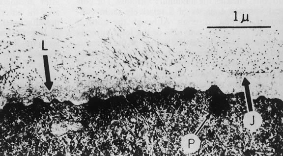
| Fig. 2. Electron micrograph of the vitreoretinal junction (J) 1 mm posterior to the equator of the eye. This demonstrates the insertion of the vitreous fibers to the inner limiting lamina (L). Attachment plaques (P), which anchor the inner limiting lamina to the surface glial cells, are illustrated. (Foos RY: Anatomic and pathologic aspects of the vitreous body. Trans Am Acad Ophthalmol Otolaryngol 1973;77:171) |