|
|
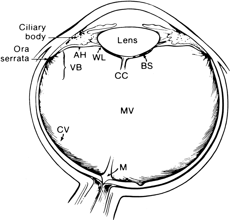
| Fig. 1. Diagram of the eye showing the relationship of the vitreous to the other intraocular structures. (AH, anterior hyaloid; VB, vitreous base; MV, medullary vitreous; WL, Wieger's ligament; BS, Berger's space; CC, Cloquet's canal; M, area of Martegiani). |