|
|
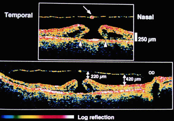
| Fig. 18. Stage 3 hole. Top. Optical coherence tomogram (3 mm long) shows the hyperreflective operculum (arrow) next to the minimally reflective membrane corresponding to the posterior hyaloid. The edges of the hole are thickened by cystic spaces and detached from the retinal pigment epithelium by 850 μm (arrowheads). Bottom. Composite optical coherence tomogram shows the detachment of the posterior hyaloid from the entire posterior hole. (OD, optic disc). |