|
|
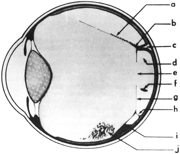
| Fig. 23. Vitreous detachment in proliferative diabetic retinopathy. a. blood deposited on the detached posterior surface of the formed vitreous after hemorrhage into the posterior fluid vitreous; b. neovascular and fibrous proliferations creating a tight vitreoretinal adhesion, which pulls the retina forward and holds the formed vitreous posteriorly; c. localized collection of subretinal fluid; d. curved upper surface of a “mushroom” of formed vitreous extending posteriorly to the retina through a “hole” in the posterior vitreous surface; e. hole in the posterior vitreous surface; f. blood collected in the dependent portion of the mushroom of vitreous after hemorrhage into the formed vitreous; g. posterior vitreous surface; h. a single new vessel stretching between the retina and proliferations on the detached posterior vitreous surface without traction retinal detachment; i. blood pooling between the retina nd the posterior vitreous surface at the inferior limit of vitreous detachment after hemorrhage into the posterior fluid vitreous; j. blood settled out in the inferior part of the formed vitreous. (Davis MD: Natural course of diabetic retinopathy. In Kimura SJ, Caygill WM [eds]: Vascular Complications of Diabetes Mellitus. St Louis, CV Mosby, 1967) |