Chapter 53
Primary Angle-Closure Glaucoma
RUTHANNE B. SIMMONS, MARTHA H. MONTENEGRO and RICHARD J. SIMMONS
Main Menu   Table Of Contents

Search

NOSOLOGY AND TERMINOLOGY
EPIDEMIOLOGY
FACTORS CAUSING PRIMARY CLOSURE OF THE ANGLE
CLINICAL PRESENTATIONS
ERRONEOUS DIAGNOSIS IN ACUTE ANGLE-CLOSURE GLAUCOMA
TREATMENT OF ACUTE ANGLE-CLOSURE GLAUCOMA
TREATMENT OF SUBACUTE ANGLE-CLOSURE GLAUCOMA
CHRONIC ANGLE-CLOSURE GLAUCOMA
TREATMENT OF CHRONIC ANGLE-CLOSURE GLAUCOMA
COMBINED MECHANISM GLAUCOMA
SPECIAL SITUATIONS
PRIMARY ANGLE-CLOSURE GLAUCOMA WITHOUT PUPILLARY BLOCK
INCISIONAL SURGERY FOR NANOPHTHALMOS
LASER CYCLODESTRUCTIVE SURGERY
CLASSIC MALIGNANT GLAUCOMA
NONPHAKIC MALIGNANT GLAUCOMA
MALIGNANT-LIKE GLAUCOMA
REFERENCES

Angle-closure glaucoma must be given a high priority among ocular diseases because its effects can be devastating. Bilateral blindness can result in 2 to 3 days from onset. Angle-closure glaucoma can, however, be dramatically eliminated forever in a given eye with proper recognition and therapy. Few conditions in ocular disease offer such a great opportunity for relief of human suffering. There is a dramatic difference between the devastation of untreated angle-closure glaucoma and its permanent prevention with early recognition and appropriate treatment.

The first type of glaucoma recognized by early physicians was probably angle-closure glaucoma. Total blindness invariably followed recurrent attacks of severe eye pain. Most diagnoses were made at a late stage, where advanced visual loss, iris atrophy, and fixed mydriasis were present. Banister, in 1622, was the first Western physician to document elevated eye pressure.1 Von Graefe attributed the pressure rise in acute glaucomas to some form of inflammation, and in 1857 described iridectomy as its surgical treatment.2 In 1897, the concept of angle closure was introduced to modern ophthalmology by Czermack.3 It was not until the beginning of the 20th century that the pathophysiology of angle-closure glaucoma was understood. Curran4 in 1920, Banziger5 in 1922, Sugar6 in 1941, and Chandler7 in 1952 accurately described the anatomic and pathophysiologic basis of angleclosure glaucoma.

These workers and others established criteria for diagnosis and therapy for angle-closure glaucoma. As a result of such early work and teaching thereafter by Chandler and Grant,8 Shaffer,9 and others, the incidence and the resultant morbidity from primary angle-closure has dramatically decreased over the past 35 years. For example, in the northeastern United States in the early 1960s, glaucoma consultants saw a high incidence of angle-closure glaucoma. In addition, such cases were frequently referred after late diagnosis and after great damage had been done to one or both eyes of a patient. Surgery often involved attempts to salvage an eye after the occurrence of extensive synechial closure of the angle, disc damage, permanently reduced vision, and optic atrophy. Surgical therapy of such eyes always involved incisional surgery, often followed by devastating sequelae such as flat anterior chamber, malignant glaucoma, and other difficult problems. In the same region some 35 years later, the incidence of angle-closure cases requiring referral to a glaucoma specialist is markedly decreased. The disease is recognized much more widely by general ophthalmologists who employ appropriate diagnostic measures including correct gonioscopy and early recognition of potentially closable angles. Laser therapy is safely used by a large number of practitioners, often near the patient's community so that therapy can be closely monitored. Therefore, at least as seen in this region of the United States, it appears to us that the clarification and teaching of angle-closure glaucoma and its sequelae by the early workers and their disciples has resulted in a marked improvement in therapy of this disease and a marked reduction in detrimental social and personal consequences. Even problems such as malignant glaucoma, which were very frequent 30 to 35 years ago in consultation practices, are relatively rare today. These problems are now frequently recognized and dealt with appropriately by general ophthalmologists, thereby reducing to a relatively small number those requiring referral to consultants because of unremitting and uncontrolled progression.

Back to Top
NOSOLOGY AND TERMINOLOGY
The three clinical types of primary angle-closure glaucoma, defined on the basis of symptoms alone are: (1) acute angle-closure glaucoma, which occurs as a dramatic, violent attack with closure of the entire angle; (2) subacute angle-closure glaucoma, which occurs with intermittent milder attacks, with only part of the angle closing; and (3) chronic angle-closure glaucoma, which is a silent, gradual closure of the angle, often confused with open-angle glaucoma. The latter term is reserved for this third clinical type of primary angle-closure glaucoma and is not a synonym for permanent peripheral anterior synechiae, which can be caused by a wide variety of ocular conditions, in addition to primary angle-closure glaucoma. Permanent peripheral anterior synechiae should be designated as such. When possible, the term should be modified by the causative condition when known, for example, “permanent peripheral anterior synechial closure secondary to postoperative flat anterior chamber” or “secondary to uveitis,” and so on.

In the acute form of primary angle-closure glaucoma, a severe attack of angle-closure may occur rapidly. Onset may occur in 30 to 60 minutes with very high intraocular pressure, corneal epithelial edema, pain and congestion of the eye, and markedly blurred vision. The symptoms are severe, persistent, and obvious; the entire angle usually closes. In the subacute form, the angle closes only in part of the circumference. The angle usually opens spontaneously giving rise to symptoms including some or all of the following: intermittent pain, headache, ocular erythema, and visual phenomena, such as blurring and halos. In subacute angle-closure glaucoma, the symptoms are more mild than in the acute form; are spontaneously relieved after one or more hours; and may recur frequently. In the chronic form of primary angle-closure glaucoma, the patient may or may not experience symptoms. The glaucoma is usually discovered on routine examination when elevated in-tra-oc-u-lar pressure, optic disc changes, or visual-field loss become apparent. In this form, gradual closure of part of the circumference of the angle occurs over months or years and the pressure slowly rises as more and more of the angle becomes closed. In all three clinical types, the sole cause of the rise in intraocular pressure is closure of the angle. The closure may be temporary, reversible appositional closure of iris to cornea, or it may become permanent due to formation of peripheral anterior synechiae.

Primary angle-closure glaucomas may also be classified based on the mechanism involved. By far, the most common mechanism is relative pupillary block. The pupillary iris, in apposition to the anterior lens surface, causes the aqueous humor to be partially trapped behind the iris, leading to anterior bowing of the peripheral iris and closure of the anterior chamber angle. Hence, the term pupillary block glaucoma. More rarely, angle closure can be caused by an anatomic configuration called plateau iris, in which the peripheral iris may occlude the trabecular meshwork without pupillary block. These mechanisms will be discussed in more detail later in the chapter.

Lastly, the term combined mechanism glaucoma refers to the coexistence of primary angle-closure glaucoma and primary open-angle glaucoma in the same eye. In such cases of glaucoma, after elimination of angle closure with iridotomy and where the angle is open without peripheral anterior synechia, we prefer to use the term residual open-angle glaucoma.

Back to Top
EPIDEMIOLOGY
The prevalence of primary angle-closure glaucoma varies among different populations. The prevalence of angle-closure glaucoma is as low as 0.09% in the general British population,10 whereas the prevalence is 0.5% in the general Eskimo population. This latter rate is as high as 11.7% if one considers Eskimo women over the age of 60 years.11 According to Alper, acute angle-closure glaucoma is rare in African-Americans.12 In a series of 350 consecutive cases of glaucoma, Neumann did not find a single acute angle closure among Liberians.13 Angle-closure glaucoma has a reduced prevalence in American Indians.14 The meticulous slit lamp observation of 2000 angles led van Herick to establish a 2% incidence of narrow angles, of which 1.64% were actually occludable.15
Back to Top
FACTORS CAUSING PRIMARY CLOSURE OF THE ANGLE

ANATOMIC FACTORS

Several anatomic features predispose an eye to primary angle-closure glaucoma. These relate to the size and shape of the eye, its cornea, anterior chamber, angle, lens, iris, and ciliary body.

The Eye

Ultrasonographic measurements have shown that eyes with primary angle-closure glaucoma have an average axial length 1 mm shorter than normals.16,17 Hyperopes are therefore more predisposed than emmetropes or myopes to angle closure. However, myopia does not preclude the development of angle closure.18

The Cornea

The corneal radius of curvature and diameter are usually smaller than average in primary angle-closure glaucoma eyes. There is a tendency for corneae with smaller diameters to have steeper curvatures, which partially compensate for the change in refractive power.19

The Anterior Chamber

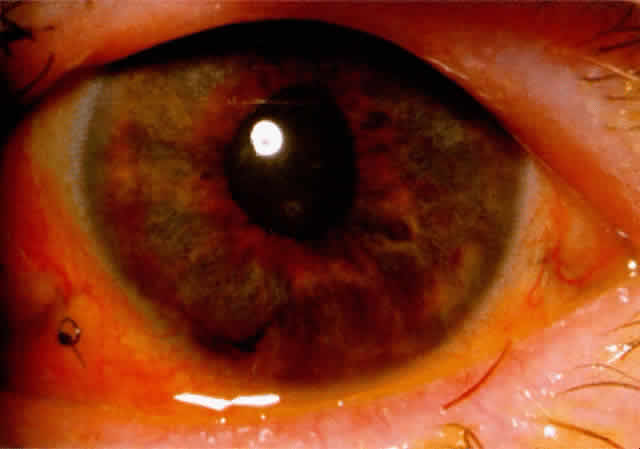
The central anterior chamber in eyes with angle closure appears to be 1 mm shallower, on average, than that in normal eyes. It seems that the 1 mm shallowing of the anterior chamber is caused by increased volume and forward positioning of the lens.20 Eyes with anterior chamber depth greater than 2.5 mm seldom develop angle closure. According to Barkan,21 the central anterior chamber depth was less than 1.5 mm in 75% of angle-closure glaucoma cases.

It should be emphasized that the depth of the anterior chamber can vary in a given individual and in a given eye. Generally, the anterior chamber becomes more narrow with advancing age. A patient seen with a moderately narrow angle at age 50 is likely to have a much more narrow angle at age 60, and perhaps a dangerously narrow angle at age 70 or 80. This appears to be due to increasing size of the crystalline lens with age and perhaps to increasing laxity of the zonules, which allows the lens-zonular diaphragm to move anteriorly.

In addition, variations in angle depth can occur as aqueous production alters. Chandler and Grant frequently observed that an angle that is anatomically extremely narrow or closed could be opened and appear wide and nonoccludable when aqueous production was markedly reduced, such as when carbonic anhydrase therapy was instituted. Thus, a patient treated for angle-closure glaucoma in one eye might be misdiagnosed as having a nonoccludable angle in the fellow eye when, in fact, the fellow eye was extremely narrow before treatment with carbonic anhydrase inhibitors. There may be variation in the angle depth with any therapeutic agent that alters aqueous production. Similar effects could be produced as a result of other variations in aqueous production, such as the reduction in aqueous humor production after contusion to the eye, choroidal separation, or chronic uveitis.

The Lens

The average axial lens thickness is 0.6 mm greater in angle-closure glaucoma eyes compared with normals. Axial thickening of the lens and its forward displacement due to laxity of the zonules increases with age. The lens size relative to the anterior chamber volume may bring about shallowing of the anterior chamber. An eye with a normal size anterior segment may develop anterior chamber shallowing with an unusually large lens. This same effect may be seen with a normal size lens in a small anterior segment. In addition, ultrasonographic measurements of the radius of curvature of the anterior lens surface is on average 2.3 mm steeper in angle-closure glaucoma eyes than normals.22

The Angle

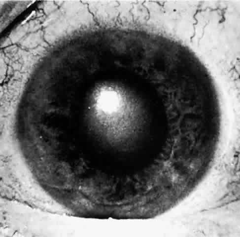
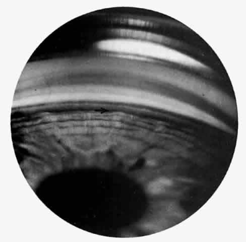
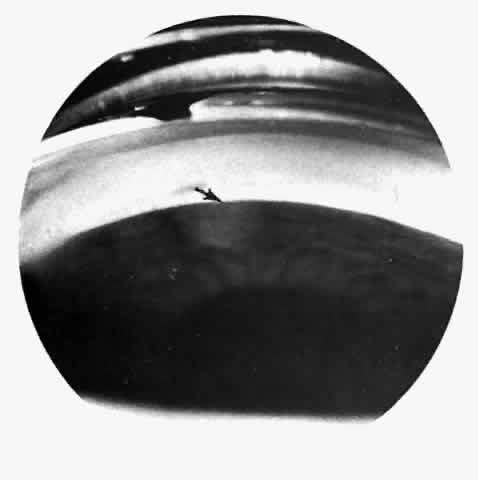
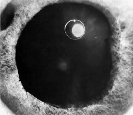
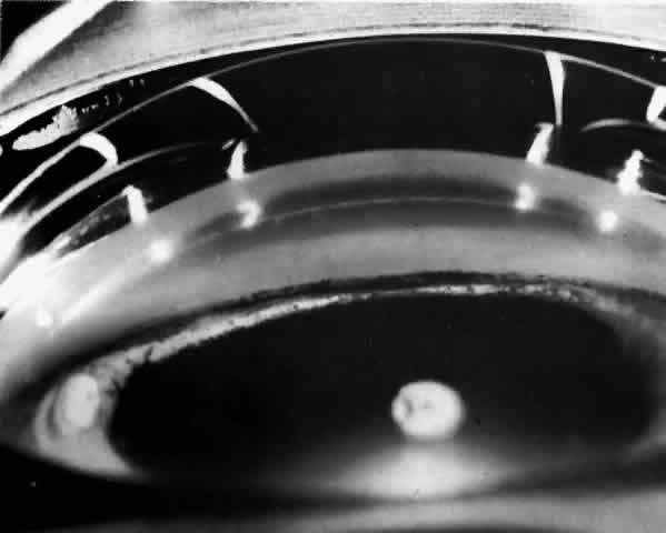
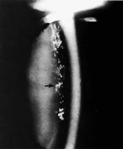
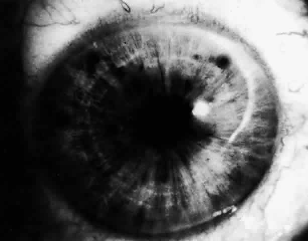
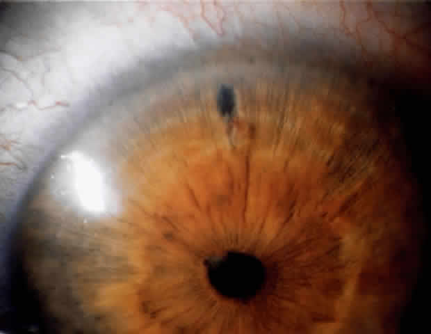
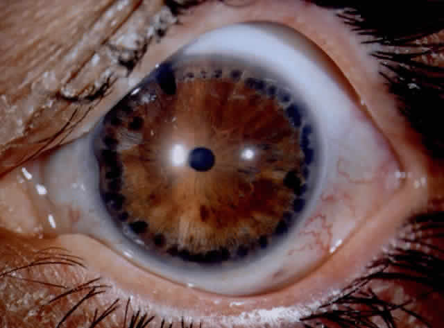
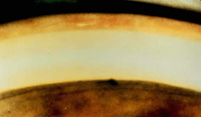
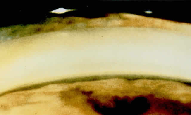
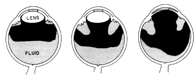
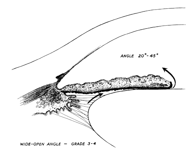
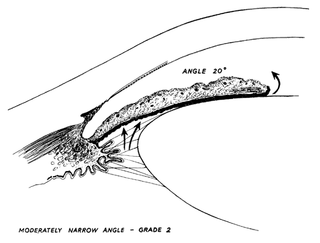
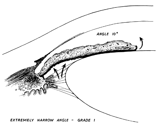
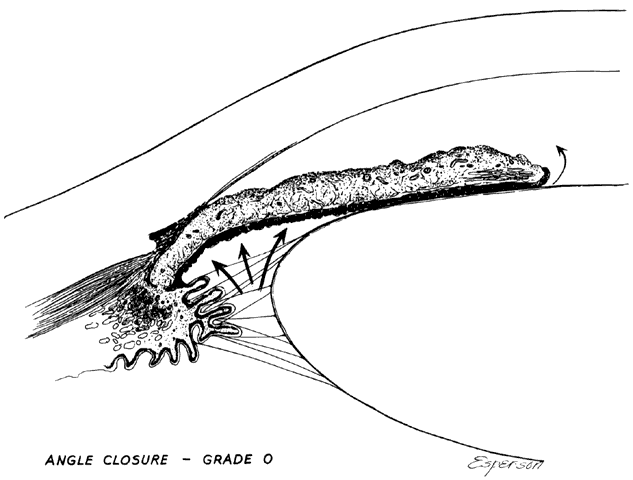
There are several classifications that have been developed in an attempt to identify those patients at risk for angle closure. Van Herick15 developed a slit lamp grading system based on the depth of the peripheral anterior chamber as measured by the adjacent corneal thickness seen in the slit lamp beam directed at the limbus. In van Herick's system, a grade 1 depth is the narrowest and a grade 4 depth is a deep anterior chamber. When a grade 1 or 2 depth is present, that is, peripheral anterior chamber depth less than one half corneal thickness, gonioscopy should be performed to rule out occludable angles. Scheie's23 grading system is based on the depth of angle structures visible on gonioscopy with a grade IV angle being narrow and visible only to Schwalbe's line. Shaffer developed the following classification for describing angle width and the likelihood of angle closure. If an angle recess width is 20 degrees or more, angle closure is improbable (Fig. 1). Angles between 10 and 20 degrees have an increased risk of developing angle closure (Fig. 2). Below 10 degrees, angle closure is probable or actually present (Figs. 3 and 4).24 When evaluating an angle, however, it is also necessary to consider the peripheral iris configuration and iris root insertion.25 Spaeth developed a gonioscopic classification system based on three elements: (1) the angle width in degrees; (2) the configuration of the peripheral iris; and (3) the insertion of the iris root.26

Fig. 1. Anterior chamber angle width. Cross-sectional diagram of a deep anterior chamber with a nearly flat iris plane, minimal apposition between the iris and lens at the pupil, and a wide-open angle entrance to the filtration area. In this type of eye, aqueous passes easily from the posterior chamber through the pupil into the anterior chamber and out through the filtration area. (Kolker AE, Hetherington J Jr: Becker and Shaffer's Diagnosis and Therapy of the Glaucomas, p 42. St. Louis, CT Mosby, 1970)

Fig. 2. Anterior chamber angle width. A Shallow anterior chamber with a more anteriorly located lens than is shown in Figure 1. Apposition of the lens and iris in the pupillary zone is increased, producing a relative pupillary block that interferes with aqueous flow from the posterior to the anterior chamber. The pressure differential between the posterior and anterior chambers is increased. (Kolker AE, Hetherington J Jr: Becker and Shaffer's Diagnosis and Therapy of the Glaucomas, P 42. St. Louis, CV Mosby, 1970)

Fig. 3. Anterior chamber angle width. Extremely narrow anterior chamber angle entrance with a mid-dilated pupil and a lax peripheral iris. Increased pressure in the posterior chamber pushes the peripheral iris forward where it lies near the filtration area; at this stage, it does not block outflow of aqueous or increase intraocular pressure. (Kolker AE, Hetherington J Jr: Becker and Shaffer's Diagnosis and Therapy of the Glaucomas, p 43. St. Louis, CV Mosby, 1970)

Fig. 4. Anterior chamber angle width. The angle has closed and the iris now lies against the trabecular meshwork, obstructing outflow. If closure has occurred around a significant portion of the circumference of the angle, intraocular pressure will rise. (Kolker AE, Hetherington J Jr: Becker and Shaffer's Diagnosis and Therapy of the Glaucomas, p 43. St. Louis, CV Mosby, 1970)

This large number of classification systems can generate confusion in communication between observers. By one grading system, a dangerously narrow angle may be described as grade IV. By another system, the same angle might be labeled grade I. The diversity of the grading systems requires both the retranslation from grade to anatomic interpretation and the identification of whose grade I or IV is being used. It seems that with frequent modern international communication of medical information, it is simpler and safer to use straightforward scientific English prose to describe angle anatomy. Chandler and Grant8 prefer to describe each angle verbally and to state whether it is judged closable or not.

The Iris and Ciliary Body

In some emmetropic and hyperopic, but rarely myopic eyes, the anterior chamber is shallow in structure with a narrow angle. This configuration can be related to heredity, a large lens, a structurally crowded anterior segment, or other anatomic characteristics. In some eyes, especially hyperopic eyes, the iris root inserts more anteriorly on the anteromedial surface of the ciliary body. The iris plane is typically convex. In addition, the ciliary body is more developed in hyperopic eyes, perhaps due to increased accommodative effort.27 These factors lead to increased narrowing of the angle recess.

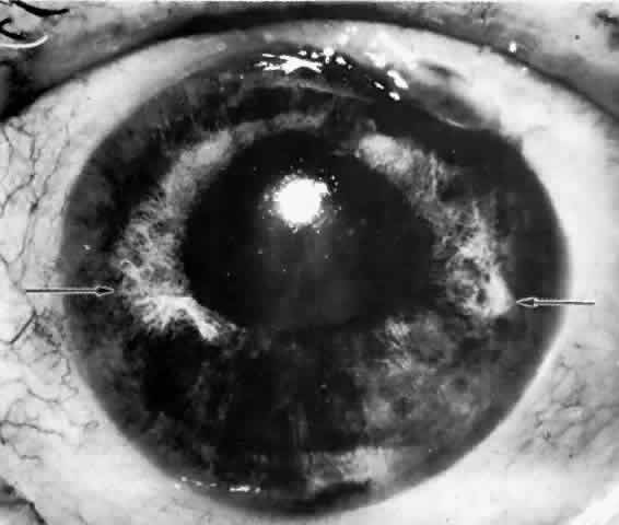
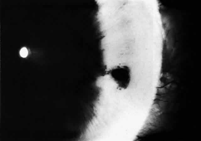
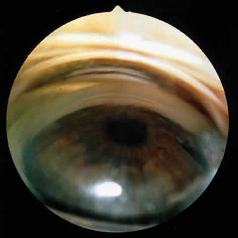
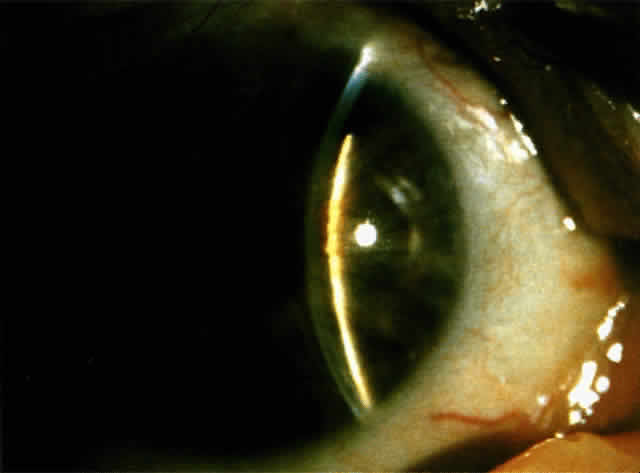
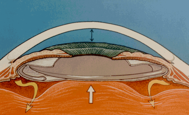
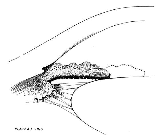
Another anatomic configuration, the so-called plateau iris configuration, may predispose to closure of the angle. This is a relatively rare anatomic configuration of the peripheral iris and was first described by Tornquist.28 In plateau iris configuration eyes, the anterior chamber appears to have normal axial depth and the iris plane remains flat. The chamber angle may be narrow, however, due to the location of the iris plane adjacent to anterior chamber structures. In the far periphery, the iris drops abruptly to a more posterior plane to create a very narrow recess over the filtration meshwork. In a somewhat different anatomic description, Barkan stated that the peripheral iris appears to be redundant with marked folds crowding the angle.29 This particular arrangement of the anterior iris plane gives the angle an appearance resembling a deep valley surrounding a flat-topped hill, thus the designation “plateau” (Fig. 5). Such eyes are susceptible to closure of the angle when the pupil dilates physiologically or pharmacologically.

Fig. 5. Plateau iris configuration. The anterior chamber is deep axially and the iris plan is flat. The angle is open but is narrow when the pupil is small (dotted line) and closed when the pupil is dilated. (Kolker AE, Hetherington J Jr: Becker and Shaffer's Diagnosis and Therapy of the Glaucomas, p 198. St. Louis, CV Mosby, 1970)

These anatomic factors may be said to predispose to closure of the angle but other factors, either physiologic or pharmacologic, must be superimposed to bring about actual closure of the angle.

PHYSIOLOGIC FACTORS

In an anatomically predisposed eye, several physiologic mechanisms can trigger angle-closure glaucoma. Of particular interest are the iris-lens interaction, pupil size, accommodation, and uveal vascular status.

Relative Pupillary Block

IRIS-LENS INTERACTION.Aqueous humor flows from the posterior chamber, between the posterior iris surface and anterior lens capsule, around the pupil edge and into the anterior chamber. In a normal eye, the iris is in contact with the lens only in the immediate vicinity of the pupillary margin. In eyes with shallow anterior chambers, the forward position of the lens brings the lens-iris diaphragm forward relative to the iris insertion plane. In addition, the anterior lens surface's greater curvature increases the area of apposition of the posterior iris surface to the lens. The increased surface apposition of the iris to lens causes an increased resistance to forward aqueous flow causing a pressure differential between the anterior and posterior chamber. The slightly greater pressure in the posterior chamber leads to forward bowing of the peripheral iris. This mechanism is known as relative pupillary block and it depends greatly on pupillary size and rigidity of the peripheral iris. Under normal circumstances, the iris tends to hang lax in the fluid medium and is sensitive to any pressure differential between the two chambers. Some degree of relative pupillary block is constantly present under normal circumstances, but eyes with a greater extent of lens-iris apposition have more relative pupillary block and are the ones most vulnerable to angle closure.

PUPIL SIZE. One of the most important factors in closing the angle in an anatomically predisposed eye is dilation of the pupil. Dilation leading to closure of the angle may occur as a result of a variety of causes including darkness, emotion, and both topical and systemic dilating drugs. Iris-lens contact is maximal with pupil sizes between 3 and 4.5 mm, enhancing the relative pupillary block mechanism.30 In mid-dilation (3 to 4.5 mm), peripheral iris laxness combined with persistent relative pupillary block leads to closure of a narrow angle by the peripheral iris in predisposed eyes. Wide and rapid dilation minimizes the relative pupillary block phenomenon since iris-lenticular contact is reduced, and the aqueous passes forward through the widely dilated pupil easily. Dilation with sympathomimetic agents, alone or with cycloplegics, tends to decrease peripheral iris laxity by contracting the dilator fibers and thus decreases the likelihood of angle closure while the pupil is rapidly dilating. However, when the iris and ciliary body are slowly recovering from the drug effects and the pupil is gradually becoming smaller in size, the iris at mid-dilation reaches its worst relationship with the lens; relative pupillary block is maximal and the peripheral iris is lax. In this position of mid-dilation following wide dilation, an attack of acute angle closure commonly occurs.

Topical anticholinergics are safe in the majority of eyes but they also increase the risk of angle closure in predisposed eyes. Cycloplegia results in a posterior displacement of the ciliary body, tightening of the zonules, flattening of the lens, and theoretical deepening of the anterior chamber. Mydriasis increases the lens-iris contact. When the pupil is in mid-dilation, the lens-iris contact is maximal thereby increasing relative pupillary block. In addition, cycloplegic mydriasis increases peripheral laxity of the iris since the dilator fibers are not contracted by cycloplegics alone and this further increases the likelihood of closure of the angle. Cycloplegia deepens the anterior chamber in a normal eye, but in a small anterior segment with a relatively large lens bearing a steeper than normal anterior curvature, the slight benefit of a retroplacement of the lens is overcome by the increased relative pupillary block related to midmydriasis. Pure miosis, as seen after instillation of the alpha adrenergic blocking agent thymoxamine, can break angle closure by relieving iris crowding of the angle.31 Thymoxamine hydrochloride acts by inhibition of the iris dilator muscle and has no effect on the ciliary muscle and therefore on accommodation.

ACCOMMODATION. In situations where miosis, whether physiologic or pharmacologic, is seen concurrently with an angle-closure attack, the mechanism appears to be more related to accommodation than to the pupil size. Contraction of the circular muscle of the ciliary body relaxes the zonules and allows forward displacement of the lens. During accommodation, the central portion of the lens becomes more curved while the peripheral zone of the lens surface flattens. In young people, the radius of curvature of the central anterior surface of the lens changes from 10 mm at rest to 5.3 mm in full accommodation.32 There is a slight increase in the axial length of the lens due to the forward bulge of the anterior surface. The coexistence of the aforementioned physiologic mechanisms increases the lens-iris surface contact and enhances the pupillary block phenomenon in predisposed eyes.

UVEAL VASCULAR STATUS. Some authors postulate that the iris-lens diaphragm may suddenly move anteriorly under the influence of increased choroidal blood volume. This mechanism is uncertain and any relationship is unproven. While this may explain an acute angle-closure attack following severe emotional stress, a more plausible explanation relates it to mydriasis due to increased sympathetic tone.

PATHOPHYSIOLOGIC FACTORS

According to Goldmann's equation, assuming constant aqueous production, the pressure driving fluid through the trabecular meshwork out of the eye (perfusion pressure) is directly proportional to the resistance to flow in the eye. The perfusion pressure is the result of intraocular pressure minus episcleral venous pressure. Chandler and Grant concluded that in angle-closure glaucoma, the rate of the rise of intraocular pressure is directly proportional to the degree of obstruction to outflow. A normal eye has excess capacity for outflow through the trabecular meshwork to withstand partial angle closure without significant rise in intraocular pressure. The unaffected trabecular meshwork increases its filtration and keeps the intraocular pressure from rising significantly.

In eyes with anatomically narrow but open angles without glaucoma, the filtration apparatus is normal and therefore the intraocular pressure remains at a normal level. As in normal eyes, there is asymmetry in the width of the angle in different quadrants. The angle appears more narrow in the upper than in the lower quadrants. This observation is constant despite the type of gonio lens used to visualize the angle. It presents even with Koeppe lens direct gonioscopy with the patient in the supine position. The closure of the angle probably evolves gradually from above downward. As closure develops throughout the angle, more and more of the trabecular meshwork is obstructed and an increase in intraocular pressure occurs.

In primary angle-closure glaucoma, obstruction of outflow starts by apposition of the peripheral iris to the trabecular meshwork. There may be temporary apposition of the iris to trabecular meshwork, or the closure may become permanent due to formation of peripheral anterior synechiae. Goniosynechiae tend to occur earlier and more frequently superiorly than inferiorly. The extent of closure at any given time determines the reduction of outflow facility and the level of elevation of the intraocular pressure. Furthermore, in partially closed angles, the efficiency of the remaining functional angle will determine the facility of outflow and pressure elevation.

How the closure of the angle evolves over time and in extent will determine the actual clinical presentation of each particular eye.

Back to Top
CLINICAL PRESENTATIONS
Classically, three types of angle-closure glaucoma have been described: acute, subacute, and chronic. We prefer the Chandler-Grant classification based on symptoms alone, which correlates well with the pathophysiology and clinical presentation.

ACUTE ANGLE-CLOSURE GLAUCOMA

Symptoms

This entity presents with a rapid onset of severe, deep aching pain that may be localized to the eye or may follow the trigeminal distribution (Table 1). It may be specifically identified by the patient as eye or frontal pain, but may be vaguely diffuse or ignored by the patient who may stress other symptoms. The attack may evolve in 1 hour or less and quite often marked pain may be accompanied by a vagal response with nausea and vomiting. While patients may complain of varying severity of pain, blurred vision is a frequent symptom. Additional visual symptoms described as “colored halos” or “rainbows” around lights and total “greyouts” or “blackouts” of the involved eye may be present.


TABLE 1. Diagnosis of Acute Angle-Closure Glaucoma

  History

  Acute onset


  Symptoms

  Pain
  Halos
  Visual loss
  Nausea
  Emesis


  Clinical Signs

  Intraocular pressure (usually high during attack but low after attack in recovery)
  Mid-dilated, unreactive pupil
  Ciliary-conjunctival injection
  Corneal edema
  Engorged iris vessels
  Cells in aqueous (but no keratic precipitates)
  Closed angle gonioscopically (may be open but narrow and “closable” angle after attack)
  Fellow eye-narrow angle judged “closable” by gonioscopy
  Iris atrophy
  Posterior synechiae
  Glaukomflecken of Vogt
  Optic atrophy


Signs

On early examination, the cardinal sign of primary acute angle-closure glaucoma is present: a fixed mid-dilated pupil. There is profound reduction in central visual acuity. Photophobia and conjunctival hyperemia are striking external findings (Fig. 6). Intraocular pressure usually exceeds 40 mmHg and may be as high as 70 or 80 mmHg during the attack. On biomicroscopy, perilimbal hyperemia with a ciliary flush is accompanied by peripheral conjunctival and episcleral congestion. The peripheral iris bulges anteriorly and comes in close contact with the peripheral corneal endothelium for 360 degrees. Early microcystic epithelial corneal edema evolves to stromal edema. The anterior chamber appears shallow but formed centrally with cells, pigment and aqueous flare. Sustained high intraocular pressure affects the iris sphincter muscle, as well as its blood supply.33 Ischemia of sectors of the iris develop, probably due to compression of nutrient radial iris vessels causing infarction of the compromised iris and possibly also the ciliary body. This results in the release of debris and proteins, causing an aseptic anterior uveitis.

Fig. 6. A most reliable sign in angle-closure glaucoma is the unreactive pupil in the mid-dilation. Conjunctival and ciliary vascular engorgement, as well as epithelial corneal edema, is present. In this eye, no atrophy of the iris is visible, but the iris sphincter is unreactive, presumably as a result of ischemia caused by the elevated intraocular pressure. If high pressure is relieved before permanent atrophy of the iris occurs, the pupil will again become reactive. (Courtesy of Ayerst Laboratories and Dr. H. Saul Sugar)

If gonioscopy is obscured by corneal epithelial edema, application of topical 100% glycerol on the anesthetized cornea will often clear the edema sufficiently to permit adequate visualization of the peripheral anterior chamber and angle structures. During an attack, one sees only the cornea and iris meeting directly, with no evidence of the angle entrance or angle structures (Figs. 7 and 8). Indentation gonioscopy, introduced by Forbes,34 may be useful to differentiate between appositional and synechial closure of the angle. Central compression with a four mirror gonioscopy lens on the central cornea pushes the central aqueous to the peripheral anterior chamber. This maneuver pushes the peripheral iris posteriorly and opens an appositionally closed angle. If peripheral anterior synechiae are present, the angle will not be opened with indentation. The lower the initial pressure, the easier the examination. If the pressure is extremely high, indentation gonioscopy is extremely difficult, painful, and the cornea may be too edematous to allow visualization. If despite corneal dehydration with glycerol, edema of the affected eye precludes clear gonioscopy, examination of the angle structures of the fellow eye is often helpful. An extremely narrow closeable angle is usually present in the fellow eye giving strong evidence to the diagnosis of angle closure of the affected eye. Funduscopic examination is usually of little value because it is commonly precluded by corneal edema. When possible, early ophthalmoscopy shows pulsation of the retinal arterioles.

Fig. 7. In this goniophotograph, the angle is moderately wide open. As one looks just anterior to the edge of the peripheral iris (arrow), one sees the dark ciliary band, white scleral spur, and trabecular meshwork, which is moderately dark and pigmented in this eye. Schwalbe's line anterior to the trabecular meshwork is not visible in this photo. Such an angle is much too wide to be susceptible to primary angle-closure glaucoma.

Fig. 8. In contrast to Figure 7, this angle is closed. At the arrow, one sees the edge of the peripheral iris. The ciliary band, scleral spur, and trabecular meshwork are covered by peripheral iris, and one sees only cornea anterior to it. This could be reversible appositional closure of the angle of permanent synechial closure.

When a patient is seen after an attack that has been in progress for several hours or even days, the eye usually will display decreased vision. The pupil is irregular in shape, nonreactive to light, and resistant to the effects of miotics or mydriatics. It is a common misconception that the intraocular pressure remains elevated in angle-closure glaucoma. In some cases, low tension may be found after aqueous production has decreased while the angle remains closed. If the eye has recovered spontaneously from an attack, the intraocular pressure may be very low due to reduced aqueous formation with or without an open angle. It is important to realize that low pressure following elevated pressure due to acute angle-closure glaucoma does not necessarily mean that the angle-closure attack is over. The pressure will rise as soon as aqueous production resumes, which may be a few hours to many weeks later. The cornea may show Descemet's folds and later recover its transparency but occasionally the corneal edema may persist for weeks after reduction of intraocular pressure. A prolonged iris stromal edema encourages the formation of posterior synechiae in the pupillary area and peripheral anterior synechiae at the level of the angle structures. The destruction of iris tissue results in grayish patches of atrophic iris stroma that may extend to the periphery (Figs. 9 through 11). Tiny white-gray spots with discrete edges may be seen in the pupillary zone. These anterior subcapsular lenticular opacities, best known as glaukomflecken, appear to be the result of damage to the anterior lens epithelium from very high intraocular pressure (Fig. 12).35 Glaukomflecken may result from very high intraocular pressure of any etiology, but are especially characteristic of previous attacks of angle-closure glaucoma.

Fig. 9. Iris atrophy, presumably due to iris ischemia, commonly occurs in sever and prolonged acute angle-closure glaucoma. In this eye, gray, thin, depigmented areas of atrophy of the iris, most marked at the arrows, are present around the pupillary margin clockwise from 8 o'clock to 3:30. Iris atrophy from high intraocular pressure in angle-closure glaucoma usually involves irregular radial sectors of the iris and is most marked near the pupil. In these sectors, the pupil is usually permanently fixed and dilated as a result of the atrophic process.

Fig. 10. In this eye, iris atrophy due to neglected angle-closure glaucoma is so extensive that the pupil is fixed in wide dilation. Some pigment granules from the atrophic iris lie scattered about on the iris stroma. There is a posterior synechia at 2 o'clock at the iris margin. Glaukomflecken are very numerous in this eye but are not visible in this photo because oblique slit illumination is not used; they are visible in Figure 12, in which proper illumination is used. (Photographed by Dr. David Donaldson)

Fig. 11. Same eye as is shown in Figures 10 and 12. This gonioscopic view shows much pigment debris released by the atrophic process in the iris on the angle wall. The pigment seen lies on the angle wall between Schwalbe's line anteriorly and the synechial attachment of the iris. Throughout the portion of the angle in this photo, synechiae attach at the level of the anterior trabecular meshwork, covering the ciliary band, scleral spur, and posterior trabecular meshwork and obstructing all useful outflow from this portion of the angle. (Photographed by Dr. David Donaldson)

Fig. 12. Same eye as shown in Figures 10 and 11. With oblique slit-lamp illumination, glaukomflecken of Vogt appear as pleomorphic gray-white patches just beneath the anterior lens capsule. These are presumed to be areas of necrotic lens tissue caused by very high pressure. With time, they lie deeper and deeper beneath the anterior lens capsule as new lens fibers are laid down, but they will persist permanently as silent records of previous high intraocular pressure. (Photographed by Dr. David Donaldson)

A neglected acute angle-closure attack may show a striking gonioscopic picture. Occasionally, if the attack resolved spontaneously, or if it was medically broken, the angle reopens to such an extent that it may be difficult to believe that angle closure was responsible for the attack. In such a case, the widening of the previously closed angle is due to the reduction in aqueous production, which may follow an acute attack. Some peripheral anterior synechiae may persist after the resolution of the acute phase and may bear witness to the fact that the angle was previously closed. In the fundus, peripapillary hemorrhages, exudates, and disc edema may follow resolution of an acute attack with high pressure. In severe relapsing attacks, the anterior chamber angle often is closed permanently by peripheral anterior synechiae around all or part of the angle circumference. Although the symptoms may improve with decreased tension due to decreased aqueous production, the eye may develop high pressures when normal aqueous production resumes. The natural course of the disease shows that delayed treatment of an acute episode may lead to permanent synechial angle closure, and even permanent blindness in 2 to 3 days.

Differential Diagnosis

Any painful congested eye may mimic acute angle-closure glaucoma to some degree, but usually such common disorders as keratoconjunctivitis, corneal abrasion or foreign body, and trauma are readily distinguished by careful history and examination. Certain other disorders may mimic acute angle-closure glaucoma more closely, however.

NEOVASCULAR GLAUCOMA

Neovascular (hemorrhagic) glaucoma often presents as a painful congested eye with high intraocular pressure, corneal edema and cells, and flare in the anterior chamber. There may be an associated history of diabetes mellitus or retinal vascular occlusion with sudden painless loss of vision months before inflammation of the eye occurred. On examination, this entity is distinguished by rubeosis, a fine network of abnormal arborizing vessels on the iris surface, which extends over the angle structures. One need see only a few arborizing vessels on the meshwork to make this diagnosis. Broad peripheral anterior synechiae are often associated with a mat-like membrane with blood vessels, which gradually progresses and contracts to close the entire angle, sometimes obscuring the extensive network of new vessels that initiated angle closure.

ACUTE IRITIS

Acute iritis with secondary glaucoma may cause blurred vision with halos, occasionally moderately elevated intraocular pressure, and cells in the anterior chamber. The onset is usually more gradual than that in acute angle-closure glaucoma. Often, keratic precipitates are present and the pupil is miotic. Most importantly, the anterior chamber usually is not abnormally shallow and gonioscopy shows an open angle. If the view is obscured, gonioscopy of the fellow eye will usually reveal a wide open angle.

GLAUCOMATOCYCLITIC CRISIS

Glaucomatocyclitic crises or Posner-Schlossman's syndrome usually presents with recurrent attacks of visual blur, halos, cells in the aqueous, and an elevated intraocular pressure. This can often be distinguished by the presence of a few keratic precipitates, which never occur in primary angle-closure glaucoma itself, and by an open angle.

PHACOLYTIC GLAUCOMA

Phacolytic glaucoma also may produce a painful congested eye with blurred vision and high intraocular pressure. However, in addition to keratic precipitates, the angle is open, there is a cataract, and a hypopyon is sometimes present. The cataract is usually very advanced or mature, but in rare cases, an immature cataract may be present.

PHACOMORPHIC GLAUCOMA

Phacomorphic glaucoma presents as asymmetric central shallowing of the anterior chamber in the presence of a unilateral advanced or mature intumescent cataract. The intraocular pressure is elevated but there is no evident inflammatory reaction in the aqueous. The fellow eye shows a normal anterior chamber depth and an open angle on gonioscopy. This secondary angle-closure glaucoma is caused by enhanced pupillary block due to lens enlargement. Increased lens apposition to the iris results. Once the diagnosis is made, initial treatment with mydriatics and cycloplegics may be sufficient to deepen the anterior chamber and relieve the relative pupillary block. Laser iridotomy may then be performed as a temporary measure followed by cataract removal.

CYSTS OR TUMORS OF THE IRIS AND CILIARY BODY

Cysts or tumors of the iris and ciliary body cause secondary closure of the angle. The most common of these are intraepithelial cysts of the iris and ciliary body. When these are present, they may narrow the angle and predispose to acute angle closure. When they are present around much of the circumference of the angle, the entire angle may close; when they are present segmentally, they cause only partial angle closure. One should suspect cysts or a tumor if there is abrupt localized bulging of the iris in the periphery in one or more areas. If the pupil can be widely dilated, one can sometimes see the cysts in the posterior chamber by using a gonioscopy lens. If these cysts are closing the angle, or if they enlarge such that they obscure the visual axis, they can usually be collapsed using laser to puncture them. This can be performed directly through a dilated pupil via the transpupillary route with a mirrored gonioscopy lens using the argon laser. When the cysts cannot be seen through the dilated pupil, they may be collapsed by penetrating the cyst anteriorly through the anterior iris stroma with a Nd:YAG laser. Laser therapy is a simple, noninvasive, and atraumatic technique, which we believe to be the procedure of choice for treatment of these cysts.

Back to Top
ERRONEOUS DIAGNOSIS IN ACUTE ANGLE-CLOSURE GLAUCOMA
Acute angle-closure glaucoma, a great mimic, may in its various phases be mistaken for other ocular, neurologic, and medical diseases (Table 2).8,36 Examples of a few of the clinical situations that sometimes lead to mistaken diagnoses are described; all of these examples are real and have been actually encountered in sophisticated medical centers.


TABLE 2. Conditions Mimicked by Symptoms of Acute Angle-Closure Glaucoma

  Uveitis

  Cells, flare, fibrin
  Erythematous, congested globe


  Ocular Vascular Occlusion

  Papilledema
  Retinal hemorrhages


  Neurologic Disease

  Fixed dilated pupil
  Papilledema
  Headache


  Gastrointestinal Disease

  Nausea and emesis


  Sinus Disease

  Headache-may be recurrent
  Malaise


DIAGNOSIS: UVEITIS

A patient may give a history of acute onset of pain and blurring of vision of 1 or more days' duration; later the symptoms became less severe. When the patient presents for examination, the eye is somewhat congested with many folds in Descemet's membrane, cells in the aqueous, a moderately dilated pupil, and normal or subnormal intraocular pressure. Keratic precipitates are not present. There may be corneal edema and posterior synechiae. Gonioscopy in the affected eye may be difficult because of the condition of the cornea but the angle in the fellow eye is shown to be narrow. This situation is often misdiagnosed as acute iritis. However, acute iritis never has the precipitous onset characteristic of acute angle-closure glaucoma, which can occur suddenly in 30 to 60 minutes. Nor does one see the semidilated pupil in cases of iritis; with iritis the pupil is characteristically smaller than in the fellow eye. Only one set of circumstances explains the situation just described. It represents spontaneous recovery from an attack of acute angle-closure glaucoma. This diagnosis is attested to by the history of acute onset of symptoms and the finding of a semidilated pupil; the absence of keratic precipitates; folds in Descemet's membrane; and the narrow angle in the fellow eye. Keratic precipitates commonly occur in uveitis but never occur as a result of primary angle-closure glaucoma.

DIAGNOSIS: OCULAR VASCULAR OCCLUSION

A patient presents and gives a history of vague headache and decreased vision. On ophthalmoscopy there is disc edema, engorged retinal venules, with hemorrhage and exudates around the optic disc in the affected eye. These findings are interpreted as central retinal vein occlusion with engorged vessels, peripapillary hemorrhages, and edema. The patient may be referred to a retinal specialist for retinal circulatory evaluation, when, in fact, the diagnosis is a spontaneously resolved episode of acute angle-closure glaucoma with suddenly decreased intraocular pressure and subsequent disc edema and vascular engorgement.

DIAGNOSIS: NEUROLOGIC DISEASE

A patient presents to a primary care physician and gives a history of headache and malaise. On examination, the pupil is fixed and mid-dilated and there is disc edema. The findings are interpreted as neurologic disease with papilledema. The patient is referred for neurologic evaluation and undergoes extensive evaluation for intracranial disease. No intracranial disease is found. Finally, the patient reports decreased vision. An ophthalmologist is called. Evaluation shows the presence of angle closure and the episode represents spontaneous recovery from an attack of acute angle-closure glaucoma. The ophthalmoscopic findings result from reactive hyperemia of the previously ischemic optic nerve head that can occur following a dramatic change in intraocular pressure from high levels to subnormal levels. The fixed dilated pupil is, of course, the prime and cardinal sign of primary angle-closure glaucoma either in an attack or in spontaneous recovery.

DIAGNOSIS: GASTROINTESTINAL DISEASE

An elderly patient may be brought to an emergency facility with vomiting, dehydration, and coma. The patient is admitted to a medical service and treated for these problems empirically with intravenous rehydration, restoration of electrolyte balance, and full gastrointestinal studies. When the patient recovers consciousness, the patient reports previous vague headaches and later mentions that he or she can't see. Finally, an ophthalmologist is consulted. On evaluation, the patient is found to have angle-closure glaucoma. Angle-closure glaucoma can, through its secondary effects, mimic gastrointestinal disorders

DIAGNOSIS: SINUS DISEASE

The patient presents emergently with frontal pain and malaise. No ocular symptoms are mentioned by the patient. A diagnosis of sinus infection is made and the patient is sent home with systemic antibiotics. The patient again presents with bilateral frontal pain and is again sent home with different systemic antibiotics and analgesics. When the patient next presents, he or she is bilaterally blind. An ophthalmologist is called and angle-closure glaucoma is finally diagnosed too late.

These situations emphasize that angle-closure glaucoma is a great mimic of other ocular, neurologic, and medical diseases and must always be suspected if any of the characteristic signs are present.

Back to Top
TREATMENT OF ACUTE ANGLE-CLOSURE GLAUCOMA
Early diagnosis and prompt treatment are essential to improve the likelihood of a favorable outcome to this potentially blinding disease. Initial medical treatment should be started immediately upon diagnosis in an office or emergency room to try to rapidly reduce the intraocular pressure and reopen the angle. Immediate lowering of the intraocular pressure prevents optic nerve damage and iris ischemia and increases the chances of reversing the attack. The pupillary response can provide some guide as to the response to medical therapy. If the pupil is reactive when the patient is first seen, the eye will likely show a quick response to medical therapy. Frequently, the pupil is fixed in mid-dilation, slowly starts to become miotic, and then rapidly becomes miotic as the angle reopens and the pressure drops precipitously to normal or subnormal levels. The opening of the angle signifies that the attack is broken but definitive treatment requires laser or incisional surgery.

In refractory cases, the pupil does not become miotic. Rather, the angle remains closed and, although the pressure may fall due to drug-induced decreased aqueous production, the pressure will rise, often to dangerous levels, in hours or days.

MEDICAL THERAPY

Oral osmotic agents are effective in quickly lowering the intraocular pressure and should be administered first if the patient is not experiencing emesis. Antiemetics may help prevent nausea from progressing to emesis. Oral osmotic agents can be cold but should not be diluted with fluid or ice lest their osmolality and hence their efficiency be reduced. Glycerin 1 to 1.5 g/kg in a 50% solution is a time-honored effective agent. A maximum decrease in intraocular pressure is obtained 30 to 90 minutes later and may remain low for the ensuing 5 to 6 hours. Since glycerin causes hyperglycemia and dehydration, extra caution is needed in administering it to diabetics and elderly patients with renal failure or cardiovascular disease. Nausea and vomiting can be precipitated by this agent. Isosorbide, the anhydride form of mannitol, is a powerful oral osmotic diuretic, which has the advantage that it can be used safely in diabetic patients because it is not metabolized. The recommended dose is 1 to 2 g/kg of body weight of a 50% solution. The peak ocular hypotensive effect is seen in 1 to 3 hours and lasts 3 to 5 hours.

If gastric intolerance and nausea preclude the use of oral agents, mannitol may be given intravenously in a 20% solution 2 g/kg of body weight over 30 minutes. Mannitol, with a high molecular weight, penetrates the eye poorly and effectively reduces intraocular pressure. The maximal reduction in pressure is obtained 1 hour after intravenous administration of mannitol. Intravenous urea, a formerly widely used osmotic agent, has a small molecular weight. Urea penetrates the eye more easily and therefore is not as effective as mannitol in lowering the pressure. Although it is an alternative, urea should be used with extreme care to avoid cardiovascular complications.

Carbonic anhydrase inhibitors are available for treatment of elevated intraocular pressure and should be used in maximum dose by either the intravenous, oral, or topical routes. Acetazolamide is most widely selected for the emergency treatment of angle-closure glaucoma. Its pressure-lowering effect is due to its reduction of aqueous production, and it is very useful in rapidly lowering intraocular pressure using either an oral or intravenous dose. An initial dose of 2 × 250 mg orally of acetazolamide can be used if the patient has adequate gastric tolerance. The alternative intravenous dosage of a 500-mg bolus is rapidly effective for patients with nausea. Additional full doses of acetazolamide can be given 4 to 6 hours later to lower the intraocular pressure. Topical carbonic anhydrase inhibitors may be used initially, particularly if a patient has emesis. At the time of this writing, they appear to be as effective as oral carbonic anhydrase inhibitors with little or no systemic side effects, but more experience with their use in acute angle-closure glaucoma is needed.

The initial use of strong miotics such as 2% or 4% pilocarpine every 15 minutes for four doses is indicated to try to break an early angle-closure attack. Thereafter, its use may prove ineffective in an attack longer than 1 or 2 hours. The pupillary sphincter muscle becomes ischemic in an attack and thereby becomes unresponsive to repeated further doses of pilocarpine. After 1 to 2 hours of pilocarpine therapy, a drop can be given every 30 minutes. An irregular, nonresponsive pupil is common in prolonged attacks as the result of an atrophic sphincter muscle due to ischemia.

Beta blockers have been an effective adjunctive therapy in dealing with angle-closure attacks. Beta blockers reduce intraocular pressure by decreasing aqueous production. Timolol, an effective nonselective beta blocker, reaches a high concentration and activity in the posterior chamber 30 to 60 minutes after topical application. A drop of a nonselective beta blocker can be administered twice at a 30-minute interval as part of the initial treatment. A drop may be repeated 4, 8, and 12 hours later.

Apraclonidine is an effective alpha2 agonist ocular hypotensive agent. Apraclonidine works by reducing aqueous humor production with little or no effect on outflow. Apraclonidine, both 0.5% and 1%, have been shown to be equally effective in achieving a mean 34% intraocular pressure reduction 5 hours after topical application. Apraclonidine can be useful in angle-closure glaucoma in conjunction with other medical therapy.

Once intraocular pressure lowering or miosis of the pupil is achieved, topical therapy with pilocarpine, beta-blockers, carbonic anhydrase inhibitors and apraclonidine should be continued until definitive surgical treatment is performed or reopening of the angle is assured. Repeat gonioscopic examination should be undertaken. If necessary, a drop of topical glycerin can be used to dehydrate the cornea sufficiently to allow for gonioscopy. At this point, indentation gonioscopy, by pushing the central aqueous to the periphery, may open an appositionally closed angle. This technique may be tried as a therapeutic means to break an angle-closure attack. Even if successful in reopening the angle, indentation gonioscopy does not replace the use of definitive treatment: peripheral iridotomy.

LASER SURGICAL TREATMENT

Regardless of the appearance of the angle on gonioscopy, all eyes that have suffered a primary acute angle-closure attack should have a peripheral iridotomy. The purpose of a peripheral iridotomy is to reestablish aqueous flow between the posterior and anterior chambers. Furthermore, it allows an equilibrium between anterior and posterior chamber pressures, and if done before peripheral anterior synechiae develop, may be curative.

Laser Iridotomy

Until the late 1970s, the procedure of choice for most primary angle-closure glaucoma cases was a surgical iridectomy. However, even if successful, this intraocular procedure is not without its risks and complications. The safety, ease, and efficacy of laser procedures made argon iridotomy a preferred alternative to surgical iridectomy. More recently, Nd: YAG laser when available or, in some unusually thick irides, combined argon and Nd:YAG laser iridotomy, has superseded the argon laser alone.

The laser iridotomy technique involves several preparatory steps. The intraocular pressure should be lowered as much as possible before laser iridotomy. Premedication with 2% pilocarpine 15 to 30 minutes before the procedure stretches the peripheral iris and reduces the pupil size. Topical anesthesia is usually adequate. A drop of glycerin can help in dehydrating and clearing a cloudy cornea. Most ophthalmologists prefer to use a contact lens when performing laser iridotomies. The most commonly used is the Abraham iridotomy lens, which is a modified fundus lens with a + 66 diopter planoconvex button bonded to its anterior surface. It reduces by half the laser spot size and increases by four times the power density of the laser at the iris. The lens is also helpful in keeping the eyelids open and the eye free of movement.

The best location for an iridotomy is in the superior quadrants close to the limbus where it will be totally covered by the superior lid (Fig. 13). Careful observation of the superior lid tear meniscus is helpful to avoid optical aberrations that can develop if the iridotomy is placed in this area. The appropriate settings for laser iridotomy depend on the laser used, the color and thickness of the iris, and the physician's experience (Tables 3 and 4). A deep crypt in the iris, if present, may be chosen as the site for iridotomy to enhance effective penetration.

Fig. 13. An argon laser iridectomy made in the palpebral fissure enlarged over time, causing diplopia. The 2 o'clock and 10 o'clock positions are preferable, since they are usually hidden by the upper lid.


TABLE 3. Argon Laser Iridotomy Parameters


StepSpot Size (μm)Power (mW)Time (seconds)Applications
Step 1: to stretch the iris2001000.24
Step 2: to accomplish the iridotomy505000.51 or more
Step 3: to remove residual pigment505000.05Variable


TABLE 4. Nd: YAG Laser lridotomy


StepsPowerBurstsApplications
One step7.4 mJ31 or more


The postoperative results of argon laser iridotomy are comparable to Nd: YAG laser iridotomy with regard to visual acuity and post-laser pressure elevation. The photodisruptive action of the Nd:YAG laser requires fewer applications to achieve a patent iridotomy, often one or two, compared with argon photocoagulation. Minute, inconsequential bleeding from the iridotomy site is relatively frequent with Nd:YAG photodisruption.

The Nd:YAG laser causes less iritis and corneal endothelial damage than the argon laser. There is less risk of posterior synechiae with the Nd: YAG laser due to the lesser degree of post-laser inflammation. While Nd: YAG laser iridotomies are generally smaller than argon laser iridotomies, the former have a much lower risk of closure. It is therefore recommended that two iridotomies be performed superiorly when the argon laser is used (Fig. 14) and only one be performed when the Nd:YAG laser is used (Fig. 15). Nd:YAG laser iridotomy is preferred in cases of aphakic or pseudophakic pupillary block because it inhibits vitreous occlusion of the iridotomy by often penetrating the anterior vitreous face, as well as the iris. Finally, Nd:YAG laser iridotomy takes less time, which makes the procedure easier for the patient. The Nd: YAG laser iridotomy is the procedure of choice when available (Table 5).37,38 In cases where a thick, heavily pigmented, dark iris makes Nd: YAG laser iridotomy more difficult, one may employ argon laser with benefit to thin the iris stroma followed by Nd: YAG laser to penetrate. All laser iridotomies are followed by some inflammation and should be treated with topical anti-inflammatory agents for 5 to 7 days. Some surgeons prefer to dilate the pupil temporarily after iridotomy but we do not usually do so since dilation increases the chance of post-laser pressure elevation.

Fig. 14. Two argon laser iridectomies were performed in this phakic eye for primary angle closure. These are done at the 2 o'clock and 10 o'clock positions, avoiding 12 o'clock in order that bubbles and dispersed pigment would not obscure visibility during the procedure. Two procedures, rather than one, are done so that in case one happens to become occluded, the other remains to prevent pupillary block and angle closure.

Fig. 15. Nd: YAG laser iridotomy performed superiorly. Only one Nd: YAG iridotomy is necessary in routine, non-inflamed cased because they are less likely to close than argon iridotomies. (Photograph courtesy of C. Davis Belcher, III, MD)


TABLE 5. Argon Versus Nd:YAG Iridotomy

  Argon Laser Iridotomy

  Two iridotomies superiorly under upper lid recommended

  Increased risk of closure
  Increased post-laser inflammation
  Frequent posterior synechiae
  Much safer than surgical iridectomy


  Nd: YAG Laser Iridotomy

  One iridotomy superiorly under upper lid recommended

  Low risk of closure
  Rare minimal post-laser inflammation
  Posterior synechiae uncommon
  Safest, simplest method, preferred


Laser Pupilloplasty

Some authors have used argon laser pupilloplasty as a temporary means to disrupt pupillary block. Multiple spots of argon laser are applied to the iris collarette region creating stretch burns that distort the pupil border, pull the pupil away from the crystalline lens, and thereby break the pupillary block. In some cases where patient cooperation is impossible or the cornea is hazy, this may be simpler to employ than iridotomy.

Laser Gonioplasty

Laser gonioplasty is used to stretch the peripheral iris and temporarily open the angle in an otherwise unresponsive acute angle-closure attack39 or when an iridotomy cannot be achieved due to inadequate energy transmission to the iris, for example, when the cornea is edematous. Laser burns are placed on the peripheral iris circumference. The argon laser parameters for gonioplasty are outlined in Table 6. When the acute episode resolves and the peripheral cornea clears, an iridotomy can be attempted.


TABLE 6. Argon Laser Settings for Gonioplasty (Peripheral Iridoplasty) in Angle-Closure Glaucoma


ParameterSetting
Spot size250/μm
Power50–300 mW: inversely proportional to iris pigment
Time0.2 second
ApplicationsApproximately 2–5 per clock hour (30°), treat 1 quadrant (10–15 burns) per session


There are few relative contraindications to laser iridotomy. In the presence of severe corneal edema unresponsive to topical glycerin or other corneal opacification, laser cannot be effectively used. Poorly cooperative patients who cannot maintain fixation may present an undue hazard for the procedure. An incisional surgical approach is best for some such cases.

INCISIONAL SURGERY FOR PRIMARY ACUTE ANGLE-CLOSURE GLAUCOMA

There are special circumstances where laser iridotomy is not feasible and an incisional surgical approach is required. These include: (1) an inability to visualize the iris due to corneal edema or opacity, which can persist in some patients for 4 to 8 weeks after a severe attack of acute angle-closure glaucoma; (2) a shallow or flat anterior chamber with broad iridocorneal contact; (3) an uncooperative patient; and (4) unavailable laser equipment. The techniques of anterior chamber deepening with operative gonioscopy, incisional surgical iridectomy, and goniosynechialysis are presented here for those rare circumstances in which one might need to employ them. Filtering surgery may ultimately be needed when other measures are inadequate.

Anterior Chamber Deepening and Operative Gonioscopy

Local anesthesia is adequate and effective for the procedure. No corneal or muscle bridle suture is necessary. A lateral beveled corneal paracentesis 2 to 3 mm wide is performed to facilitate drainage of aqueous from the eye. The posterior lip of the incision is then repeatedly depressed, gradually draining fluid from the posterior chamber to the anterior chamber and out the paracentesis. By repeatedly depressing the posterior lip of the paracentesis, as much aqueous as possible is drained from the eye. When no more aqueous can be evacuated, the anterior chamber is filled with an easily removable viscoelastic.

A four-mirror indirect gonioscopy lens is used with the surgical microscope to evaluate the angle. With the anterior chamber deepening and operative gonioscopy technique, the amount of angle closure is documented. Often this procedure results in opening of surprisingly extensive areas of the angle. Ninety additional degrees of open angle were found in 67% of acute angle-closure glaucoma cases when compared with the preoperative gonioscopy.40 Gonioscopic evaluation is facilitated with this preliminary step and the correct selection of the next step for each case can be made.

If less than 180 degrees of synechial closure is seen, the procedure of choice is a peripheral iridectomy. If more than 240 degrees are closed, the appropriate procedure is a filtering operation, such as trabeculectomy. If one finds from 180 to 240 degrees of angle closure, the surgical decision depends on the patient's age and optic nerve cupping. Older patients may do well with an iridectomy while younger patients, with more years of glaucoma control anticipated, may be judged to require trabeculectomy. Severe cupping may sway the surgeon's judgment to choose trabeculectomy because lower pressures will likely be needed.

Incisional Surgical Iridectomy

If an incisional surgical iridectomy is chosen, the pupil is made miotic if possible with miotic drops or acetylcholine intracamerally. A 3-mm superior peritomy may be performed although some surgeons choose omission of a peritomy. A 3-mm corneal or scleral incision is then performed at or 1 mm behind the surgical limbus. The incision should be at right angles to the limbal tangent and beveled slightly anteriorly and aimed just anterior to the iris insertion to allow the iris to prolapse. The posterior lip of the incision is depressed and the peripheral iris will almost always prolapse into the incision. An iridectomy is then performed. The posterior lip of the wound is again depressed allowing repositioning of the margins of the iridectomy. The corneal incision is closed with one or more sutures and the anterior chamber is reformed with balanced salt solution through the paracentesis. At the end of the procedure, 2% fluorescein is routinely used to rule out the presence of a leak because a leak would give rise to complications, such as a flat or shallow anterior chamber.

Goniosynechialysis

In past decades, the high incidence of complications and low success rate of goniosynechialysis was discouraging. The procedure was performed in the 1950s and 1960s, but with only balanced salt solution available to maintain the anterior chamber and to control bleeding, it was technically difficult.41 This procedure was recently re-evaluated by Campbell and Vella,42 introducing the technical advantage of the newly developed viscoelastic substances. By keeping the anterior chamber formed and inhibiting bleeding mechanically, viscoelastics facilitate the surgical reopening of angles with extensive synechial closure. They recommend anterior chamber deepening using a viscoelastic substance. The peripheral anterior synechiae are then separated from the trabecular meshwork using a smooth-tipped cyclodialysis spatula. Good candidates for this procedure are eyes with fairly healthy optic discs and less than 6 months of synechial closure.

Filtering Surgery

Trabeculectomy may be required if the angle remains significantly closed with peripheral anterior synechiae and the pressure remains elevated despite a patent iridotomy or iridectomy.

SUBACUTE ANGLE-CLOSURE GLAUCOMA

This variant is more common than the acute form. Most patients complain of recurrent episodes of dull eye ache and blurred vision that subside spontaneously. Typically, blurred vision presents with colored halos around lights. Symptoms develop more often in the evening when reduced light allows relative dilation of the pupil precipitating angle closure. The attack may resolve during sleep due to sleep-induced miosis. As the episodes are self-limited, examination is usually done when the eye is asymptomatic and visual acuity is normal. The intraocular pressure may be low if an acute episode subsided recently, or it may be normal to slightly increased. On van Herick's estimation, the peripheral anterior chamber is shallow, and gonioscopy shows an irregular angle width. Some sections of the angle may be extremely narrow and apposed but extensive peripheral anterior synechiae are not present. When found, peripheral anterior synechiae are proof of previous episodes of angle closure. In some such cases, sector iris atrophy and glaukomflecken may be present as well.

Back to Top
TREATMENT OF SUBACUTE ANGLE-CLOSURE GLAUCOMA

LASER IRIDOTOMY

Laser iridotomy is the most appropriate long-lasting therapy for subacute angle-closure glaucoma.

LASER GONIOPLASTY

Argon laser gonioplasty may be used in an attempt to break recently formed synechiae. This technique widens the angle in subacute and angle-closure glaucomas. The procedure is done with a Goldmann goniolens, aiming the laser burn to the base of recently formed synechiae (see Table 6). A visible sustained iris contracture and flattening of the surrounding iris pull the iris away from the cornea. This phenomenon deepens the peripheral anterior chamber. Gonioplasty is less permanent than iridotomy and retreatment may be necessary.

FILTRATION SURGERY

If synechiae are so extensive in subacute angle-closure glaucoma that inadequate pressure control cannot be achieved with standard antiglaucomatous medications, then filtration surgery such as trabeculectomy can be performed.

Back to Top
CHRONIC ANGLE-CLOSURE GLAUCOMA
Chronic angle-closure glaucoma may present as a silent insidious disease, which mimics open-angle glaucoma. The process may be symptomless and have a prolonged onset over months or years. Slit lamp examination discloses shallow anterior chambers. Gonioscopy shows appositional or synechial closure of a majority of the angle. The apposition is most common in the superior angle. Typically, areas of synechial closure are continuous and their transition to areas of open angle is gradual rather than scattered and abrupt as may be seen after uveitis. Because inflammation is absent, synechiae may be less likely to develop despite extensive appositional areas. The intraocular pressure rises to a level proportional to the degree of closure. Perhaps because intraocular pressure elevates gradually, as in primary open-angle glaucoma, no corneal edema, perilimbal injection, or symptoms are present. The optic nerve head shows various degrees of cupping commensurate with the degree and duration of intraocular pressure elevation.

Inflammatory glaucoma with late secondary angle closure may mimic a chronic angle-closure glaucoma. Inflammation can produce angle-closure glaucoma without pupillary block when precipitates or exudates in the angle organize and contract to form peripheral anterior synechiae. These synechiae are usually intermittent and abrupt, not contiguous and gradual in contour. This can occur in entities such as sarcoidosis, ankylosing spondylitis, pars planitis, and juvenile rheumatoid arthritis. The diagnosis depends on the presence of active inflammation in the anterior chamber and systemic accompanying symptoms of the disease. Treatment includes anti-inflammatory drugs, such as topical steroids, as well as antiglaucoma medications.

Back to Top
TREATMENT OF CHRONIC ANGLE-CLOSURE GLAUCOMA

LASER IRIDOTOMY

Laser iridotomy is the first step in the treatment of chronic primary angle-closure glaucoma.

LASER GONIOPLASTY

Laser gonioplasty can be successful in separating weak or early synechiae from the trabecular mesh-work. After eliminating pupillary block with laser iridectomy, it is advisable to consider gonioplasty to try to separate persistent peripheral anterior synechiae. This offers the possibility of partially restoring function of the trabecular meshwork since weak, recently formed synechiae may be opened by this procedure.

FILTRATION SURGERY

The same sequence of therapy should be employed as in subacute angle-closure glaucoma, namely, the sequence of laser iridotomy, possible laser gonioplasty, conventional antiglaucomatous medications and, if needed to control intraocular pressure, incisional filtration surgery, such as trabeculectomy.

Back to Top
COMBINED MECHANISM GLAUCOMA
Primary angle-closure glaucoma coexisting in an eye with chronic open-angle glaucoma is termed combined mechanism glaucoma. In an eye with open-angle glaucoma but a narrow closable angle one should suspect combined mechanism glaucoma. Periodically, repeated gonioscopy should be performed in such an eye. One should try to identify areas of closed angle or peripheral anterior synechiae, which would result in the recognition that one is dealing with combined mechanism glaucoma. If the angle is found to close, develop even a single synechia, or be judged closable by gonioscopy, a laser iridotomy should be performed to eliminate the threat of further or future angle-closure glaucoma. Therapy can then be directed solely to the remaining disorder of open-angle glaucoma.
Back to Top
SPECIAL SITUATIONS

PROVOCATIVE TESTS

Occasionally patients consulting for routine examination and having narrow angles may present a particularly difficult therapeutic decision for the ophthalmologist. Patients predisposed to angle-closure glaucoma often may be recognized before angle closure occurs. A family history of angle-closure glaucoma and hyperopia are risk factors for angle-closure glaucoma. The gonioscopist must judge whether the narrow angle is occludable and whether the patient is thereby at risk of developing angle-closure glaucoma. When in doubt, an angle judged potentially occludable by gonioscopy should be treated with Nd:YAG laser iridotomy prophylactically in view of the marked safety of the procedure and the potentially devastating consequences of later damage from severe angle-closure glaucoma.

Provocative tests today have become largely obsolete because they are not reliable and because laser iridotomy is effective in eliminating the future possibility of primary angle closure in suspected cases. In past years when the diagnosis of occludable angles required a surgical iridectomy with all the inherent risks of intraocular surgery, provocative tests were used in an attempt to verify the ophthalmologist's clinical judgment of occludability of the angles. A negative provocative test is inconclusive and does not assure that angle closure cannot develop. Angles can vary in depth depending on the degree of aqueous production, which may vary depending on many factors. Angles will become more narrow with increased aqueous production and wider with decreased production. Fluid intake can increase aqueous production, which, in turn, can increase angle narrowness. Also, angle depth can vary with the size of the pupil at the times of examination indicating variation in the degree of relative pupillary block resulting from different pupillary sizes. In addition, angles often become more narrow with advancing age. Positive provocative tests can occur after several negative ones and need to be repeated periodically. Prophylactic iridotomy should be done if a high risk of developing acute angle-closure glaucoma exists, despite a negative provocative test. This is especially true in a noncompliant patient, a potentially unavailable patient, a patient with poor access to glaucoma care, and a patient who is physically or mentally disabled.

Therefore, provocative tests do not, with sufficient accuracy, predict future angle-closure cases. The medical literature supports their inaccuracy, and this is supported by the distressingly large number of anecdotal reports of patients with serious angle-closure attacks days or weeks after negative provocative tests. In addition, provocative tests carry the real risk of iatrogenically precipitating angle closure, which then does not respond to therapy.

Provocative tests are presented here for those rare situations where a laser iridotomy is impossible due either to the lack of equipment or the inability of the patient to cooperate with laser treatment. Several pharmacologic or physiologic provocative tests have been used to produce angle closure.

The Prone Dark Room Provocative Test

The most widely used and most physiologic test is the prone dark room test, which simulates situations experienced in normal life. It is also the most easily reversed and, therefore, the safest test. Gonioscopy is performed before the test in both eyes. The test is designed to produce maximal normal dilation of the pupils and allow enough time for angle closure and increased intraocular pressure to develop. After recording initial intraocular pressure, the patient is placed in a dark room with patches or a blindfold covering both eyes. The patient is then asked to sit at a table with head facing down on the table in a dark room for at least 1 hour. The patient must stay awake during the test to avoid sleep-induced miosis. The face down position is important because it promotes forward displacement of the lens and may increase any tendency toward angle closure. Sixty to ninety minutes later, the pressure is measured and gonioscopy is repeated. It is important to minimize exposure to light before performing post-dark room examination.

An increase in intraocular pressure of 8 mmHg or more is considered positive. On gonioscopy, crowding of the angle by the iris sustains the diagnosis and calls for prophylactic laser iridotomy. An asymmetric increase in intraocular pressure even lower than 8 mmHg, consistent with an asymmetry in the angle width between the two eyes, should be considered positive. More than 50% of these tests may yield negative results. This does not rule out the possibility of developing angle-closure glaucoma, and patients should be informed of the symptoms and asked to have yearly examinations.

The Mydriasis Test

The mydriasis test, a pharmacologic test, can be performed by instilling a drop of 0.5% or 1% tropicamide or other mydriatic agent in an eye that has previously been tested for pressure and angle appearance. When mid-dilation is achieved 5 to 10 minutes later, tonometry and gonioscopy are done and repeated at 15-minute intervals for 1 hour after the initial pretest pressure. As with the prone-dark room test, a positive test is indicated by a rise in pressure of 8 mmHg or more and angle closure evident on gonioscopy. Some cases of open-angle glaucoma may yield false-positive results due to a decreased facility of outflow with cycloplegia and mydriasis. Therefore, gonioscopy is necessary to confirm the diagnosis. A negative test is inconclusive. The following precautions are necessary when using this test because one may precipitate a severe angle-closure attack that may require prompt surgical intervention: (1) test one eye only at a session and (2) use a weak mydriatic agent, such as 0.5% or 1% tropicamide with a relatively short duration of action. The patient should be warned and instructed to recognize acute angle closure symptoms since they may appear after the patient has returned home and the pupil recovers from dilation. The mydriasis test is nonphysiologic because it involves the use of physician-introduced pharmacologic agents.

The Triple Test

A provocative test proposed by Kirsch43 involves using a water load, followed by a mydriatic agent, followed by a miotic.

THE FELLOW EYE IN ANGLE-CLOSURE GLAUCOMA

In angle-closure glaucomas, the fellow eye has a 75% chance of developing angle closure.44 If gonioscopy reveals a similarly narrow angle in the fellow eye, a prophylactic laser iridotomy should be performed in that eye. The procedure is safe and will prevent angle closure in almost all cases.

In some instances, the fellow eye in angle-closure glaucoma may be evaluated while the primary eye is under treatment with carbonic anhydrase inhibitors and hyperosmotic agents. In this situation, the anterior chamber angle of the fellow eye may not appear to be as narrow as is expected. The observer may be puzzled by the difference between the two eyes and may be led to conclude that the fellow eye is not capable of angle closure and is not suitable for prophylactic iridotomy. Later, when medical therapy for the primary eye is withdrawn, the anterior chamber angle of the fellow eye may be found to be narrow and closure may actually occur. Usually, the explanation for the temporary discrepancy between the angles in the two eyes is that in the fellow eye the anterior chamber angle was widened artificially by the medical therapy used for the primary eye. It is important to be aware of the potential deception and to not be misled by it.

MULTIPLE OCULAR DISEASES

In complex cases with multiple ocular diseases where the angles are narrow, it is wiser to eliminate the possibility of angle-closure glaucoma as a complicating factor. Examples include eyes about to undergo scleral buckling surgery; narrow angles in the presence of central retinal vein occlusion; and eyes with uveitis where miosis and posterior synechiae might precipitate angle-closure glaucoma. In such cases where the angles appear narrow and are judged occludable by the gonioscopist, Nd: YAG laser iridotomy should be performed to eliminate the possibility of angle-closure glaucoma.

Back to Top
PRIMARY ANGLE-CLOSURE GLAUCOMA WITHOUT PUPILLARY BLOCK
The majority of primary angle-closure glaucomas are triggered by a relative pupillary block phenomenon in which pressure rises in the posterior chamber pushing the iris forward. A peripheral iridotomy equalizes the pressure in the anterior and posterior chambers and eliminates relative pupillary block. The iris moves slightly back and the angle reopens.

Infrequently, eyes with well-documented primary angle closure will not respond to peripheral iridotomy. These are rare syndromes with peculiar features deserving separate consideration.

PLATEAU IRIS CONFIGURATION AND SYNDROME

This relatively rare condition is a form of primary angle-closure glaucoma in which the plateau configuration of the iris (described earlier under anatomic considerations) is primarily responsible for the angle closure. In plateau iris, little or no relative pupillary block is present. After spontaneous or pharmacologic pupil dilation, the peripheral iris crowds and occludes the angle.

Clinically, this entity presents as acute or subacute angle-closure glaucoma in individuals of either sex. An attack of angle-closure glaucoma may follow spontaneous or pharmacologic mydriasis. Gonioscopy shows plateau iris configuration. The angle will be closed when the pupil is dilated, but the angle will reopen when the pupil is made miotic with miotic therapy or strong light. Laser iridotomy should be performed on such eyes to eliminate the presence of any degree of relative pupillary block.

In some such eyes, the angle will retain its plateau configuration but will widen enough to prevent reclosure with mydriasis after iridotomy. No further therapy is needed other than periodic observation to be sure the angles remain open as expected. We term this group of eyes as having plateau iris configuration.

Other eyes after iridotomy will not only retain their plateau iris configuration, but also retain the ability of the angle to reclose with mydriasis after iridotomy. In these eyes with plateau iris, future closure after laser iridotomy may be prevented by chronic miotic eye drops or gonioplasty, which usually lasts years or permanently, to eliminate the peripheral narrowness of the angle.45 Periodic gonioscopic examination is recommended for these eyes. We term this group as having plateau iris syndrome.46

The differential diagnosis of plateau iris syndrome includes entities with persistent elevated pressure despite iridotomy. One should consider extensive peripheral anterior synechiae, imperforate or occluded iridectomy, intraepithelial cysts of the iris and ciliary body, combined mechanism glaucoma, and malignant glaucoma.

NANOPHTHALMOS

Nanophthalmos, or dwarf eye, is a rare, refractory, and potentially devastating form of primary angle-closure glaucoma. It is a rare congenital malformation resulting from arrest in development after closure of the embryonic fissure. The nanophthalmic eye is grossly normal but reduced to two thirds of normal ocular volume on average. It is a bilateral condition usually without coexisting systemic or ocular abnormalities.47 The axial length is between 15 and 20.5 mm with reduced equatorial and transverse diameters. The crystalline lens, however, is normal in size leading to a crowded, shallow anterior chamber. The lens to eye volume ratio is four to eight times larger in nanophthalmic eyes as compared with normal eyes.48 High hypermetropia is the rule; however, emmetropia and, rarely, myopia have been described.49

The smaller the eye, the more likely it is to have difficulty from the typical nanophthalmic sequellae of angle-closure glaucoma, uveal effusion, and exudative retinal detachment, and the more likely it is to exhibit a poor response to conventional therapy for angle-closure glaucoma. Eyes close to the borderline range for axial length in nanophthalmos (e.g., 19 to 22 mm) do not all behave with the adverse characteristics of nanophthalmos. However, any eye with a short axial length should be carefully observed for stigmata of nanophthalmos to attempt to avoid the disastrous consequences of characteristic nanophthalmic ocular behavior.

Histologically, the sclera is abnormally thick and the collagen fibrils are irregularly arranged.50,51 There is an increased pulse amplitude in nanophthalmic eyes perhaps due to impaired drainage through the vortex veins.48,52 The choroid is thick and tends to thicken with time, often eventually progressing to uveal effusion with ballooning choroidal elevation and nonrhegmatogenous exudative retinal detachment. The thickened abnormal sclera shows reduced permeability to proteins causing an osmotic gradient, which further contributes to the tendency toward uveal effusion.53 The fundus may rarely demonstrate macular hypoplasia and retinal pigment epithelial changes but is usually normal.54,55

Nanophthalmos may be sporadic but can be inherited as an autosomal recessive or autosomal dominant trait.47,56,57 Frequently, many patients within one family are bilaterally blind from angle-closure glaucoma. In our experience, many of our patients have a parental family history of consanguinity.52,57 Systemic abnormalities are usually absent. However, one case each of cryptorchidism and Hallermann-Streiff syndrome coexisting with nanophthalmos have been reported.58,59

Nanophthalmic patients usually display normal ocular function, other than hyperopia, with an open angle and relatively deep anterior chamber during the early years of life. Later, in the third to sixth decades of life, the anterior chambers tend to shallow. Several factors may contribute to this shallowing, including crystalline lens enlargement with consequent anterior chamber crowding. The iris takes on a classic “vesuvian” configuration named for its appearance, which is reminiscent of a volcano (Fig. 16). This is secondary to enlargement and anterior displacement of the lens. Also, choroidal thickening develops often with resulting choroidal effusions occupying posterior volume in the small globe. This further causes shallowing of the anterior chamber. Choroidal engorgement may be due to impairment of venous drainage through the vortex veins, which worsens with advancing years because of increasing inelasticity of the thickened sclera.60,61 Reduced scleral permeability may also lead to impairment of transscleral exit of choroidal fluid, also contributing to increased intraocular pressure and shallow anterior chamber.53,62 The result is the gradual onset of angle-closure glaucoma often in the third to sixth decades of life in nanophthalmic patients. Angle closure develops, either acute, subacute, or chronic, with subsequent synechial angle closure.

Fig. 16. Goniophotograph of an eye with nanophthalmos showing the marked anterior convexity of the iris or what is termed vesuvian iris.

Advances in evaluation of these patients have occurred in the past two decades. Of value is the added information from refined waterbath B scan ultrasonography, high resolution anterior segment ultrasound biomicroscopy, and high resolution MRI. The waterbath B scan ultrasound allows evaluation of the choroid, the sclera, and the posterior structures. High resolution anterior segment B scan biomicroscopy allows remarkably detailed images of the peripheral iris, ciliary body, and other anterior structures in nanophthalmos. High resolution MRI produces extremely detailed differentiation of the choroid, sclera, and anterior ocular structures in nanophthalmos. Hopefully these instruments will further elucidate the diagnosis and treatment of this disease.

If nanophthalmic patients are treated without recognition of the special nanophthalmic form of angle-closure glaucoma present, disastrous results can occur. Such patients develop a flat anterior chamber resistant to all forms of therapy; gradual opacification of the cornea and crystalline lens due to apposition of these structures in the anterior segment; large space-occupying posterior choroidal effusions; and exudative retinal detachments. The common course is blindness and pain from elevated pressure, unresponsiveness to all conventional forms of therapy, and the occurrence of similar events in the fellow eye resulting in bilateral blindness.

Once angle-closure glaucoma has occurred, if conventional surgery such as incisional surgical iridectomy or a filtering procedure is employed to lower the intraocular pressure, there is usually a marked increase in choroidal effusion and exudative retinal detachment. In some such cases, the retina is found the next day in a Y-fold behind the crystalline lens (Fig. 17). In other cases, the crystalline lens has been forced by the massive choroidal effusion through the filtering sclerostomy into the subconjunctival space.63 Once the eye has reached this condition, the prognosis for salvage is poor but occasionally successful in the hands of an experienced retinal surgeon.

Fig. 17. Nanophthalmic eye status-posttrabeculotomy with the retina visible behind the lens on postoperative day 1.

For the ophthalmologist, it is important to identify these small eyes as nanophthalmic before angle closure occurs and treat them with a specific series of special approaches. Initially, we believe that nanophthalmic eyes should be treated at the first sign of significant narrowing of the anterior chamber with Nd: YAG laser iridotomy to eliminate any current or future element of pupillary block. This should be performed bilaterally. As further progressive narrowing of the anterior chamber occurs, as it will even in the absence of pupillary block, gonioplasty can be used to widen the peripheral angle and prevent closure of the angle (Fig. 18; see Table 6). As the angle becomes progressively more and more narrow, gonioplasty can be applied to the narrowest quadrants of the angle, often at separate sessions, before closure actually occurs to widen the angle and delay the onset of closure (Figs. 19 and 20).

Fig. 18. Nanophthalmic eye with laser iridectomy at the 11 o'clock position and multiple circumferential gonioplasty burns used to widen the anterior chamber angle successfully.

Fig. 19. Goniophotograph of a nanophthalmic angle before treatment with laser gonioplasty. Note the absence of the angle structures visible. (Belcher CD, Thomas JV, Simmons RJ: Photocoagulation in glaucoma and anterior segment disease, p 133. Baltimore, Williams & Wilkins, 1984)

Fig. 20. Goniophotograph of a nanophthalmic angle after treatment with laser gonioplasty. Note the widening of the angle with structures now visible to the scleral spur. (Belcher CD, Thomas JV, Simmons RJ: Photocoagulation in glaucoma and anterior segment disease, p 133. Baltimore, Williams & Wilkins, 1984)

In months or years, in most cases, further shallowing of the anterior chamber will gradually occur despite the aforementioned laser therapies. Further intervention must then be entertained for imminent angle closure and rising intraocular pressure. Medical therapy can be employed to lower intraocular pressure. These include beta-blockers, alpha2 agonists, and topical or oral carbonic anhydrase inhibitors; however, medical therapy is usually only temporarily effective. Miotics and mydriatics are usually ineffective in altering the anterior chamber depth or altering the anterior chamber angle width.

Back to Top
INCISIONAL SURGERY FOR NANOPHTHALMOS
Incisional surgery in nanophthalmos is usually precipitated by one of two events occurring simultaneously or separately: pressure elevation due to angle-closure glaucoma and posterior uveal thickening and effusion with exudative retinal detachment. Several forms of scleral surgery have been recommended for these two indications. They are all designed to try to avoid shallowing of the anterior chamber, choroidal thickening, choroidal effusion, and exudative retinal detachment by improving transscleral fluid outflow or improving vortex vein outflow.

INCISIONAL SURGERY FOR POSTERIOR EFFUSION IN NANOPHTHALMOS

Brockhurst first recommended posterior scleral surgery for choroidal effusions in nanophthalmos involving a wide area of scleral thinning with decompression of the vortex veins.61 It was hoped that improved outflow of blood through the vortex veins and improved transscleral flow through the thinned sclera would allow decompression of the posterior segment and resolution of the choroidal and subretinal exudation in these cases. Some of these cases so treated had deepening of the anterior chamber and widening of the angle, as well as resolution of choroidal thickening and exudative retinal detachment.61 Whether this improvement was due to the vortex vein decompression or to the accompanying scleral thinning is uncertain.

Gass,53 in treating uveal effusion syndrome, observed that vortex vein decompression was difficult to accomplish technically. He found success similar to that of Brockhurst with resolution of uveal effusions and exudative retinal detachment by employing scleral thinning in the posterior segment with a small full-thickness sclerectomy in the thinned scleral beds without decompressing the vortex veins.

Brockhurst64 currently believes that the inclusion of vortex vein decompression as part of the scleral surgery may produce longer lasting improvement in these posterior segment complications but this issue remains unresolved.

INCISIONAL SURGERY FOR GLAUCOMA IN NANOPHTHALMOS

All nanophthalmic eyes do not develop the typical adverse events following incisional surgery for glaucoma but it is currently impossible to predict which eyes will display this detrimental course. We therefore chose to perform scleral surgery on all cases requiring incisional surgery for glaucoma, either as a separate session before glaucoma surgery or in one session as anterior sclerotomies or sclerectomies at the time of glaucoma surgery.

Scleral Surgery at a Separate Session Before Trabeculectomy

When the glaucoma is sufficiently controlled to allow a delay, it is our preference to perform scleral surgery at a preparatory session 4 to 8 weeks before trabeculectomy. This delay allows response to the scleral surgery and allows inflammation to subside before trabeculectomy. For these cases requiring trabeculectomy for uncontrolled intraocular pressure, the preparatory scleral surgery consists of extensive posterior scleral thinning behind the equator in two 6- by 10-mm areas, with the addition of full-thickness sclerectomies in the posterior scleral beds.

When posterior segment scleral surgery is performed weeks to months before intended glaucoma surgery, the pressure may fall in some cases without the actual performance of the anticipated trabeculectomy. This has been observed by Wax and co-workers65 and in our experience as well. Such observations may support the concept of separating the sclerectomies in time from the trabeculectomy since the trabeculectomy may be avoided or delayed for months or years if sufficient improvement in intraocular pressure occurs. In many cases, however, we have observed that, months or years later, shallowing of the anterior chamber, angle closure, and elevation of pressure occurs in spite of an initial fall in pressure following the scleral surgery.

Anterior Sclerotomies or Sclerectomies at the Time of Trabeculectomy for Nanophthalmos

In some eyes, uncontrolled glaucoma in nanophthalmos does not allow a delay of 4 to 8 weeks for a separate preparatory scleral surgery session. In such cases, inferior sclerotomies are performed before creating the superiorly placed trabeculectomy at the same surgical session. Our technique has been to use two large full thickness scleral “V” shaped flaps. These flaps are 4 mm on a side and are made entirely through the sclera exposing the uvea. The posterior edge of the flap is allowed to remain hinged. The apex of the V points toward the limbus and is approximately 4 mm behind the limbus. This scleral dissection is performed in both anterior lower quadrants. If the chamber is easily formed at surgery and there are no signs of posterior effusion or shallowing of the chamber, the scleral flaps are allowed to remain unsutured and attached to the globe by the posterior hinged edge of the inverted V. In cases where shallowing of the anterior chamber, evidence of choroidal effusion, or difficulty in forming the anterior chamber is encountered during trabeculectomy, 4- by 4- by 4-mm triangles of sclera are excised by severing the posterior hinged edge of the V-shaped scleral flap. The conjunctiva is closed over the sclera with a carefully performed watertight closure.

Anterior opening of the sclera in the two inferior quadrants at the time of modified trabeculectomy hopefully increases uveoscleral outflow. In some cases, bleb-like elevations of conjunctiva appear over the sclerectomies, signifying fluid outflow through the sclerectomies. In some cases, this succeeds in allowing deepening of the anterior chamber with no choroidal effusions. In others, there is a gradual decrease in the function of these sclerotomies possibly due to fibrosis over the scleral windows and later, weeks and months in the future, choroidal effusion, choroidal thickening, shallowing of the anterior chamber, and elevation of pressure occurs. Performing the scleral surgery, while valuable and necessary, does not guarantee that difficulties will not arise.

Jin and Anderson62 have reported that sclerectomies anteriorly in one or two lower quadrants will greatly aid in preventing flat anterior chamber and posterior choroidal effusion at the time of trabeculectomy for angle-closure glaucoma in nanophthalmos.

Future experience will elucidate whether preceding trabeculectomy with separate scleral surgery is beneficial or whether scleral surgery can be performed at the same time as incisional glaucoma surgery for nanophthalmos.

Modification of Trabeculectomy in Nanophthalmos

When we preform trabeculectomy in nanophthalmos we modify the procedure. At the outset of the operation, we fill the chamber with a viscoelastic substance through a paracentesis incision to help prevent hypotony during the procedure. In addition, we employ preplacement of sutures in the external scleral flap to immediately close the external flap upon opening the inner sclerostomy. This will reduce the period of time the eye is hypotonus. We then suture the external trabecular flap tightly, later using laser suture lysis as needed to restore flow through the trabeculectomy. It is hoped that by this technique and by the use of viscoelastics in the anterior chamber, hypotony can be avoided soon after filtration surgery. Thus, shallowing and flattening of the anterior chamber and posterior choroidal effusion related to the hypotony may be minimized or avoided.

Back to Top
LASER CYCLODESTRUCTIVE SURGERY
Laser cyclodestructive surgery with the Nd: YAG or diode lasers can be effective in reducing pressure in nanophthalmos and may be particularly valuable in nanophthalmic patients because it avoids the inherent risks of incisional surgery. The timing of this procedure remains to be elucidated. At the present time, we employ it as a last resort when other surgeries have failed.

Therapy for nanophthalmos continues to be extremely serious and dangerous. While successful in some cases, it may be ineffective in others. Further improvements in diagnosis, evaluation, understanding, and therapy of nanophthalmos is eagerly awaited.

THE MALIGNANT GLAUCOMAS

Malignant glaucoma was first described by yon Graefe in66 and since that time has been recognized as one of the most serious of the glaucomas. The classic syndrome presents with elevated intraocular pressure and a flat or shallow anterior chamber following intraocular surgery without pupillary block. Malignant glaucoma is named for its poor response to conventional treatments. No neoplastic process is suggested. More recently, as the pathophysiology has become better understood, several other names such as ciliary block glaucoma,67,68 aqueous misdirection syndrome, posterior aqueous diversion syndrome, and direct lens block glaucoma69 have been recommended. The term malignant glaucoma is still widely used and understood by clinicians world-wide and we have retained the accepted traditional terminology here.

Several related malignant glaucoma syndromes have been described, which are outlined here under the heading of the malignant glaucomas. Classic malignant glaucoma has been used for those cases of secondary glaucoma following incisional surgery for angle-closure glaucoma in phakic eyes. In aphakic and pseudophakic eyes, the term nonphakic malignant glaucoma has been proposed. Malignant-like glaucoma has been used for all other cases where the etiology is not posterior diversion of aqueous but there is marked anterior displacement of the lens-iris diaphragm and the vitreous. These cases also present with elevated intraocular pressure and a flat or shallow anterior chamber, which resembles the clinical picture of malignant glaucoma.70

Cyrlin71 has concisely organized the occurrence of the malignant glaucomas. We employ, with modification, a similar organization (Table 7). It describes the occurrence of classic malignant glaucoma, nonphakic malignant glaucoma, or malignant-like glaucoma under a variety of circumstances.


TABLE 7. The Malignant Glaucomas

  1. After incisional surgery
    1. Glaucoma surgery (classic malignant glaucoma)
      1. Surgical iridectomy
      2. Filtering surgery
        1. full thickness
        2. guard sclerostomy (trabeculectomy)


    2. Cataract surgery (nonphakic malignant glaucoma)
      1. Intracapsular cataract extraction
        1. aphakic
        2. pseudophakic

      2. Extracapsular cataract extraction/phacoemulsification
        1. aphakic
        2. pseudophakic


  2. After laser surgery in glaucoma patients (classic malignant glaucoma)
    1. Following laser iridotomy
      1. Narrow angle70
      2. Acute angle closure116
      3. Chronic angle closure117

    2. Following laser suture lysis after trabeculectomy118,119
    3. Following transscleral Nd: YAG laser cyclophotocoagulation120

  3. After miotics (malignant-like glaucoma)
    1. With prior surgery
      1. Iridectomy121
      2. Filtration103

    2. Without prior surgery102

  4. Following trauma69 (malignant-like glaucoma)
  5. Associated with retinal disease (malignant-like glaucoma)
    1. Retinopathy of prematurity122,123
    2. After retinal detachment surgery124
    3. After central retinal vein occlusion125
    4. After pan-retinal photocoagulation126

  6. Associated with inflammation69 (malignant-like glaucoma)
  7. Associated with infection (malignant-like glaucoma)
    1. Fungal keratomycosis115
    2. Nocardia asteroides127

  8. Spontaneously101,128 (malignant-like glaucoma)


Back to Top
CLASSIC MALIGNANT GLAUCOMA

CLINICAL FEATURES

Malignant glaucoma is, fortunately, rare and has been reported in the literature as occurring in 2% to 4% of patients operated on for angle-closure glaucoma. It may occur after surgery for angle-closure glaucoma regardless of the type of operation performed and after all three clinical types of angle-closure glaucoma. It may also be seen in other postoperative settings.

Malignant glaucoma is characterized by shallowing or flattening of the anterior chamber and elevation of intraocular pressure following surgery. It is unresponsive to conventional medical and surgical treatment for glaucoma and occurs despite the presence of an open surgical coloboma in the iris (Fig. 21). Initially, one may observe normal intraocular pressure but eventually, if the condition persists, the tension will rise. This condition may be noted immediately after surgery for angle-closure glaucoma, on the first postoperative day or any time during the early weeks or months following surgery. In some instances, the condition has occurred a year or more after surgery. It has been observed72 that the onset often coincides with the cessation of cycloplegic drops or the initiation of miotic therapy after surgery for angle-closure glaucoma.

Fig. 21. Malignant glaucoma eye. The peripheral anterior chamber is flat despite a patent iridectomy.

The fellow eye in malignant glaucoma is strongly predisposed to develop the condition. If the same clinical situation, such as closure of the angle, exists in the fellow eye at the time of surgery, one may expect a similar malignant course. In patients in whom the lens has been removed as therapy of malignant glaucoma, the condition may persist after cataract surgery. The features and behavior of the malignant glaucoma in aphakic and pseudophakic eyes are similar to those in phakic eyes. The level of intraocular pressure at the time of surgery in an attack of angle-closure glaucoma is a poor guide to the likelihood of the occurrence of malignant glaucoma because the tension may be normal or subnormal as a result of the effects of preoperative medication or of reduced aqueous formation, yet malignant glaucoma may follow.

Malignant glaucoma is unlikely when the angle has been entirely opened with medical therapy before surgery for angle-closure glaucoma. However, when the angle is partially or totally closed at the time of laser or conventional surgery, the incidence of malignant glaucoma is increased.

PATHOPHYSIOLOGY

Observations concerning the relationship of the anterior vitreous, ciliary processes, and peripheral lens, and of the vitreous structure, as well as other information, has lead to acceptance of the hypothesis proposed by Shaffer.73 Shaffer proposed that malignant glaucoma is caused by posterior diversion of aqueous flow from the posterior chamber into the vitreous in place of the normal route from posterior to anterior chamber (Fig. 22).

Fig. 22. Left. Diagram of the working hypothesis of fluid trapped in or behind the vitreous body in a phakic eye with malignant glaucoma. Center. Fluid may occur at various sites in the posterior segment, according to this hypothesis. Fluid could in fact be diffused throughout the vitreous, but this is not shown in the diagram. Right. Diagram of the working hypothesis of fluid trapped in or behind the vitreous body in an aphakic eye with malignant glaucoma. (Simmons RJ: Malignant glaucoma. Br J Ophthalmol 56:263, 1972)

In malignant glaucoma, several anatomic observations have been made. Grant74 has carefully observed the relationship of the anterior vitreous, the ciliary processes, and the periphery of the lens. He noted that the tips of the ciliary processes may touch and overlap the equator of the lens when viewed through an iris coloboma. The ciliary processes are frequently pressed anteriorly and their tips are sometimes flattened by the periphery of the lens. In several patients, Grant observed the tips of some of the ciliary processes firmly adherent to the lens after an attack of malignant glaucoma had subsided. The space between the ciliary processes, however, can be seen to be open, and through the spaces one can see the vitreous behind. In phakic eyes with malignant glaucoma, the anterior vitreous face can sometimes be seen abnormally far forward behind the ciliary processes. In aphakic eyes, Grant has observed the vitreous face abnormally far forward in contact and adherent to the ciliary processes.

Other interesting anatomic observations have been made with regard to the vitreous body in malignant glaucoma. In a few instances in which the eye was unusually clear, a smooth vitreous face was seen in the eye anterior to the middle of the vitreous cavity. This has been interpreted as a detached posterior hyaloid membrane. In some patients, this face was smooth and seemingly bowed forward and an optically clear space was present behind it, suggesting that fluid was trapped behind a detached vitreous body. Examination of the vitreous in certain eyes with malignant glaucoma has revealed optically clear areas within the vitreous cavity felt to be pockets of fluid. For both eyes of one patient, Tolentino made detailed vitreous drawings after Chandler and Simmons had successfully treated the patient medically several times for recurrent attacks of malignant glaucoma.75 These showed areas of fluid within the vitreous cavity that communicated with the posterior chamber of the eye.

Epstein and colleagues76 further support Shaffer's hypothesis with perfusion studies in enucleated human and calf eyes. They found that increasing the pressure in the space posteriorly to the posterior hyaloid face in a posterior vitreous detachment decreased fluid movement through the vitreous gel. Further, they found that this resistance to flow could be further increased by decreasing the surface area of the anterior hyaloid membrane by apposing the peripheral anterior hyaloid to the peripheral lens and ciliary body. Fatt77 also found that increasing the pressure on the vitreous body caused decreased fluid conductivity through dehydration of the vitreous gel. This may explain Chandler's observation that lowering the intraocular pressure preoperatively in angle-closure glaucoma reduced the risk of developing malignant glaucoma.

Quigley78 postulated the following sequence of events responsible for initiating malignant glaucoma: (1) posterior pressure is placed on the vitreous gel by fluid in the space posterior to the detached vitreous; (2) there is increased aqueous flow through the vitreous anteriorly, but some compaction or dehydration occurs; (3) this dehydration decreases fluid conductivity through the vitreous, further increasing the posterior pressure and decreasing fluid conductivity; and (4) the compacted vitreous gel is anteriorly displaced by the posterior-to-anterior pressure differential, which causes shallowing of the anterior chamber. This may explain why osmotic agents, which decrease the fluid in the vitreous cavity, are effective in malignant glaucoma.

Therefore, many postulates point to an adverse cycle in which increased intraocular pressure with decreased aqueous outflow contributes to posterior diversion of aqueous. This leads to increased pressure in the vitreous and retrovitreal space and a subsequent decrease in vitreal permeability, forward movement of the vitreous, and increased apposition of the anterior hyaloid membrane to the lens and ciliary processes. This causes further decreased vitreous permeability, increasing vitreal expansion, and increasing posterior pressure, all of which leads to shallowing of the anterior chamber, further decreasing aqueous outflow and increasing intraocular pressure. Luntz and Rosenblatt79 have outlined this cycle in a flow chart (Fig. 23).

Fig. 23. Suggested pathogenesis of malignant glaucoma, emphasizing the role of the vitreous in maintaining the disease process. The final common pathway of malignant glaucoma involves a self-perpetuating increased posterior pressure from expansion of the vitreous. Contributing factors include decreased permeability of the anterior hyaloid; decreased surface area available for flow throughout the vitreous; and posterior aqueous diversion. Treatment is aimed at short-circuiting the cycle of vitreous expansion and rising intraocular pressure. (Luntz MH, Rosenblatt M: Malignant glaucoma. Surv Ophthalmol 32:73, 1987)

It should be remembered that the these postulates are considered reasonable working hypotheses at the present time, but that gradual progress in observation and science will undoubtedly modify these scenarios in the future.

DIFFERENTIAL DIAGNOSIS

Before final acceptance of the diagnosis of malignant glaucoma, several entities should be considered (Table 8).


TABLE 8. Differential Diagnosis of Malignant Glaucoma


 Malignant GlaucomaChoroidal SeparationPupillary BlockSuprachoroidal Hemorrhage
Anterior chamberFlat or shallowFlat or shallowFlat or shallowFlat or shallow
Intraocular pressureNormal or elevatedSubnormalNormalNormal or elevated
Fundus appearanceNormal; no choroidal elevationLarge, smooth, light brown choroidal elevationsNormal; no choroidal elevationDark brown or dark red choroidal elevations
Suprachoroidal fluidAbsentStraw-coloredAbsentLight red or dark red blood present
Relief by drainage of suprachoroidal fluidNoYesNoYes
Relief by iridotomy/iridectomyNoNoYesNo
Patent iridectomy presentYesYesNoYes
OnsetAt surgery or first 5 days postoperatively (but sometimes weeks to months postoperatively)First 5 days postoperatively, occasionally laterEarly or late post-operativelyAt surgery or first 5 days postoperative (rarely later); associated with sudden severe pain and sudden increase in external congestion


Pupillary Block

Pupillary block with a flat or shallow anterior chamber should be ruled out as a possibility before accepting the diagnosis of malignant glaucoma. If there is no patent coloboma in the iris, there is no way to distinguish malignant glaucoma with certainty from cases of pupillary block. Therefore, it is mandatory that a patent iridotomy be demonstrated, and if none can be demonstrated in the iris, a new iridotomy must be performed. In patients in whom a laser iridotomy or a surgical iridectomy has been attempted, one must be sure to visually demonstrate its patency. On occasion if a surgical iridectomy is performed, the wound of entry for the iridectomy may be too far posterior and a piece of uveal tissue may be removed from the ciliary body instead of from the peripheral iris. While the surgeon believes a peripheral coloboma in the iris has been created, the pupillary block continues relentlessly because there is no communication between the posterior and anterior chambers. To avoid this, it is important that the iridectomy be demonstrated in the anterior chamber by visualization with either slit-lamp biomicroscopy or gonioscopy. For some patients, a surgical iridectomy may have been performed successfully but the opening in the iris is obstructed by incarceration in the wound. In some cases, a laser or surgical iridectomy is inadvertently made only through the anterior stroma of the iris, and the posterior pigment epithelium of the iris is intact. The result is a coloboma that appears normal until closer examination shows that it is not patent. If this is noticed preoperatively, the posterior pigment layer can easily be opened with the Nd:YAG laser. If one cannot be sure that a patent coloboma exists in the iris, another should be made. If pupillary block is present, the anterior chamber will readily deepen after iridotomy is performed and this deepening confirms that the correct diagnosis was pupillary block, not malignant glaucoma.

Another variant of pupillary block has been described by Shrader and co-workers and others.80,81 They described pseudophakic eyes, with iris bombé despite the presence of a patent iridotomy, which were treated successfully with photodisruption of the vitreous face (Fig. 24). They believe these cases demonstrate iridovitreal block where the etiology is apposition or adherence of the anterior hyaloid face to the posterior iris surface. This causes the aqueous to be trapped behind the anterior hyaloid face due to lack of free access of aqueous humor from the posterior to the anterior chamber. Iridovitreal block has also been described in aphakic eyes.

Fig. 24. Artist's rendition of malignant-like glaucoma in a pseudophakic eye with a large intraocular lens (such as a lens with a 7-mm optic) placed within the capsular bag. Crowding of the anterior segment occurs particularly in hyperopic eyes with creation of an iatrogenic posterior diversion of aqueous. Note posterior diversion and shallowing of the anterior chamber with pseudolenticular-ciliary block. (Reed JE, Thomas JV, Lytle RA, Simmons RJ: Malignant glaucoma induced by an intraocular lens. Ophthalmic Surg 21:177, 1990)

Iridovitreal block is distinctly different pathophysiologically from malignant glaucoma. In iridovitreal block, there is no communication between the posterior and anterior chambers, while in malignant glaucoma no such obstruction is present. In iridovitreal block, there is trapping of aqueous in the posterior chamber only, in this case behind the iris-anterior hyaloid diaphragm, which is in apposition or adherent. This acts like the iris itself in pupillary block. In malignant glaucoma, there is posterior diversion of aqueous into or behind the vitreous body. Iridovitreal block is relieved by simple Nd: YAG laser disruption of the anterior hyaloid membrane and malignant glaucoma is not.

Early recognition of iridovitreal block is often missed because the intraocular pressure is often initially normal. Any degree of iris bombé in the presence of patent iridotomies is a clue to this diagnosis. The position of the anterior hyaloid face in relation to the iridotomies must be carefully studied with high power slit-lamp magnification. Definitive diagnosis is made by Nd: YAG laser to the anterior hyaloid face either through the iridotomy or pupil.

Choroidal Separation

Choroidal separation should be distinguished from malignant glaucoma. Choroidal separation with a flat or shallow anterior chamber commonly occurs following filtering surgery. It is usually easily differentiated from malignant glaucoma by the presence of hypotony in choroidal separation. In addition, in most patients, one can see the choroidal separation as large smooth elevations in the fundus periphery with direct and indirect ophthalmoscopy. In some patients, choroidal separation can be present but shallow and in the periphery and not distinguishable by ophthalmoscopy. Choroidal separation may also be distinguished preoperatively by ultrasound. At surgery, choroidal separation can be further differentiated from malignant glaucoma by making sclerotomies into the suprachoroidal space to look for the presence of suprachoroidal fluid. Suprachoroidal fluid is almost never present in malignant glaucoma (only one eye in approximately 70 has had a small amount of suprachoroidal fluid).82 If the characteristic straw-colored fluid of variable viscosity is found, the diagnosis is almost always choroidal separation. A fiat or shallow anterior chamber associated with hypotony and choroidal separation persisting longer than a few days should be treated by drainage of the suprachoroidal fluid and reformation of the anterior chamber with air, saline, or possibly viscoelastic.

Suprachoroidal Hemorrhage

Hemorrhage in the suprachoroidal space should be ruled out in addition to choroidal separation and pupillary block. In some instances, hemorrhage into the suprachoroidal space can cause shallowing of the anterior chamber and a normal or elevated pressure at or after surgery for angle-closure glaucoma, mimicking the clinical picture of malignant glaucoma. The onset is sudden and painful, occurring at surgery or within 48 hours of initial surgery, often associated with sudden marked erythema and congestion of the globe. In suprachoroidal hemorrhage, indirect ophthalmoscopy performed at or after surgery usually shows a dark brown or dark red elevation of the choroid in the fundus periphery. If direct visualization is impossible, a mass may be demonstrated by ultrasound. If suprachoroidal hemorrhage is present, sclerotomy into the suprachoroidal space will reveal blood. In instances of flat anterior chamber and elevated intraocular pressure from suprachoroidal hemorrhage, the blood should be removed through scleral openings in the inferior two quadrants and the anterior chamber should be formed with air, saline, or viscoelastic (Fig. 25).

Fig. 25. When suprachoroidal hemorrhage is present, it will be found on entering the suprachoroidal space during the confirmation procedure. When present, it should be fully drained from the two sclerostomies with repeated reformation of the anterior chamber until further blood cannot be obtained.

Rarely, the hemorrhage may be intrachoroidal with little or no hemorrhage in the suprachoroidal space. The clinical presentation is the same as with suprachoroidal hemorrhage but there is no free blood found on incision into the suprachoroidal space. Drainage is not possible but spontaneous absorption will occur with time.

MEDICAL TREATMENT OF CLASSIC MALIGNANT GLAUCOMA

In the past, miotics were frequently used in the medical treatment of malignant glaucoma. Their use, however, has been uniformly ineffective, and often the malignant glaucoma seems to have been made worse by miotic therapy. Acetazolamide was tried in conjunction with miotics but equally poor results were observed. In 1962, however, Grant conceived the idea of mydriatic-cycloplegic drops for use in malignant glaucoma, and Chandler and Grant72 reported eight successive patients in whom mydriatic-cycloplegic therapy had been successfully used to deepen the anterior chamber and relieve malignant glaucoma. However, with further experience it became apparent that all patients with malignant glaucoma could not be improved with mydriatic-cycloplegic therapy. The use of acetazolamide in combination with mydriatic-cycloplegic drops appears to be effective in some patients in whom mydriatic-cycloplegic drops alone were ineffective. Further reports83–87 called attention to the value of hyperosmotic agents in malignant glaucoma. Based on these reports, the use of mydriatic-cycloplegic therapy plus carbonic anhydrase inhibitors and hyperosmotic agents, begun simultaneously and continued concurrently, was tried and found effective.88 This combined medical regimen, in addition to beta blockers, alpha2 agonists, and possibly topical carbonic anhydrase inhibitors, is now considered optimal medical therapy for malignant glaucoma89 (Table 9). This combined therapy is used for approximately 4 to 5 days, if tolerated, and all parts of the combined regimen are used simultaneously and concurrently. It is currently unclear whether topical and oral carbonic anhydrase inhibitors may be used concomitantly without increased side effects. It is also not known whether topical carbonic anhydrase inhibitors are equally efficacious as oral agents. Approximately 50% of the patients with malignant glaucoma may be relieved with this program of medical therapy. In pseudophakic and aphakic eyes, the same medical regimen has been used and appears to be equally effective. Juzych and associates90 recommend injecting viscoelastic into the anterior chamber concurrent with medical therapy to protect the lens and corneal endothelium and to inhibit peripheral anterior synechiae formation during this initial phase of medical management. They stated that reforming the anterior chamber with viscoelastics was of therapeutic benefit. We have no experience with this therapy as of this writing.


TABLE 9. Optimal Medical Therapy for Malignant Glaucoma

  1. Mydriatic-cycloplegic drops: 2.5% Phenylephrine, 1 drop qid
    Plus
    1% Atropine, 1 drop qid
  2. Beta-adrenergic inhibitors, 1 drop bid
  3. Alpha2- adrenergic agonists: 1/2% Apraclonidine, 1 drop tid
  4. Carbonic anhydrase inhibitors: Topical carbonic anhydrase inhibitors: 2% Dorzolamide, 1 drop tid
    And/or*
    Oral carbonic anhydrase inhibitors: Acetazolamide, 250 mg, orally, qid
    Or
    Intravenous carbonic anhydrase inhibitors: Acetazolamide 500 mg, IV
  5. Hyperosmotic agents: 45% Isosorbide solution, 1.5 g/kg body weight, po, twice daily if tolerated or maximum tolerated frequency
    Or
    50% Glycerin solution, 1–1.5 g/kg body weight, po, twice daily if tolerated or maximum tolerated frequency
    Or
    20% Mannitol solution, 2 g/kg body weight, IV, once or twice daily

* At the time of publication, it is unknown if topical and oral carbonic anhydrase inhibitors are additive with respect to either clinical efficacy or side effects.
† Isosorbide is recommended due to its greater safety and the fact that it is not metabolized.


If the malignant glaucoma is relieved by this treatment and the anterior chamber forms, some components of the regimen may be gradually withdrawn. Observation is necessary to be sure that the condition does not recur as the medical therapy is decreased. One may first eliminate the hyperosmotic agents, then the acetazolamide, then the phenylephrine, next the alpha2 agonist, and then the beta-blocker, but the atropine drops are continued indefinitely. In some instances when atropine has been eliminated, the malignant course has recurred. When medical therapy was reinstituted, the disease no longer responded to medical therapy. For this reason, it is wise to consider continuance of the topical atropine indefinitely after the successful medical treatment of malignant glaucoma. Because the use of miotics after successful medical relief of malignant glaucoma may precipitate a recurrence, it is wise to avoid the future use of miotics in such eyes. Further knowledge may clarify whether they can be safely employed in some instances after successful medical treatment despite the fact that they appear to be safe after surgical treatment.

For patients in whom medical therapy does not result in reformation of the anterior chamber and relief of the malignant glaucoma within approximately 4 to 5 days, one must turn to surgical treatment.

SURGICAL TREATMENT OF CLASSIC MALIGNANT GLAUCOMA

History

Many operations have been recommended for the relief of classic malignant glaucoma but conventional glaucoma operations have been almost completely ineffective (Table 10). Simple posterior sclerotomy was recommended in 1877 by Weber91 and, although there have been some successful reports of its use, it has generally been an unreliable operation for malignant glaucoma.


TABLE 10. Surgical Therapy for Malignant Glaucoma

  Conventional glaucoma operations (unsuccessful)
  Simple posterior sclerotomy (Weber, 1877)
  Lens extraction (Rheindorf, 1887; Chandler, 1950)
  Lens extraction plus deep incision into the vitreous (Shaffer, 1954)
  Perilenticular incision of the vitreous (Chandler, 1964)
  Chandler's vitreous operation (Chandler, Simmons, and Grant, 1965)
  Chandler's vitreous operation with automated vitrectomy


Lens extraction has been widely used for malignant glaucoma since it was first recommended in 1877,92,93 but the extraction is usually difficult and vitreous is usually lost anteriorly. Chandler94 reported the successful use of lens extraction in six patients in 1950 and urged its further use. It is of interest that cataract extraction was uniformly successful in relieving malignant glaucoma when vitreous was lost anteriorly; however, when vitreous was not lost anteriorly at the time of cataract surgery, the malignant glaucoma usually continued. These observations suggested that the vitreous plays an important role in malignant glaucoma. In 1954, Shaffer73 reported on the importance of the role of the vitreous in malignant glaucoma in both phakic and aphakic eyes. He used a posterior sclerotomy to soften eyes with malignant glaucoma before lens extraction. At the time of cataract extraction, there was no loss of vitreous anteriorly, and frequently the malignant glaucoma continued. When the malignant glaucoma continued, he recommended incision through the pupil into the vitreous with a knife. In some eyes in which the anterior hyaloid was incised by an incision into the anterior portion of the vitreous, the malignant course was relieved. In others, it was necessary to reoperate, incising more deeply into the vitreous to relieve the malignant glaucoma. Shaffer postulated that in malignant glaucoma the aqueous was trapped in or behind the vitreous body. He further postulated that incision into the vitreous opened the anterior hyaloid, and, in some instances, the posterior hyaloid. Fluid trapped behind the anterior or posterior hyaloid membranes was thereby released (see Fig. 22).

In 1964, Chandler, bearing in mind both his own experience and that of Shaffer, which suggested that the vitreous body had an important role in malignant glaucoma, devised a procedure directed at the vitreous itself rather than at the lens. The rationale for this procedure was to release fluid that, according to Shaffer's hypothesis, was trapped in or behind the vitreous body. In 1964, he gave a preliminary report95 of a series of six successive patients with malignant glaucoma in whom medical therapy had failed but who were successfully relieved with this new surgical procedure. The procedure consisted of passing an 18-gauge needle between the ciliary processes and the lens into the vitreous cavity, a distance of 17 mm. Fluid was allowed to escape from the needle, then the needle was withdrawn and the scleral wound was closed. The anterior chamber was then deepened with air. Cycloplegic drops were continued indefinitely. Although all six eyes treated by Chandler in this manner were successfully relieved of the malignant glaucoma, lens opacity occurred in all; therefore, Chandler revised the procedure.

Chandler's Malignant Glaucoma Procedure

In 1965, Chandler devised and first used the procedure currently recommended for malignant glaucoma, now with the addition of automated vitrectomy. Later, Chandler, Simmons, and Grant reported88,96 the uniformly successful use of this procedure in a series of instances in which medical therapy had failed to relieve malignant glaucoma. Chandler's malignant glaucoma operation consists of two parts: (1) a preliminary confirmation procedure; and (2) a vitreous operation for malignant glaucoma.

CHANDLER'S PRELIMINARY CONFIRMATION PROCEDURE. The procedure, which includes initial steps that we call “Chandler's preliminary confirmation procedure” definitively rules out other possibilities in the differential diagnosis before this vitreous surgery for malignant glaucoma. If there is any doubt about the presence of a patent coloboma in the iris at the time the patient is brought to the operating room, an additional peripheral iridectomy should be performed initially. If pupillary block is present, aqueous will be released from the posterior chamber and the anterior chamber can then be formed immediately with irrigating fluid. If pupillary block is not present, the anterior chamber will remain shallow or flat despite the presence of a new surgical coloboma in the iris. The surgeon must proceed immediately with other surgical measures.

When pupillary block has been ruled out, sclerotomies into the suprachoroidal space in both lower quadrants of the eye should be made to determine if suprachoroidal fluid or blood is present and to drain it if necessary. If suprachoroidal fluid is present, the diagnosis is almost always choroidal separation rather than malignant glaucoma. The suprachoroidal fluid is removed from both lower quadrants, the anterior chamber is filled with irrigating fluid, air, or both, and the operation is terminated. If blood is obtained through the sclerotomies, the diagnosis is suprachoroidal hemorrhage. The blood is drained and the anterior chamber is filled with saline and air.

If one finds no blood or fluid in the suprachoroidal space when the two sclerotomies are performed at the initiation of the surgical procedure, one may close the wound in the lower nasal quadrant and proceed with surgery for malignant glaucoma through the scleral wound in the lower temporal quadrant (Fig. 26). This allows for operative confirmation of malignant glaucoma before performing the operation for treatment of malignant glaucoma.

Fig. 26. Malignant glaucoma eye after Chandler's procedure. Note the anterior location of the two sclerotomies inferiorly, closed by fine sutures.

CHANDLER'S VITREOUS OPERATION FOR MALIGNANT GLAUCOMA. Chandler's vitreous operation88,89 (Fig. 27) for malignant glaucoma was originally designed before automated vitreous suction cutters (AVSC) were available. The AVSC instrument is now preferred. First, however, the original non-AVSC technique will be described in a step-wise fashion.

Fig. 27. Chandler's vitreous operation. A. After a conjunctival incision, a radial scleral incision is centered 3.5 mm behind the external limbus. Diathermy (not shown) is then applied around the scleral wound. B. A Wheeler knife is used to pierce the uvea and enter the vitreous cavity. The knife is kept away from the lens by aiming it toward the optic nerve head. C. An 18-gauge needle is inserted 12 mm into the eye. (Hemostat guard to control needle depth is not shown.) D. A syringe is attached and 1 to 1.5 mL of fluid is aspirated. E. A very large air bubble is placed in the anterior chamber to deepen it to abnormal depth. The small air bubble shown will be enlarged by continued injection. (Simmons RJ: Malignant glaucoma. Br J Ophthalmol 56:263, 1972)

Original Nonautomated Technique

A beveled incision is made in the peripheral cornea for the later injection into the anterior chamber. If one is not sure that a patent coloboma is present in the iris, a peripheral iridectomy is performed. A radial conjunctival-tenon's capsule incision is made 3 to 5 mm behind the external limbus in both lower quadrants, and a radial incision 4 mm in length is made in its base through the sclera into the suprachoroidal space in each lower quadrant. This incision is centered a measured distance of 3.5 mm behind the external limbus (Fig. 28A). The scleral incisions are opened to be sure that no fluid or blood is present in the supracilliary or suprachoroidal space. Because suprachoroidal fluid or blood can occasionally be present but not flow freely from a sclerotomy, a smooth spatula, such as a standard cyclodialysis spatula, should be carefully inserted into the lips of the sclerotomy and passed in circumferentially both clockwise and counterclockwise in the suprachoroidal space. The spatula must be pressed against the inner scleral surface to avoid damage to the underlying choroid. In the inferonasal quadrant, the scleral wound is left unsutured and the conjunctival-tenon's capsule opening is then closed with a single suture. At the lower temporal scleral wound, a ring of surface diathermy is placed around the scleral opening, using diathermy with sufficient power to produce brown discoloration of the sclera. This diathermy is used in an attempt to prevent later bleeding from the underlying uvea, and it also serves to widen the scleral opening slightly. A thin, sharp blade approximately 1 mm in width and 10 mm in length (similar to a Wheeler knife; see Fig. 28B) is then inserted into the vitreous cavity to a depth of approximately 10 mm through the pars plicata anterior to the vitreous base. The knife is aimed toward the optic nerve to avoid contact with the crystalline lens. The wound in the uvea is enlarged slightly anteriorly and posteriorly to a total length in a radial direction of 3 mm, with its center 3.5 mm behind the external limbus.

Fig. 28. A suction-cutter device such as the Ocutcome (without infusion) is inserted through the needle track into the vitreous cavity to remove fluid vitreous or vitreous. (Maestre FA, Simmons RJ: Malignant glaucoma. In Ritch R, Shields MB, Krupin T (eds): The Glaucomas. St. Louis, CV Mosby, 1995)

A hemostat is then placed on an ordinary 18-gauge needle as a guard or hilt exactly 12 mm from the point of the needle. The 18-gauge needle (see Fig. 28C) is passed 12 mm into the vitreous cavity in the direction of the optic nerve. The hemostat serves as a guard on the needle to prevent its penetrating beyond the intended 12 mm and also serves as a handle to allow careful control of the needle. When the needle is fully inserted, the tip is moved from side to side a few millimeters in an attempt to open more widely the pathway of the needle through the vitreous body and through the hyaloid membranes. Fluid is allowed to escape from the needle and the wound spontaneously. Whether or not fluid escapes, a syringe is attached to the needle (see Fig. 28D) and 0.5 to 1.5 ml of fluid, or vitreous, or both is aspirated.

Current Automated Technique

With the advent of automated vitreous suction cutters, the original procedure described by Chandler was made safer. The vitrector is used instead of the needle to remove a central core of vitreous and possibly to aspirate any loculated collection of aqueous in the vitreous cavity (see Fig. 28). It is important to follow details of Chandler's effective original procedure, particularly the use of diathermy and the exact location of the entry into the globe, to avoid complications and retain efficacy. The anterior entry, 3.5 mm behind the limbus, recommended by Chandler allows disruption of the anterior hyaloid with the vitrectomy and greatly increases the reliability of the procedure. Standard pars plana vitrectomy with a more posterior incision into the vitreous body is less effective. Byrnes and co-workers97 reported on the use of pars plana vitrectomy in 21 patients with malignant glaucoma. They had an initial success rate of 70%. Eighty-three percent of the eyes that failed were phakic and they attributed these failures to the technical difficulties in perforating the anterior hyaloid face in phakic patients without damaging the lens. They again emphasized the importance of reestablishing anterior aqueous flow by disrupting the anterior hyaloid in addition to performing a vitrectomy.

A small amount of saline is injected into the anterior chamber through the previously made paracentesis wound in the cornea to partially restore the shape of the semicollapsed eye, but the eye is not filled out completely to normal contour and tensions. One should avoid excessive saline injection into the anterior chamber because, in some eyes, saline will flow posteriorly. The saline will become trapped in or behind the vitreous body and the eye will then again become firm, with vitreous, iris, and lens pushed tightly into the anterior segment, as was the case before surgery.

The anterior chamber is then filled with a very large air bubble that artificially deepens it and pushes the iris, lens (when present), and anterior vitreous posteriorly. (In Fig. 28E, the early stages of the air injection are represented; the injection is continued until the air bubble is much larger than is shown in the figure.) The anterior chamber is deepened to a depth that is both greater than that of a normal myopic eye and much greater than the original shallow depth of the anterior chamber of eyes predisposed to primary angle-closure glaucoma and malignant glaucoma. At the end of the procedure, the eye should be very soft. If the pressure is measurable at the end of the procedure, too much saline has been injected into the eye. It is tempting, with the advent of viscoelastic agents, to recommend filling the anterior chamber with such agents at the end of the procedure. Substances injected into the anterior chamber in such cases have a tendency to divert posteriorly into or behind the vitreous body, recreating the original problem. For this reason, we currently do not recommend the injection of viscoelastics into the anterior chamber at the end of this procedure.

In pseudophakic and aphakic eyes with malignant glaucoma, the procedure is the same with a few exceptions. The entry wound is made at the limbus since there is no lens to be injured, the vitrector is passed through an existing or newly made coloboma of the iris, and the vitrector is advanced into the eye a distance of 15 mm instead of 12 mm because the incision is more anterior. Atropine is instilled and is continued regularly from the time of the operation for an indefinite period. It is not known whether in all cases cycloplegics can later be discontinued and whether miotics can later be used without danger of precipitating a recurrence of the malignant glaucoma. Our experience so far suggests that cycloplegics can be later discontinued and that miotics can be employed safely after this procedure has relieved malignant glaucoma.

Chandler's procedure has been tried extensively since 1965. The procedure has been shown to be simple, regularly effective, and safe, apparently much more so than extraction of a lens from an eye with malignant glaucoma, which has been recommended in past years. Even if a cataract suitable for removal is present in the eye at the time of the malignant glaucoma attack, it is probably safer to first carry out Chandler's procedure on the vitreous to relieve the malignant glaucoma. Later when the eye is quiet after relief of the malignant glaucoma, lens extraction can be performed under better circumstances.

Experience with this procedure has shown surprisingly few complications.88,89 Small transient punctate fundus hemorrhages have been encountered in a few patients, and transient choroidal separation has occurred in approximately 33% of the eyes. However, there have been few major permanent complications attributable to the procedure when it is carried out precisely as recommended. When complications have been reported verbally to us, in each case, departure from the original protocol was evident. It is important, therefore, that the details of the procedure be followed precisely, since it is not known with certainty how far one can depart from the original protocol and still retain the important features of efficacy and safety.

At present, the success and relative safety of this operation make it the surgical treatment of choice for malignant glaucoma when a full trial of medical therapy has been unsuccessful (Table 11).


TABLE 11. Recommended Protocol for Treatment of Malignant Glaucoma

  1. Diagnosis of malignant glaucoma; choroidal separation, pu-pillary block, and suprachoroidal hemorrhage ruled out.
    Then
  2. Argon laser treatment of ciliary processes if visible,
    And
  3. Nd: YAG laser anterior hyaloidotomy if visible,
    And
  4. Maximal tolerated medical therapy for 4–5 d.
    If unresponsive, then
  5. Chandler's surgical confirmation technique with vitreous surgery and automated vitrectomy but adherence to details of Chandler's technique as outlined here followed by medical therapy.
    If necessary,
  6. Repeat Chandler's vitreous surgery with automated vitrectomy.
    If all of above are unsuccessful, consider,
  7. Conventional pars plana vitrectomy, with/or without lens extraction
  8. Transscleral cyclophotocoagulation


LASER THERAPY FOR THE MALIGNANT GLAUCOMAS

Argon Laser

Herschler has reported on the use of the argon laser to relieve attacks of malignant glaucoma.98 He has achieved success in five of six initial patients. This technique involves the direct laser treatment to shrink or ablate two to four ciliary processes through a peripheral iridectomy. The patients treated in this manner had been unresponsive to 2 to 3 days of the standard full medical regimen. Their chambers subsequently deepened to full depth over the next 3 to 5 days with continued medical therapy. The usefulness of this technique, while very attractive in its simplicity and safety, is limited since the cornea is often too hazy to employ it. When visibility is adequate, however, we see no reason not to try Herschler's suggestion.

Neodymium: Yttrium Aluminum Garnet Laser

In nonphakic malignant glaucoma, neodymium:yttrium aluminum garnet (Nd: YAG) laser iridotomy/ vitrotomy should be attempted first if the media are adequately clear. The Nd: YAG laser can accomplish an incision in the anterior hyaloid through a peripheral iridectomy or through the pupil of selected pseudophakic or aphakic eyes.99 When needed, the Nd: YAG can create a new iridotomy combined with anterior hyaloidotomy by additional laser bursts behind the iridotomy. If it can be accomplished technically through adequately clear media, this procedure may deepen the anterior chamber and resolve the malignant glaucoma.

The role of the Nd:YAG laser to disrupt the hyaloid membrane in the treatment of phakic malignant glaucoma is less well defined. Theoretically, if the media are clear and all intravitreal structures clearly visible, deep incision into the vitreous could possibly be accomplished in these patients using Nd:YAG energy. Unfortunately, such clarity is rare in these eyes. The use of the Nd: YAG laser or other future lasers to perform Chandler's vitreous procedure may be possible in the future.

MANAGEMENT OF THE FELLOW EYE IN CLASSIC MALIGNANT GLAUCOMA

The fellow eye is markedly predisposed to malignant glaucoma. One must recall that in the century before current therapies, cases of malignant glaucoma were almost all bilaterally blinded by this condition. If the same condition exists in the fellow eye at the time of surgery for primary angle-closure glaucoma, especially closure of the chamber angle, one can reliably expect the occurrence of malignant glaucoma in the fellow eye also, and careful attention should be paid to it. While medical therapy is being administered to the primary eye involved with malignant glaucoma, it is wise to perform a prophylactic Nd: YAG laser iridotomy on the fellow eye. If this can be accomplished when the angle is open and before an attack of angle-closure glaucoma in the fellow eye, it is unlikely that malignant glaucoma will follow. For this reason, it is advisable to begin medical treatment on the eye involved with malignant glaucoma and then prepare to perform a prophylactic Nd: YAG iridotomy on the fellow eye within 1 to 48 hours.

If the prophylactic iridotomy is not performed early, it is possible that because of systemic absorption, the intensive mydriatic-cycloplegic therapy for the eye with malignant glaucoma might cause enough mydriasis in the fellow eye to precipitate an attack of angle-closure glaucoma in that eye. So-called prophylactic miotic drops should not be given to the fellow eye before iridotomy because it is possible that these miotic drops might themselves precipitate angle-closure glaucoma or contribute to the development of malignant glaucoma.

As is true in ordinary primary angle-closure glaucoma, in malignant glaucoma when the anterior chamber angle of the fellow eye is evaluated, one should bear in mind that the angle may be artificially widened temporarily by any agent that reduces aqueous production, such as carbonic anhydrase inhibitors and hyperosmotic agents used for the primary eye. One should be aware of the possible effect and should avoid a false conclusion that the fellow eye is not a potential candidate for angle closure and malignant glaucoma.

After laser iridotomy, the fellow eye should be observed closely to be sure that the signs of malignant glaucoma, should they appear, are recognized and dealt with early. The use of mydriatic-cycloplegic drops, such as 2.5% phenylephrine and 1% cyclopentolate, each instilled four times daily, is desirable during the initial postoperative care of the fellow eye. These drops not only tend to prevent posterior synechiae by dilating the pupil but also tend to minimize the possibility of malignant glaucoma.

Back to Top
NONPHAKIC MALIGNANT GLAUCOMA
It is important to realize that conditions similar to classic malignant glaucoma have been identified after cataract extraction with aphakia94 or pseudophakia (large 7 mm posterior chamber intraocular lenses).81 Malignant glaucoma has also been described in the presence of an anterior chamber lens secondary to posterior displacement of the iris against the anterior hyaloid face.100 The therapy is similar to that described under classic malignant glaucoma.
Back to Top
MALIGNANT-LIKE GLAUCOMA
Many ophthalmologists have observed conditions that seem very similar to malignant glaucoma in their appearance and behavior. Some ophthalmologists have chosen to call all these conditions malignant glaucoma per se, but our preference is to consider them malignant glaucoma-like phenomena or malignant-like glaucoma70 because these atypical expressions of a malignant glaucoma-like mechanism do not necessarily have the same pathophysiology or treatment. Each entity should be evaluated individually and treated based on its own pathophysiology.

Malignant-like glaucoma has been described in eyes without preexisting glaucoma or miotic therapy69,101 and in eyes with miotic therapy without prior surgery.102 In addition, some observers have observed malignant glaucoma-like phenomena in eyes operated on for open-angle glaucoma and in eyes which were operated on for open-angle glaucoma and then subsequently treated with miotics.103

Phelps reported angle-closure glaucoma secondary to ciliary body swelling.104 Levene69 identified such phenomena in eyes where trauma and inflammation were present. Anterior displacement of the iris-lens diaphragm has been described after central retinal vein occlusion. In such cases, it was suggested that the etiology was accumulation of fluid leaking from the occluded vessels into the retrovitreal space.105 Fluorescein angiography has shown that such leakage does occur.106 In addition, in retinopathy of prematurity, a high percentage of eyes eventually develop angle-closure glaucoma said to be related to anterior displacement of the lens-iris diaphragm. Lens extraction has been advocated, as well as iridectomy, for these cases.107–109

Shallowing of the anterior chamber after scleral buckling surgery is a common phenomena. It appears to be related to crowding of the posterior segment due to the presence of the scleral buckle, as well as choroidal detachment and rotation of the ciliary body around the scleral spur bringing the iris forward. Indeed, in the laboratory, one is able to induce shallowing of the anterior chamber by compressing the equator of the globe, simulating a scleral buckling procedure.110,111 It was demonstrated by Grant that by injecting fluid into the suprachoroidal space, closure of the angle can be induced.112

Inflammation and infection can also precipitate a malignant-like glaucoma. With severe endophthalmitis, particularly fungal endophthalmitis, posterior diversion of aqueous can occur secondary to synechiae and debris at the pupillary margin.113 In addition, it has been speculated that a vitreous abscess might induce anterior chamber shallowing and produce a phenomenon similar to classic malignant glaucoma.114,115–128

Back to Top
REFERENCES

1. Duke-Elder S: Diseases of the lens and vitreous: glaucoma and hypotony. In Duke-Elder S (ed): System of Ophthalmology, Vol 11, p 381. St. Louis, CV Mosby, 1969

2. von Graefe A: Die iridectomie bei amauros mit sehnerven-excavation. Archiv F Ophthalmol 3(II):546, 1857

3. Czermack W: Einiges zur lehre von der Enstehung und dem verlaufe des prodromalen und acuten glaukomfalles. Prag Med Wschr 22:1, 1897

4. Curran EJ: A new operation for glaucoma involving a new principle in the etiology and treatment of chronic glaucoma. Arch Ophthalmol 49: 131, 1920

5. Banzinger TH: The mechanism of acute glaucoma and the explanation for the effectiveness of iridectomy for the same. Ber Deutsch Ophthal Ges 43:43, 1922

6. Sugar HS: The mechanical factors in the etiology of acute glaucoma. Am J Ophthalmol 24:851, 1941

7. Chandler PA: Narrow-angle glaucoma. Arch Ophthalmol 47:695, 1952

8. Chandler PA, Grant WM: Lectures on Glaucoma. Philadelphia, Lea & Febiger, 1965

9. Kolker AE, Hetherington J Jr: Becker-Shaffer's Diagnosis and Therapy of the Glaucomas. St. Louis, CV Mosby, 1976

10. Hollows FC, Graham PA: Intraocular pressure, glaucoma and glaucoma suspects in a defined population. Br J Ophthalmol 50:570, 1966

11. Drance SM: Angle-closure glaucoma among Canadian Eskimos. Can J Ophthalmol 8:252, 1973

12. Alper GM, Laubach JL: Primary angle-closure glaucoma in the American Negro. Arch Ophthalmol 79:663, 1968

13. Neumann E, Zauberman H: Glaucoma survey in Liberia. Am J Ophthalmol 59:8, 1965

14. Wilensky J: Racial influences in glaucoma. Ann Ophthalmol 9: 1545, 1977

15. van Herick W, Shaffer RN, Schwartz A: Estimation of width of anterior chamber. Incidence and significance of the narrow angle. Am J Ophthalmol 68:626, 1969

16. Lowe RF: Causes of shallow anterior chamber in primary angle-closure glaucoma. Ultrasonic biometry of normal and angle-closure glaucoma eyes. Am J Ophthalmol 67:87, 1969

17. Storey JK, Phillips CI: Ocular dimensions in angle-closure glaucoma. Br J Physiol Opt 26:228, 1971

18. Hagan JC III, Lederer CM Jr: Primary angle closure glaucoma in a myopic kinship. Arch Ophthalmol 103:363, 1985

19. Philips CI: Etiology of angle-closure glaucoma. Br J Ophthalmol 56:248, 1972

20. Gorin G: Diagnosis and classification of angle-closure glaucoma. In Henkind P (ed): Clinical Glaucoma, Ophthalmology Series, pp 171–208. New York, Marcel Dekker, 1977

21. Barkan O: Narrow-angle glaucoma. Pupillary block and the narrow-angle mechanism. Am J Ophthalmol 37:332, 1954

22. Gernet H, Jurgens V: Echographische befunde beim primarcronischen glaukom. Graefes Arch Clin Exp Ophthalmol 168:419, 1965

23. Scheie HG: Width and pigmentation of the angle of the anterior chamber: a system of grading by gonioscopy. Arch Ophthalmol 58:510, 1957

24. Shaffer R: Gonioscopy, ophthalmoscopy, and perimetry. Trans Am Acad Ophthalmol Otolaryngol 64:112, 1960

25. Spaeth GL: Distinguishing between the normally narrow, the suspiciously shallow, and the particularly pathological, anterior chamber angle. Perspect Ophthalmol 1:205, 1977

26. Spaeth GL: The normal development of the human anterior chamber angle: A new system of descriptive grading. Trans Ophthalmol Soc UK XCI:709, 1971

27. Sugar HS: Gonioscopy and glaucoma. Arch Ophthalmol 25:674, 1941

28. Tornquist R: Angle-closure glaucoma in an eye with a plateau type of iris. Acta Ophthalmol 36:419, 1958

29. Barkan O: Iridectomy in narrow angle glaucoma. Am J Ophthalmol 91:504, 1954

30. Mapstone R: Mechanics of pupil block. Br J Ophthalmol 52: 19, 1968

31. Wand M, Grant WM: Thymoxamine hydrochloride: an alpha-adrenergic blocker. Surv Ophthalmol 25:75, 1980

32. Katz M: The human eye as an optical system. In Tasman WT, Jaeger EA (eds): Duane's Clinical Ophthalmology, Vol 1, Chap 33. Philadelphia, JB Lippincott, 1981

33. Charles ST, Hamasaki DI: The effect of intraocular pressure on the pupil size. Arch Ophthalmol 83:729, 1970

34. Forbes M: Gonioscopy with corneal indentation. Arch Ophthalmol 76:488, 1966

35. Anderson DR: Pathology of the glaucomas. Br J Ophthalmol 56:146, 1972

36. Lowe R: Acute glaucoma: missed diagnoses. Med J Aust 2:782, 1963.

37. Tomey KF, Traverso CE, Shammas IV: Neodymium-YAG laser iridotomy in the treatment and prevention of angle closure glaucoma: a review of 373 eyes. Arch Ophthalmol 105:476, 1987

38. Del Priore LV, Robin AL, Pollack IP: Neodymium: YAG and argon laser iridotomy. Ophthalmology 95: 1207, 1988

39. Belcher CD, Simmons RJ, Thomas JV: Other uses of laser in the anterior segment of the eye. In Zimmerman TJ (ed): International Ophthalmology Clinics 24(3), pp 121–151. Boston, Little Brown, 1984

40. Chandler PA, Simmons RJ: Anterior chamber deepening for gonioscopy at time of surgery. Arch Ophthalmol 74: 177, 1965

41. Chandler PA, Grant WM:Glaucoma, 2nd ed. Philadelphia, Lea & Febiger, 1979

42. Campbell DG, Vela A: Modern goniosynechialysis for the treatment of synechial angle-closure glaucoma. Ophthalmology 91: 1052, 1984

43. Lowe RF: Primary angle-closure glaucoma. a review of provocative tests. Br J Ophthalmol 51:727, 1967

44. Wand M: Diagnosis and management of angle-closure glaucoma. Focal Points 1988: Clinical Modules for Ophthalmologists 6(10): 1, 1988

45. Simmons RJ, Kimbrough RL, Belcher CD, Dulit RA: Laser gonioplasty for special problems in angle closure glaucoma. In: Symposium on Glaucoma: Transactions of the New Orleans Academy of Ophthalmology, pp 220–235. St. Louis, CV Mosby, 1981

46. Wand M, Grant WM, Simmons RJ, Hutchinson BT: Plateau iris syndrome. Trans Am Acad Ophthalmol Otolaryngol 83:122, 1977

47. Duke-Elder S: Normal and abnormal development: Congenital deformities. In Duke-Elder S (ed): System of Ophthalmology, Vol 3, pp 488–495. St Louis, CV Mosby, 1964

48. Singh OS, Simmons RJ, Brockhurst RJ, Trempe CL: Nanophthalmos: a perspective on identification and therapy. Ophthalmology 89: 1006, 1982

49. Wilmer HA, Scammon RE: Growth of the components of the human eyeball. Arch Ophthalmol 43:599, 1950

50. Trelstad RL, Silbermann NN, Brockhurst RJ: Nanophthalmic sclera: ultrastructural, histochemical, and biochemical observations. Arch Ophthalmol 100: 1935, 1982

51. Yue BYJT, Duvall J, Goldberg MF et al: Nanophthalmic sclera: morphology and tissue culture studies. Ophthalmology 93:534, 1986

52. Brockhurst RJ: Nanophthalmos with uveal effusion: a new clinical entity. Trans Am Ophthalmol Soc 72:371, 1974

53. Gass JDM: Uveal effusion syndrome: a new hypothesis concerning pathogenesis and technique of surgical treatment. Retina 3:159, 1983

54. Ghose S, Sachdev MS, Kumar H: Bilateral nanophthalmos, pigmentary retinal dystrophy, and angle closure glaucoma-a new syndrome? Br J Ophthalmol 69:624, 1985

55. MacKay C J, Shek MS, Carr RE et al: Retinal degeneration with nanophthalmos, cystic macular degeneration, and angle closure glaucoma. Arch Ophthalmol 105:366, 1987

56. Cross HE, Yoder F: Familial nanophthalmos. Am J Ophthalmol 81:300, 1976

57. Kimbrough RL, Trempe CS, Brockhurst RJ, Simmons R J: Angle-closure glaucoma in nanophthalmos. Am J Ophthalmol 88:572, 1979

58. Barad RF, Nelson LB, Cowchock FS, Spaeth GL: Nanophthalmos associated with cryptorchidism. Ann Ophthalmol 17:284, 1985

59. Stewart DH III, Streeten BW, Brockhurst RJ et al: Abnormal scleral collagen in nanophthalmos: an ultrastructural study. Arch Ophthalmol 109: 1017, 1991

60. Shaffer RN: Discussion of Calhoun FP Jr: The management of glaucoma in nanophthalmos. Trans Am Ophthalmol Soc 73:97, 1975

61. Brockhurst RJ: Vortex vein decompression for nanophthalmic uveal effusion. Arch Ophthalmol 98: 1987, 1980

62. Jin JC, Anderson DR: Laser and unsutured sclerotomy in nanophthalmos. Am J Ophthalmol 109:575, 1990

63. Richardson TM, Simmons RJ: Personal communication, 1979

64. Brockhurst RJ: Personal communication, 1995

65. Wax MB, Kass MA, Kolder AE, Nordlund JR: Anterior lamellar sclerectomy for nanophthalmos. J Glaucoma 1:222, 1992

66. von Graefe A: Beltsage zur pathologie und therapie des glaucomas. Arch F Ophthalmol 15:108, 1869

67. Weiss DI, Shaffer RN: Ciliary block (malignant) glaucoma. Trans Am Acad Ophthal Otolaryngol 76:450, 1972

68. Shaffer RN, Hoskins HD Jr: Ciliary block (malignant) glaucoma. Ophthalmology 85:215, 1978

69. Levene R: A new concept of malignant glaucoma. Arch Ophthalmol 87: 197, 1972

70. Levene R: Malignant glaucoma: Proposed definition and classification. In Shields MB, Pollack I, Kolker A (eds): Perspectives in Glaucoma. Transactions of the First Scientific Meeting of the American Glaucoma Society, pp 243–350. Thorofare, NJ, Slack, 1988

71. Cyrlin MN: Malignant glaucoma. In Albert DM, Jakobiec FA (eds): Principles and Practice of Ophthalmology, Vol 3, pp 1520–1527. Philadelphia, WB Saunders, 1994

72. Chandler PA, Grant WM: Mydriatic-cycloplegic treatment in malignant glaucoma. Arch Ophthalmol 68:353, 1962

73. Shaffer RN: Role of vitreous detachment in aphakic and malignant glaucoma. Trans Am Acad Ophthalmol Otolaryngol 58:217, 1954

74. Grant WM: Personal communication, 1971

75. Tolentino F, Chandler PA, Simmons R J: Personal communication, 1974

76. Epstein DL, Hasimoto JM, Anderson PJ, Grant WM: Experimental perfusions through the anterior and vitreous chambers with possible relationships to malignant glaucoma. Am J Ophthalmol 88: 1078, 1979

77. Fatt I: Hydraulic flow conductivity of the vitreous gel. Invest Ophthalmol Vis Sci 16:565, 1977

78. Quigley HA: Editorial: Malignant glaucoma and fluid flow rate. Am J Ophthalmol 89:879, 1980

79. Luntz MH, Rosenblatt M: Malignant glaucoma. Surv Ophthalmol 32:73, 1987

80. Shrader CE, Belcher CD, Thomas JV et al: Pupillary and iridovitreal block in pseudophakic eyes. Ophthalmology 91:831, 1984

81. Reed JE, Thomas JV, Lytle RA, Simmons RJ: Malignant glaucoma induced by an intraocular lens. Ophthalmic Surg 21:177, 1990

82. Chandler PA, Grant WM: Personal communication, 1974

83. Tiberi GF, Diotallevi M: L'urea nella chirurgia del glaucoma maligno. Arch Ophthalmol 66:115, 1962

84. Weiss DI, Shaffer RN, Harrington DO: Treatment of malignant glaucoma with intravenous mannitol infusion. Arch Ophthalmol 69: 154, 1963

85. Offret G, Valletta J, Coscas G: L'hypotension oculaire provoquee par l'hexamethonium: Son interet et son emploi en ophthalmologie. Bull Soc Ophtalmol Fr 63: 140, 1963

86. Frezzotti R, Gentill MC: Medical therapy attempts in malignant glaucoma. Am J Ophthalmol 57:402, 1964

87. Kolker AE: Hyperosmotic agents in glaucoma. Invest Ophthalmol 9:418, 1970

88. Chandler PA, Simmons RJ, Grant WM: Malignant glaucoma: medical and surgical treatment. Am J Ophthalmol 66:495, 1968

89. Simmons RJ: Malignant glaucoma. Br J Ophthalmol 56:263, 1972

90. Juzych MS, Parrow KA, Shin DH et al: Adjunctive viscoelastic therapy for postoperative ciliary block. Ophthalmic Surg 23:784, 1992

91. Weber A: Die Ursache des Glaukoms. Arch F Ophthalmol 23:1, 1877

92. Pagenstecher H: Ueber Glaukom. Ber Dtsch Ophthalmol Ges 10:7, 1877

93. Rheindorf O: Ueber Glaukom. Klin Monatsbl Augenheilkd 25: 148, 1887

94. Chandler PA: Malignant glaucoma. Trans Am Ophthalmol Soc 48:128, 1950

95. Chandler PA: A new operation for malignant glaucoma: a preliminary report. Trans Am Ophthalmol Soc 62:408, 1964

96. Chandler PA, Simmons RJ, Grant WM: Minutes presented to New England Ophthalmological Society, Nov 16, 1966. Am J Ophthalmol 63: 1807, 1967

97. Byrnes GA, Leen MM, Wong TP, Benson WE: Vitrectomy for ciliary block (malignant) glaucoma. Ophthalmology 102:1308, 1995

98. Herschler J: Laser shrinkage of the ciliary processes. Ophthalmology 87:1155, 1980

99. Epstein DL, Steinert RF, Puliafito CA: Neodymium-YAG laser therapy to the anterior hyaloid in aphakic malignant (cileo-vitreal block) glaucoma. Am J Ophthalmol 98: 137, 1984

100. Hanish SJ, Lamberg RL, Gordon JM: Malignant glaucoma following cataract extraction and intraocular lens implant. Ophthalmic Surg 13:713, 1982

101. Schwartz AL, Anderson DR: “Malignant glaucoma” in an eye with no antecedent operation or miotics. Arch Ophthalmol 93:379, 1975

102. Rieser JC, Schwartz B: Miotic induced malignant glaucoma. Arch Ophthalmol 87:706, 1972

103. Merrit JC: Malignant glaucoma induced by miotics postoperatively in open-angle glaucoma. Arch Ophthalmol 95: 1988, 1977

104. Phelps CD: Angle-closure glaucoma secondary to ciliary body swelling. Arch Ophthalmol 92:287, 1974

105. Hyman SW, Neuman E: Transient angle closure glaucoma after retinal vein occlusion. Br J Ophthalmol 56:353, 1972

106. Eisner G: Microdensitometric study of fluorescein angiograms of the retinal vessels. Doc Ophthalmol 26: 146, 1969

107. Hittner HM, Rhodes LM, McPherson I: Anterior segment abnormalities in cicatricial retinopathy of prematurity. Ophthalmology 86:803, 1979

108. Kwitko M: Secondary glaucoma in infancy and childhood. Glaucoma in infants and children. New York, Appleton-Century-Crofts, 1973

109. Walton DS: Retrolental fibroplasia with glaucoma. In Chandler PA, Grant WM (eds): Glaucoma, pp 361–362. Philadelphia, Lea & Febiger, 1979

110. Sebestyen JG, Schepens CI, Rosenthal MI: Retinal detachment and glaucoma. Arch Ophthalmol 67:736, 1962

111. Francois J, Rabaey M: Studies on outflow of aqueous humor. Trans Ophthalmol Soc Aust 16:51, 1956

112. Grant WM: Experimental aqueous perfusion in enucleated human eyes. Arch Ophthalmol 69:743, 1963

113. Theodore F, Littman M, Alveda F: The diagnosis and management of fungal endopthalmitis following cataract extraction. Arch Ophthalmol 66: 163, 1961

114. McLean JM: Oculomycosis. Trans Am Acad Ophthalmol Otolaryngol 67: 149, 1963

115. Jones B: Principles in management of oculomyosis. Trans Am Acad Ophthalmol Otolaryngol 79:15, 1955

116. Brooks AM, Harper CA, Gilles WE: Occurrence of malignant glaucoma after laser iridotomy. Br J Ophthalmol 73:617, 1989

117. Robinson A, Prialnic M, Deutsch D et al: The onset of malignant glaucoma after prophylactic laser iridotomy. Am J Ophthalmol 110:95, 1990

118. DiSclafani M, Liebmann JM, Ritch R: Malignant glaucoma following argon laser release of scleral flap sutures after trabeculectomy. Am J Ophthalmol 108;597, 1989

119. Lytle RA, Singh OS, Simmons RJ: Malignant glaucoma following laser suture lysis trabeculectomy. Ophthalmology 98(suppl):162, 1991

120. Wand M, Schuman JS, Puliafito CA: Malignant glaucoma after contact transscleral Nd: YAG laser cyclophotocoagulation. J Glaucoma 2:110, 1993

121. Pecora JL: Malignant glaucoma worsened by miotics in a post-operative angle-closure glaucoma patient. Ann Ophthalmol 11:1412, 1979

122. Pollard AF: Secondary angle closure glaucoma in cicatricial retrolental fibroplasia. Am J Ophthalmol 89:651, 1980

123. Kushner B J: Ciliary block in retinopathy of prematurity. Arch Ophthalmol 100: 1078, 1982

124. Weiss IS, Deiter PD: Malignant glaucoma syndrome following retinal detachment surgery. Ann Ophthalmol 6: 1099, 1974

125. Weber PA, Cohen JS, Baker D: Central retinal vein occlusion and malignant glaucoma. Arch Ophthalmol 105: 635, 1987

126. Mensher JH: Anterior chamber depth alteration after retinal photocoagulation. Arch Ophthalmol 95:113, 1977

127. Lass JH, Thoft RA, Bellows AR et al: Exogenous nocardia asteroides endophthalmitis associated with malignant glaucoma. Ann Ophthalmol 13:317, 1981

128. Lowe RF: Malignant glaucoma related to primary angle closure glaucoma. Aust NZ J Ophthalmol 7:11, 1979

Back to Top