
|
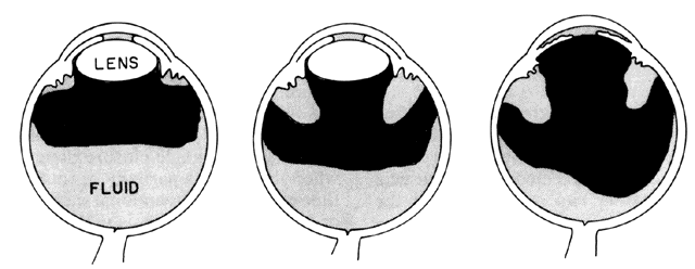
| Fig. 22. Left. Diagram of the working hypothesis of fluid trapped in or behind the vitreous body in a phakic eye with malignant glaucoma. Center. Fluid may occur at various sites in the posterior segment, according to this hypothesis. Fluid could in fact be diffused throughout the vitreous, but this is not shown in the diagram. Right. Diagram of the working hypothesis of fluid trapped in or behind the vitreous body in an aphakic eye with malignant glaucoma. (Simmons RJ: Malignant glaucoma. Br J Ophthalmol 56:263, 1972) |