|
|
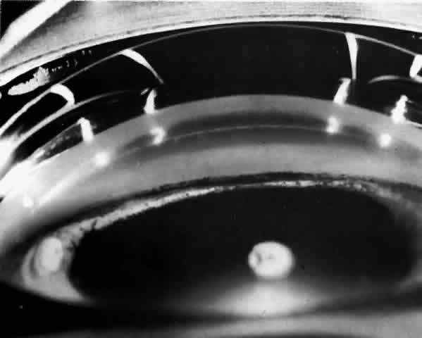
| Fig. 11. Same eye as is shown in Figures 10 and 12. This gonioscopic view shows much pigment debris released by the atrophic process in the iris on the angle wall. The pigment seen lies on the angle wall between Schwalbe's line anteriorly and the synechial attachment of the iris. Throughout the portion of the angle in this photo, synechiae attach at the level of the anterior trabecular meshwork, covering the ciliary band, scleral spur, and posterior trabecular meshwork and obstructing all useful outflow from this portion of the angle. (Photographed by Dr. David Donaldson) |