|
|
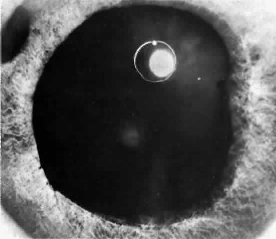
| Fig. 10. In this eye, iris atrophy due to neglected angle-closure glaucoma is so extensive that the pupil is fixed in wide dilation. Some pigment granules from the atrophic iris lie scattered about on the iris stroma. There is a posterior synechia at 2 o'clock at the iris margin. Glaukomflecken are very numerous in this eye but are not visible in this photo because oblique slit illumination is not used; they are visible in Figure 12, in which proper illumination is used. (Photographed by Dr. David Donaldson) |