Chapter 15
Venous Occlusive Disease of the Retina
GEORGE E. SANBORN and LARRY E. MAGARGAL
Main Menu   Table Of Contents

Search

CENTRAL RETINAL VEIN OCCLUSION
PAPILLOPHLEBITIS
HEMICENTRAL AND HEMISPHERIC RETINAL VEIN OCCLUSION
BRANCH RETINAL VEIN OCCLUSION
MACULAR BRANCH RETINA VEIN OCCLUSION
REFERENCES

CENTRAL RETINAL VEIN OCCLUSION
Acute occlusion of the central retinal vein presents as one of the most dramatic pictures in ophthalmology (Fig. 1); consequently, it was noted and described soon after the development of the ophthalmoscope.1,263,264 Despite many years of investigation, much of the pathophysiology of this disease remains unclear. It is second only to diabetic retinopathy as a vascular cause of visual loss,253 and the effect this condition has on visual acuity and vision-related quality of life can be significant.254

Fig. 1 A. “Blood and thunder” appearance of a central retinal vein occlusion. B. Intravenous fluorescein angiogram shows this occlusion is primarily ischemic or nonperfused. The fact that there is more nonperfusion in the inferior half of the fundus compared with the superior half is unusual.

PATHOLOGY

Occlusion of the central retinal vein is probably a result of both local and systemic causes. The actual mechanisms producing the clinical picture of central retinal vein occlusion may be roughly divided into those conditions that produce a physical blockage at the level of the lamina cribrosa, and those conditions in which hemodynamic factors result in an obstruction to the flow of blood. These mechanisms probably coexist in many patients with central retinal vein occlusion.

The pathogenesis of this condition and the underlying histopathology have remained controversial ever since Michel1 first correlated the clinical appearance with the histopathology. The fact that relatively few eyes have been histopathologically examined during the freshly obstructed stage has contributed to the problem. Many of the reported cases have involved eyes that were enucleated because of long-standing neovascular glaucoma; secondary changes that did not play a role in the original occlusion may have occurred in these eyes.

Histopathologic evaluation of eyes removed because of a central retinal vein occlusion demonstrates an occlusion at or just behind the level of the lamina cribrosa.2–7 At this location, certain anatomic factors predispose the central retinal vein to occlusion. First, the lumina of the central retinal artery and central retinal vein are narrower than they are in the orbital optic nerve, and the vessels are bound by a common adventitial sheath.8 Second, the lamina cribrosa is a sievelike, bisecting structure of connective tissue that not only provides support to the optic nerve, but also limits expansion and displacement of the optic nerve and the vessels within it.

In 1878, Michel1 found a thrombus in one patient studied. Later, both Coats2 and Harms9 believed, based on their histopathologic findings, that a primary thrombus within the intraluminal portion of the central retinal vein was the most common cause of occlusion. Verhoeff,3 however, was an early advocate of the concept that endothelial cell proliferation was the primary obstructing mechanism, and he believed that thrombosis within the vein did not occur except in patients with sepsis. He believed that most cases diagnosed as thrombosis were actually dissecting aneurysms because he found the intimal lining forced away from the venous wall by the backup of blood in a tributary vein.

Klein,5,6 who has done extensive clinical and pathologic studies of central retinal vein occlusion, believes that although primary thrombosis may occur, it is rare. She believes that thrombosis may occur more frequently as an end-stage phenomenon, complicating other initiating mechanisms in the obstructive process.

Green and co-workers7 felt that the interval between occlusion and the time of histopathologic study must be considered when interpreting the histopathology of vein occlusion. They studied 29 eyes that were enucleated 6 hours to 10 years after occlusion. As a result of this study, they hypothesized that the flow of blood through the central retinal vein becomes increasingly turbulent as the vein progressively narrows at the lamina cribrosa, where it also may be further impinged on by arteriosclerosis of the adjacent central retinal artery. This turbulence damages the endothelium in the retrolaminar vein, which exposes collagen and initiates platelet aggregation and thrombosis.7,10 Their studies show the evolution of this thrombus. Initially, the thrombus adheres where the endothelium has been severely damaged. Endothelial cell proliferation and recanalization of the vein often occur as a reparative event. Inflammation manifesting itself as phlebitis, periphlebitis, or obliterating endophlebitis is a secondary late-onset factor. Years later, a thick-walled vein with a single channel may occur (phlebosclerosis).7

In some eyes an adjacent, partially obstructed, or narrowed central retinal artery has been observed. This observation is consistent with the prevailing clinical impression that the principal condition associated with retinal vein occlusion is arteriosclerosis. Because the central retinal artery is a true artery, it may be involved in the patchy disease of larger arteries (i.e., atherosclerosis). There is an increased incidence of generalized atheromatous disease in patients who have a central retinal vein occlusion.11,12 As part of this atheromatous change, sclerosis occurs in the common adventitia, which encircles both vessels within the rather rigid support structure of the lamina cribrosa. Compression or constriction of the vein lumen and changes within the vein wall, described as phlebosclerosis, occur. As mentioned, occlusion of the central retinal vein is also influenced by the anatomic confinement of the vein and the artery within the optic nerve, as well as the compactness of the lamina cribrosa and its surrounding connective tissue.

Hayreh and co-workers13–16 have investigated the role of occlusion of the central retinal vein and central retinal artery in an animal model. They attempted to produce central retinal vein occlusion in healthy young monkeys by diathermy of the central retinal vessels in the orbit near their entry into the optic nerve sheath. Their study showed that occlusion in the orbit of the central retinal vein alone produced mildly engorged and tortuous vessels and a few retinal hemorrhages; all these conditions returned to normal in approximately 2 weeks. However, when both the central retinal vein and the central retinal artery in the orbit were obstructed simultaneously, a fundus appearance was produced that was “entirely characteristic” of central retinal vein occlusion.13 Later, histopathologic examination of these eyes showed a hemorrhagic infarct of the inner retinal layer. Hayreh and co-workers15 concluded from these experiments that concomitant arterial occlusion is essential in the production of an ischemic central retinal vein occlusion, although its occurrence is possibly only transient,16 and that the site of occlusion is important in determining both the severity and type of occlusion.16 However, this model of occlusion in the orbit of healthy young monkeys may not be comparable to the situation in the aging human, where the occlusion is located at or just posterior to the lamina cribrosa.379

Because fluorescein angiography does not typically show prolongation of arterial filling in central retinal vein occlusion, Fujino and associates17 investigated the role of arterial occlusion by producing central retinal vein occlusion in monkeys using an intravenous injection of neoprene. They were able to show that a primary and complete occlusion of the central retinal vein at the disc produces a secondary artery insufficiency. The ophthalmoscopic appearance produced in monkeys, however, is not identical to the appearance of central retinal vein occlusion in humans. This may be because this technique obstructs all the branch retinal vessels in the peripapillary region, which, in turn, may preclude collateralization.16

McLeod18 noted that in eyes with both a central retinal vein occlusion and a cilioretinal artery occlusion, there was a lack of retinal hemorrhages within the area of retina that was infarcted. He presented this an argument against the combined artery and vein occlusion hypothesis of Hayreh and colleagues.13–16 If an artery occlusion as well as a vein occlusion (combined occlusion) is necessary to produce the typical ophthalmoscopic picture of a central retinal vein occlusion, the retina should exhibit increased hemorrhage in the area supplied by the occluded cilioretinal artery.

The histopathologic picture in venous occlusion is now considerably clearer as a result of a series of experiments on branch retinal vein occlusion in the monkey.19–21 This work shows that capillary nonperfusion (ischemia) can result after isolated venous outflow occlusion without the occurrence of primary arterial inflow occlusion (ischemic capillaropathy).22 Although these experiments were performed on branch retinal vein occlusions, there is every reason to believe that the ischemia of the retina seen in central retinal vein occlusion can result from venous outflow disease alone.23

Doppler ultrasound imaging has been used to examine the blood flow in the orbit, including the optic nerve head,24,25 and has been used to examine patients with central retinal vein occlusion.26–28,256,257 As might be expected, the venous velocity in the eye of a patient with central retinal vein occlusion is markedly reduced compared either with the unaffected eye or to control eyes.24,25 There is evidence, however, that the central retinal artery blood flow is also impaired in eyes with acute central retinal vein occlusion.28 In addition, vascular resistance is slightly higher in the ophthalmic artery and short posterior ciliary arteries of both the involved and the clinically healthy fellow eye of patients with central retinal vein occlusion compared with control eyes.28 There is also a trend toward higher vascular resistance of the central retinal artery in the clinically healthy eyes of patients with central retinal vein occlusion compared with control eyes.28

The retinal pathology in an ischemic central retinal vein occlusion consists of a hemorrhagic infarction of the retina that affects primarily the inner retinal layers.29 Neovascularization of the iris and anterior chamber angle can develop; less frequently, retinal neovascularization can also occur.10 This neovascularization is likely related to the unregulated expression of vascular endothelial growth factor (VEGF) in the cells of the neurosensory retinal when affected by the hypoxia in central retinal vein occlusion.329 Later changes include thickening of the retina and reactive gliosis.30

ETIOLOGY

The precise etiology of central retinal vein occlusion is not entirely clear. There are now some clues as to the conditions associated with this condition. Many published articles have reported on the association between central retinal vein occlusion and some other condition, whether systemic or ocular. Although some of these associated conditions probably are, in some cases, related to central retinal vein occlusion, there is no way to determine in most cases whether the association is only coincidental on the basis of single-case reports.

Any study that attempts to determine either the etiology of or the features associated with central retinal vein occlusion must be a large enough prospective study that it takes into account age- and sex-matched controls and includes a comprehensive, systemic evaluation. Some reports in the literature have been retrospective,31–33 others have had no control group,33–37,258,259,260 and some have not performed a prospective, systemic evaluation.31–34

We are aware of only one prospective, large study of risk factors for central retinal vein occlusion that includes an appropriate age- and sex-matched control group and a standardized, prospective, systemic evaluation.38 The Eye Disease Case-Control Study Group examined 258 cases of central retinal vein occlusion as well as 1,142 controls. An increased risk of central retinal vein occlusion was found in patients with systemic hypertension, diabetes mellitus, and open-angle glaucoma; the risk of central vein occlusion was decreased for patients with increasing levels of physical activity and increasing levels of alcohol consumption (Table 1). For women, the risk decreased with the use of postmenopausal estrogen and increased with a higher erythrocyte sedimentation rate. The authors did attempt to divide the cases into ischemic and nonischemic central retinal vein occlusion. The following conditions all showed a significant association with ischemic cases only: cardiovascular disease, electrocardiographic abnormalities, albumin-globulin ratio, α1-globulin, history of treatment for diabetes mellitus, and blood glucose level. Both systolic and diastolic blood pressure showed significant associations with both types of central retinal vein occlusion, but the odds ratio is greater for the central retinal vein occlusion. Overall, a stronger cardiovascular risk profile was shown for the ischemic type of central retinal vein occlusion.


TABLE 1. Risk Factors for Central Retinal Vein Occlusion


Increased RiskDecreased Risk
Systemic hypertensionIncreasing levels of physical activity
Diabetes mellitus 
Increased erythrocyte sedimentation rate (women only)Increasing levels of alcohol consumption
History of glaucomaExogenous estrogens (women only)

(Data from The Eye Disease Case-Control Study Group: Risk factors for central retinal vein occlusion. Arch Ophthalmol 114:545, 1996)


A number of studies have been done to identify both genetic and acquired risk factors for large vessel venous thromboembolism. The term thrombophilia has been used to apply to those cases in which a risk factor has been identified for spontaneously occurring large-vessel venous occlusion.293,295 In a European population the three most common markers of thrombophilia are the factor V Leiden variant, hyperhomocysteinemia, and protein C deficiency.293

A number of studies have reported various hematological abnormalities in patients with a central retinal vein occlusion.39,40,41,45,260,261,262,265,295,390 These reports are difficult to interpret for a number of reasons. Many of them are single-case reports (not sited here) where the association may only be coincidental, some do not provide an appropriate control group,260 some do not separate central retinal vein occlusions from branch retinal vein occlusions,268,296 the sample size may not be large enough to pick up a significant difference,277 better and more specific tests are now available for some abnormalities that invalidate earlier results,277 and in some the parameters measured are probably the result of the occlusion and not the cause.

In some reports patients were studied at varying intervals after the occlusion, often many months after the acute event.295 The timing of laboratory investigation is important for some parameters. Williamson and co-workers showed that relative elevated blood viscosities examined within 1 month of the acute event fell significantly 1 year later.42 Iijima and co-workers found that elevated thrombin-antithrombin III complex levels measured soon after venous occlusion were not maintained, but fell markedly over several months.262

The Eye Disease Case-Control Study Group studied only a few parameters of hematologic function in their large study; these included hematocrit, erythrocyte sedimentation rate, fibrinogen, and antithrombin III.38 There was an increasing risk of central retinal vein occlusion with a calculated odds ratio for antithrombin III level and an elevated erythrocyte sedimentation rate associated with central retinal vein occlusion in women but not in men. In the multivariant logistic regression, however, the antithrombin III level did not attain significance, but for women the erythrocyte sedimentation rate was still significantly correlated.

In a number of studies comparing patients with central retinal vein occlusion to appropriately matched controls,42,43,266–276 activated protein C resistance has been found to be significantly associated with central retinal vein occlusion. This association is true both for patients younger than 50 years of age43 and for those older than 64.42 Other studies have not shown a significant association between protein C resistance and central retinal vein occlusion.288–291 A case has been reported of a 63-year-old woman who presented with a central retinal vein occlusion in one eye and a small branch retinal vein occlusion in the opposite eye; evaluation found that she was heterozygous for the factor V Leiden mutation.308 Another patient with a bilateral central retinal vein occlusion and the factor V Leiden mutation has been reported; she was also pregnant and diabetic.313

Factor V is an inactive procofactor that, when activated by thrombin producing factor Va, serves as the cofactor for factor Xa in the conversion of prothrombin to thrombin. Factor Va is inactivated by a proteolytic cleavage by activated protein C. Protein C is activated by thrombin binding to the vascular endothelium into activated protein C. Resistance to the cleavage by activated protein C is a cause for venous thrombosis.294 In almost all patients this resistance is caused by a mutation in the factor V gene (FVR506Q), which is called factor V Leiden and is present in approximately 5% of the Caucasian population. Although some patients with a central retinal vein occlusion have activated protein C resistance, a statistically significant association between the two appears not to have been proven from the available literature, and no evidence has been presented that it is a cause of central retinal vein occlusion.265,277,293,294 The fact that there is a high prevalence of factor V Leiden in the population means that any study of this factor and central retinal vein occlusion requires a large sample size to detect a significant association between the two.277,294

Another marker of thrombophilia is the presence of autoimmune antibodies reactive against cellular components of phospholipids; these include the anticardiolipin antibody or the lupus anticoagulant.46,293 The antiphospholipid–antibody syndrome often occurs in patients with systemic lupus erythematosis, but when recurrent thrombosis and antiphospholipid antibodies occur in patients without lupus it is called the primary antiphospholipid syndrome.46 Several studies have suggested an association between these antibodies and central retinal vein occlusion.277–299,301,302,384,319 Others have failed to show an association.47,381

A case have been reported of a 40-year-old woman with the primary antiphospholipid syndrome who presented with a central retinal artery occlusion in one eye and 6 years later presented with a central retinal vein occlusion in the opposite eye,301 as well as a bilateral central retinal vein occlusion associated with anticardiolipin antibodies and leukemia.316

Elevated levels of homocysteinemia are also a risk factor for vascular disease, possibly because of endothelial cell damage.305 It can be acquired in a number of ways, including smoking, increased age, insufficient intake of folic acid, renal failure, some chronic diseases, postmenopausal hormone replacement, and certain medications.293,294,305,390 It can also be inherited due to homozygous mutations on genes that encode two enzymes, methylenetetrahydrofolate reductase (MTHFR) and cystation-β-syntase (CBS), which interferes with remethylation of homocysteine.293,294

Several studies have shown an association between hyperhomocysteinemia and central retinal vein occlusion.302–307,310 However, Larsson et al. studied 116 patients with a central retinal vein occlusion who were tested for the MTHFR C677T mutation and found no statistically significant association compared to a control group.278 Some of the studies that did show an association reported on small numbers of patients,303,305,307 and another did not use a matched control group and did not take the blood samples fasting.310 Two patients have been reported with bilateral central retinal vein occlusion and elevated serum homocysteine levels311,312 and one with bilateral central retinal vein occlusion and the MTHFR 677CT mutation, although the blood homocysteine levels were not measured.317

There have been a few cases of elevated blood viscosity producing a central retinal vein occlusion. Two patients with Waldenström's macroglobulinemia presented with what appears to have been bilateral nonischemic central retinal vein occlusion, which resolved with plasmapheresis.48 A 60-year-old woman with Eisenmenger syndrome presented with a bilateral retinal vein occlusion and a secondary polycythemia.314 Multiple, bilateral retinal vein occlusions were reported in a patient with essential thrombocythemia.327

Cahill and associates have performed a meta-analysis of the published literature on total plasma homocysteine levels, serum folate and vitamin B12 levels, and homozygosity for thermolabile methylenetetrahydrofolate reductase genotype as risk factors for retinal vascular disease.390 They found that the studies in the published literature showed plasma total homocysteine levels were elevated in retinal vascular occlusion, including patients with a central retinal occlusion.390 They also found that there was a significantly low serum folate level in patients with a retinal vascular occlusion, although a separate analysis of central retinal vein occlusion was not performed.390 For those patients with a retina vascular occlusion and elevated plasma total homocysteine and a low serum folate, they recommend folate supplementation in conjunction with the patient's primary care physician.390

Except for those rare patients with bilateral retinal vein occlusion due to hyperviscosity, it is unlikely that there is a hematological cause alone for central retinal vein occlusion. Greaves has postulated that retinal vein thrombosis is a “multiple hit” phenomena in which several adverse influences affecting the composition of the blood, the vessel wall, and the blood flow produce a thrombotic event.315 Because the incidence of retinal vein occlusion increases with age, it is likely that with age there is an increased likelihood of the accumulation of a number of adverse events, only one of which may be an inherited or acquired blood disorder, that cause this occlusion.315

There is no evidence that extracranial carotid artery occlusive disease is associated with central retinal vein obstruction. Using digital subtraction angiography, Brown and associates49 studied 37 patients with central retinal vein occlusion; they found that significant ipsilateral stenosis (greater than 50%) was not higher in these patients compared with historically matched controls. They did find, however, that patients with ischemic central retinal vein occlusion had a higher incidence of overall carotid atherosclerotic obstruction (ipsilateral and contralateral) than patients with nonischemic central retinal vein occlusion.49

There appears to be no relationship between optic disc size50 and cup-to-disc ratio in central retinal vein occlusion.38,50,51 Two studies found that the axial length of patients with central retinal vein occlusion is slightly shorter than that of controls,52,318 and one did not find an association.319

CLASSIFICATION

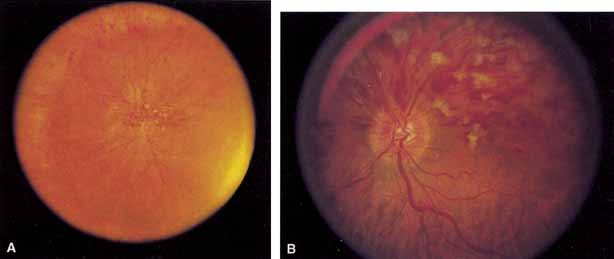
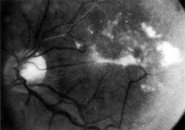
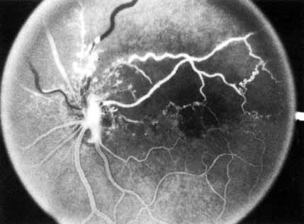
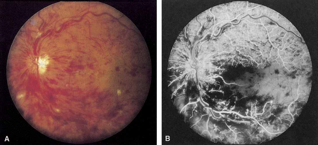
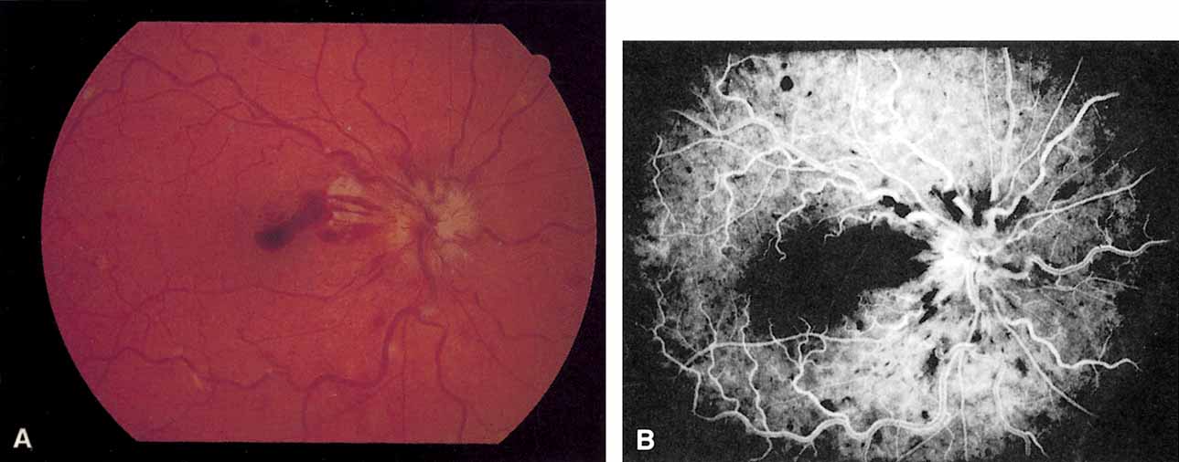
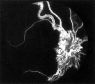
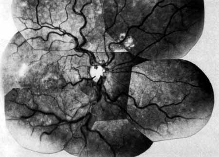
Coats55 may have been the first to suggest that patients with central retinal vein occlusion fall into two groups: one with a dramatic, “blood and thunder” ophthalmoscopic appearance, loss of vision, and a poor prognosis (see Fig. 1); and the other with mild ophthalmoscopic changes, generally good visual acuity, and a relatively good prognosis (Fig. 2). Other investigators have commented on the difference in severity among central retinal vein occlusions, relying principally on the fluorescein angiogram to assess the severity of occlusion.56–59

Fig. 2 A. Nonischemic central retinal vein occlusion. Note venous engorgement, dot, blot, and flame-shaped hemorrhages, blurring of disc margins, and a hemorrhage overlying the macula. B. Fluorescein angiogram reveals mild venous engorgement and tortuosity with virtually no capillary nonperfusion.

Hayreh60–64 also divided central retinal vein occlusion into two categories: a nonischemic type, which he called “venous stasis retinopathy,” and an ischemic type, which he called “hemorrhagic retinopathy.” Magargal and colleagues65 may have been the first to divide central retinal vein occlusion into three categories: nonperfused, which they called “hyperpermeable”; ischemic or nonperfused; and a category in which eyes could not be classified on fluorescein angiography, which they termed “indeterminate.” Hayreh and co-workers66 recently also subdivided each of these eyes into mild, moderate, and marked retinopathy based on the maximum capillary nonperfusion on the fluorescein angiogram.

Laatikainen and co-workers67 and Kohner and Shilling68 also divided central retinal vein occlusion into two groups that are similar to Hayreh's original groups.60 Magargal and associates believe that venous occlusion is a spectrum of disease with capillary nonperfusion ranging from little if any ischemia to marked ischemia, and that the amount or extent of ischemia is roughly correlated with the development of neovascular complications.65,69,70

Capillary nonperfusion following central retinal vein occlusion does appear to be a spectrum rather than one or two distinct entities that can be categorized easily. However, the recognition of the severity of capillary occlusion is clinically useful, primarily in predicting the clinical outcome. For the category with minimal to moderate capillary nonperfusion (less than 50%), nonischemic (or perfused) is a better term than venous stasis retinopathy because of the widespread (but not entirely accurate) use of the latter to refer to the retinopathy associated with chronic hypoperfusion caused by extracranial carotid artery occlusive disease.71,72

Similarly, for the category with significant capillary nonperfusion (greater than 50%), ischemic retinal vein occlusion is a better term than hemorrhagic retinopathy because neovascularization, the major complication of central retinal vein occlusion, is correlated with the degree of capillary nonperfusion69,70,73 and not with retinal hemorrhages, which change with the duration of the disease.

The amount of nonperfusion or ischemia is determined by inspecting the fluorescein angiogram. The photographer inspects not only the central 30° or 45°, but also as much of the peripheral retina as possible. The angiographer should be instructed to take photographs during the angiogram of as much of the periphery as possible (a peripheral sweep). Another method has been to classify eyes with less than 10 disc diameters of perfusion on fluorescein angiography as perfused or nonischemic, and eyes with 10 or more areas of nonperfusion as nonperfused or ischemic,74 although this method may not be accurate.320,321

It is impossible to categorize some eyes as either ischemic or nonischemic on the initial evaluation because retinal hemorrhages preclude adequate visualization of the capillary bed on fluorescein angiography.321 These unclassifiable eyes can be placed into a third category, indeterminate,65,74,75 and patients can be reevaluated when the hemorrhages begin to resolve during follow-up. Most of the eyes in this category, however, will develop ischemia (83% in the Central Vein Occlusion Study) on follow-up, and for the purposes of further evaluation, these eyes should probably be considered ischemic.74

Hayreh and associates,76 however, believe it is necessary to perform six clinical tests in order to differentiate eyes with nonischemic central retinal vein occlusion from ischemic central vein occlusion. According to them, the most reliable tests, in order, are: the relative afferent pupil defect (in unilateral central retinal vein occlusion with a normal fellow eye), the electroretinogram, perimetry, visual acuity, intravenous fluorescein angiography, and ophthalmoscopy.76 They believe that the intravenous fluorescein angiogram either provides no information at all on capillary nonperfusion or sometimes provides misleading information.76

CLINICAL CHARACTERISTICS

Nonischemic Central Retinal Vein Occlusion

Nonischemic central retinal vein occlusion is a much milder and more variable disease in appearance, symptoms, and course compared with ischemic central retinal vein occlusion. Patients with nonischemic central retinal vein occlusion are an average of 5 years younger (average age, 63 years) than those with ischemic vein occlusion.66 Complaints vary from none (i.e., condition is discovered on a routine examination) to blurred vision, which is often transient.66 The visual acuity may range from normal to counting fingers, but the majority of patients have an initial visual acuity of 20/50 or better.62

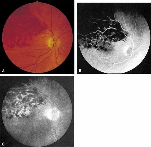
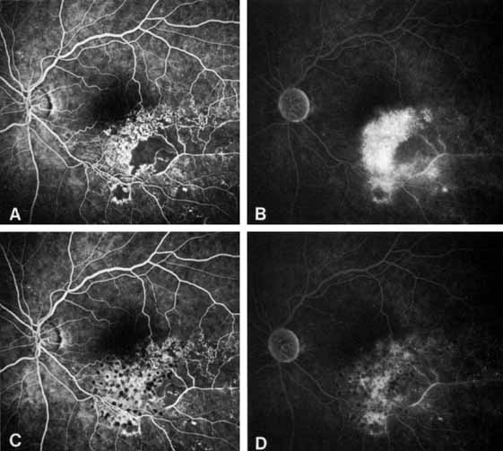
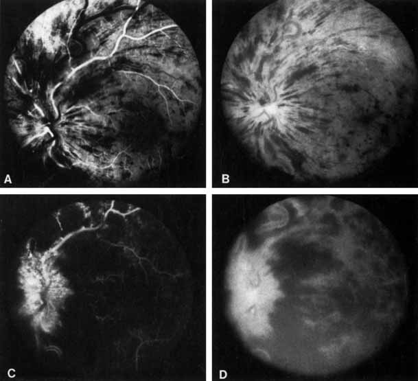
The ophthalmoscopic features of nonischemic central retinal vein occlusion are similar to those of ischemic central retinal vein occlusion, but are much less extensive (see Fig. 2; Fig. 3A and 3B). Engorgement of the venous tree (including the capillaries) is prominent; there is increased tortuosity and dilation and a darker appearance of the blood column. Retinal hemorrhages vary markedly. Sometimes they occur only peripherally; at other times, they may be rather prominent in the posterior pole.60 Cotton-wool spots are rare. Vision may be decreased because of macular edema or macular hemorrhage.

Fig. 3 A and B. Acute nonischemic central retinal vein occlusion in a 36-year-old hypertensive man. His visual acuity was 20/200. C and D. Six weeks later, he presented with eye pain, decreased vision, and neovascular glaucoma. The type of occlusion now is ischemic.

Hvarfner and Larsson have studied 74 patients with both a nonischemic and ischemic central retinal vein occlusion, 48 of whom had optic disc swelling.381 They found that optic nerve swelling was of no prognostic value in predicting neovascular complications and visual acuity 1 year after the acute event.381 Beaumont and Kang divided central retinal vein occlusion into two distinct groups based on the presence or absence of optic nerve head swelling.379 Those with optic nerve head swelling were postulated to have a venous occlusion in the retrocribrosal space and were of younger age, had less severe vascular nonperfusion, and had a better visual acuity than those without swelling who were postulated to have an occlusion at the lamina cribrosa.379

Beaumont and Kang have also identified the clinical characteristics with different sites of occlusion and proposed a new classification.382 These sites are the arteriovenous crossing, optic cup, or optic nerve; those in the nerve were further subdivided into the presence or absence of optic nerve head swelling.382 Among their findings are that primary open-angle glaucoma is significantly more common in patients with an occlusion at the optic cup and a history of smoking and hypertension is more common with occlusions at the arteriovenous crossing.383

Patchy ischemic retinal whitening located in a perivenular distribution near the macula is a transient abnormality in patients with a nonischemic central retinal vein occlusion and is associated with a generally good visual outcome.324,386 The cause is unknown.

The angiographic pattern may show little except occasionally a prolonged venous transit time. Dilation of the retinal venous circulation, mild staining of the walls of veins, and varying degrees of disc and macular edema may be present (including cystoid macular edema). Capillary nonperfusion is not a prominent feature, nor is its sequela, neovascularization. The electroretinogram is nearly normal, confirming the lack of ischemia.77 The intraocular pressure is frequently lower on the side of the occlusion.78 Synonyms for this type of central retinal vein occlusion have included partial, incomplete, imminent, threatened, incipient, or impending central retinal vein occlusion.60,76 How many central retinal vein occlusions in this category are actually incomplete or partial occlusions that then progress to a more complete occlusion is unknown. It does appear that some eyes with nonischemic central retinal vein occlusion go on to develop a more ischemic type of central retinal vein occlusion (see Fig. 3); whether this represents a progression of the vein occlusion62 or simply progressive retinal capillary nonperfusion is unknown. In series in which the incidence of conversion for the nonischemic occlusion to the ischemic type has been studied, the incidence ranges from approximately 5% to 22%, depending on the duration of follow-up, and is higher for older patients.63,74,79–81

The natural course of nonischemic central retinal vein occlusion is relatively benign, except in those who go on to develop additional ischemia. The hemorrhages, vascular congestion, and engorgement gradually resolve over several months. Some patients are left with permanent cystoid macular edema, macular cystic changes, pigmentary changes, or residual microvascular abnormalities.82 Neovascularization does not generally occur, and morbidity is generally limited to a persistent, mild decrease in visual acuity with a relative central scotoma. The majority of patients will have a final visual acuity of 20/40 or better.79

Ischemic Central Retinal Vein Occlusion

Patients with an ischemic pattern are usually aware of a sudden, painless decrease in visual acuity. Vision ranges from 20/400 to hand movements. The onset, however, is generally not as rapid or the visual loss as extensive as in central retinal artery occlusion. Exceptional cases have been noted in which patients with an acute onset had reasonably good vision and yet demonstrated a picture of ischemic central retinal vein occlusion. Patients with ischemic occlusion have an average age of 68.5 years.66 Confluent hemorrhages are the most prominent ophthalmoscopic feature of an acute ischemic central retinal vein occlusion (see Fig. 3C and 3D). These hemorrhages occur in a wide variety of shapes and sizes; they are usually concentrated in the posterior pole, but may be seen throughout the retina. Hemorrhages in the superficial retina may be so prominent about the posterior pole that the underlying retina is obscured. Many hemorrhages are flame shaped, reflecting the orientation of the nerve fibers. Dot and punctate hemorrhages are interspersed and indicate involvement of the deeper retinal layers. Bleeding may be extensive, erupting through the internal limiting membrane to form a preretinal hemorrhage or extending into the vitreous. Small dot hemorrhages may be seen either isolated or clustered around small venules. The entire venous tree is tortuous, engorged, dilated, and dark. The retina is edematous, particularly in the posterior pole; some of this edema may obscure portions of the retinal vessels. Cotton-wool patches (soft exudates) are often present.

The disc margin is blurred or obscured, and the precapillary arterioles appear engorged. Splinter hemorrhages and edema are present on the disc surface and extend into the surrounding retina. The physiologic cup is filled, and the venous pulse is absent. The arterioles, often overlooked because of the other more striking pathologic features, are frequently narrowed. Sometimes in central retinal vein occlusion of acute onset, the fundus picture is less dramatic, and all the findings previously discussed may be present, but to a lesser degree. Vision depends primarily on the extent of macular involvement.

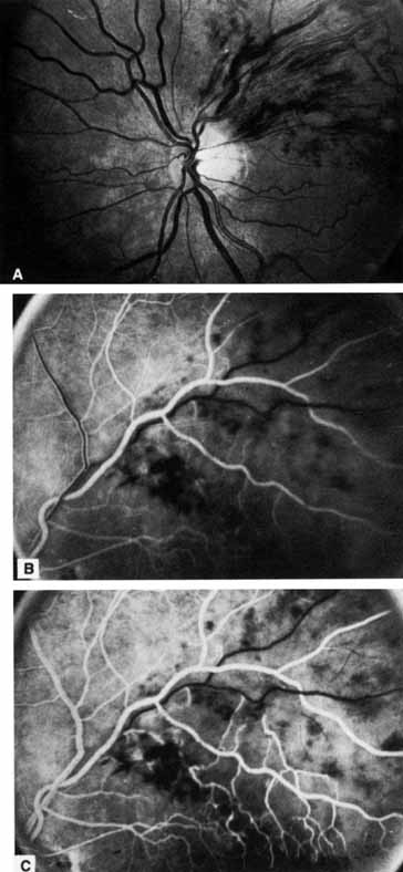
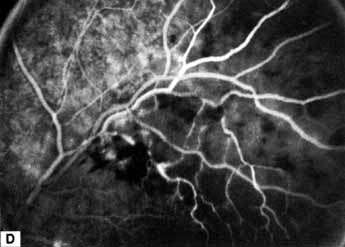
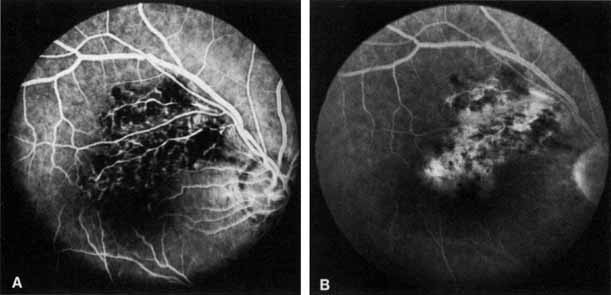
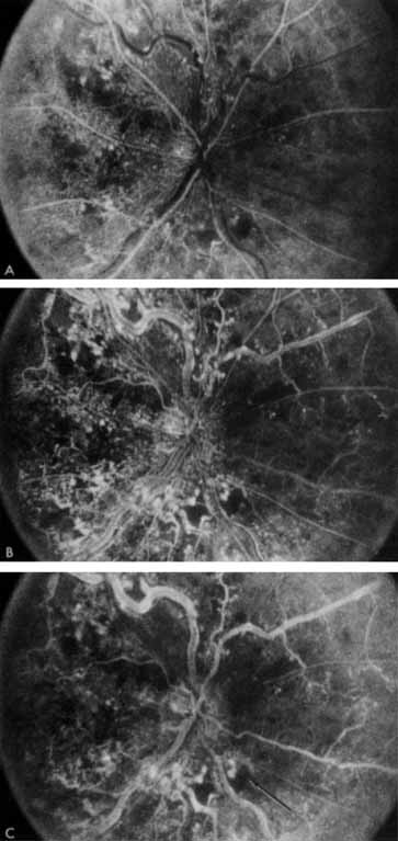
The intravenous fluorescein angiogram pattern of an ischemic central retinal vein occlusion is usually characterized by a delayed filling time of the venous tree of the retina, capillary and venous dilation, and extensive leaking of fluorescein into the retina, particularly in the macular area and in the area adjacent to the larger venous trunks and capillary nonperfusion (see Fig. 3C and 3D; Figs. 4 and 5). Microaneurysms may not be noted at the time of initial occlusion, but are usually manifest shortly thereafter. Late-phase photographs show patchy extravascular areas of fluorescence and staining of the retinal veins. Fluorescence in the macula indicates capillary leakage and edema; this not only may account for much of the initial visual loss in the acute phase, but may also eventually result in permanent structural changes. Intravenous indocyanine green videoangiography may also be helpful in showing the arterial and venous flow alterations in this condition.322

Fig. 4 Fluorescein angiogram after moderately ischemic central retinal vein occlusion. A. Early venous phase. The capillary bed is dilated and engorged. Punctate areas of fluorescence represent microaneurysms or small areas of capillary leakage. B. Midvenous phase. There is considerable delay in venous return and an increase in and coalescence of punctate areas of extravascular fluorescence. C. Late venous phase. Fluorescence staining along the vein margins and scattered areas of capillary nonperfusion (arrow) are present.

Fig. 5 Fluorescein angiogram of acute ischemic retinal vein occlusion. Capillary nonperfusion is essentially 100%.

The prognosis for ischemic central retinal vein occlusion is generally poor because of decreased visual acuity and neovascularization. Visual loss occurs because of macular edema, capillary nonperfusion, overlying hemorrhage (either retinal or vitreal), or a combination of all these. Retinal edema usually gradually subsides except in the macula, where it may persist for many months or years. Macular holes or cysts may form.83,84 Pigment clumping or fine pigment stippling and pigment atrophy are not uncommon, and persistent macular hemorrhage, even years after the occlusion, has been noted.83 Hard exudates often form an irregular circinate configuration around the macula and become more prominent months later. Occasionally an epiretinal membrane may form.

In the chronic phase, most hemorrhages gradually disappear over many months; however, scattered, flame-shaped hemorrhages and dot hemorrhages, particularly in the periphery, may be seen for years. Cotton-wool patches and microaneurysms likewise tend to disappear after several months, although in some cases the latter may persist. The venous tree becomes less tortuous and dilated. Prominent venous loops, which are collateral communications, may be observed on the surface of the disc (Fig. 6).85 These loops develop within 3 to 14 months after occlusion from the existing retinal vasculature and are collateral vessels between the obstructed disc capillaries and the unobstructed choroidal or pial capillaries.323 These retinochoroidal collateral veins, if they develop, may protect against anterior segment neovascularization,328 but may not be associated with a better visual prognosis.105 Collaterals between the central retinal vein within the globe and the patent central retinal vein behind the occlusion have not been observed.86 The extent and speed of retinal recovery probably depends to some degree on how quickly collateral vessels form, how rapidly recanalization occurs, and how adequately these compensatory mechanisms restore normal outflow. However, the exact nature and course of the collateral vessels are disputed. Anastomotic channels may develop within the retinal vasculature if pressure differentials develop between its major venous trunks. Changes in the retinal arterioles include both segmental and generalized narrowing as well as sclerosis, which is evidenced by both sheathing and widening of the light reflex. Sheathing of the veins is also common. The disc may appear nearly normal except for sheathing of the vessels in and around the papilla, and some blurring of the margins may persist. Sometimes optic atrophy is present.

Fig. 6 Fundus picture 4 years after central retinal vein occlusion. Moderate venous engorgement and small dot hemorrhages remain; microaneurysms, dot hemorrhages, and residual edema are present in the macula. Some sheathing of vessels is seen along with patchy edema residues. Note collateral channels on the retinal surface temporal to the disc and tortuous vessels on the surface of the disc representing collateral cilioretinal communications.

The fluorescein angiographic appearance varies greatly, depending on the extent of recovery. All the findings in the acute phase, consisting of venous and capillary engorgement, microaneurysms, staining of the veins, patchy extravascular fluorescence, and capillary nonperfusion, may persist indefinitely. In most instances, these findings eventually diminish so that few significant features are present on the angiogram; collateral vessels, if present, may be the only pathognomonic feature.

The most serious complication of central retinal vein occlusion is neovascularization (Table 2). Neovascularization elsewhere (NVE) occurs less frequently than neovascularization of the iris (NVI), and usually only in ischemic occlusions.66 The low incidence of retinal surface neovascularization in ischemic central retinal vein occlusion is thought to be due to the destruction of endothelial cells, which provide the source for endothelial proliferation and neovascularization.87


TABLE 2. Percentage of Ocular Neovascularization in Venous Occlusion*


 Overall Incidence of Ocular Neovascularization
Type of Vein OcclusionNVDNVENVINVG
CRVO1.1%661.6%6613%668.3–25%66,73,88
Hemi-CRVO669.3%13.4%3.2%1%
HRVO7511%9%9%3%
Quadrantic BRVO18010.6%20.7%1.6%0.8%
Macular BRVO1820.0%0.0%0.0%0.0%

*This table is for comparison of neovascularization between the types of vein occlusion. Most of the neovascularization occurs in patients with the ischemic form of the listed occlusion. CRVO, central retinal vein occlusion; HRVO, hemispheric retinal vein occlusion; BRVO, branch retinal vein occlusion; NVD, neovascularization of the disc; NVE, neovascularization elsewhere; NVI, neovascularization of the iris; NVG, neovascular glaucoma.


Neovascularization of the iris and frequently neovascular glaucoma occurs in approximately 8%62 to 25%65,66,73,88 of all central retinal vein occlusions and generally only in those eyes that exhibit an ischemic pattern of occlusion.63,65,66,68,89 Magargal and co-workers70 have shown that the incidence of neovascularization increases dramatically above approximately 50% capillary nonperfusion. The incidence of anterior segment neovascularization in nonischemic central retinal vein occlusion is approximately 1%, compared with approximately 35% to 45% for ischemic central retinal vein occlusion.63,69,70,89 Neovascularization of the iris or angle is significantly correlated with the extent of capillary nonperfusion on the fluorescein angiogram.73,89 In the series of Sinclair and Gragoudas,73 rubeosis developed in 80% to 86% of the eyes with severe nonperfusion of three to four quadrants of the posterior pole or the periphery, but in only 3% to 9% of those with less capillary nonperfusion. Abnormalities on fluorescein angiography of the iris appear not to be correlated with the development of secondary glaucoma in ischemic central retinal vein occlusion.325

Neovascularization of the iris may develop as early as 2 weeks after central retinal vein occlusion or as late as 2½years.2,65,89 Neovascularization of the iris, when it does occur, will develop in almost all patients within the first year, but usually in the first 3 months.89 Symptomatically, patients complain of tearing, irritation, pain, and further blurring of vision as the intraocular pressure in the affected eye begins to rise. The pain may become excruciating. The cornea is hazy and the pupil dilated, and a network of fine vessels is seen over the surface of the iris (rubeosis iridis) on slit-lamp examination. By the time gonioscopy reveals extension of this neovascular membrane into the trabecular network and throughout the angle, the intraocular pressure is usually markedly elevated. The angle is initially open, but later in the disease, peripheral anterior synechiae develop and the angle may become irreversibly closed, resulting in neovascular glaucoma. Large, extremely irritating bullae may form on the surface of the cornea and then break down. Dense cataracts eventually form, obscuring the fundus.

Nonperfusion in central retinal vein occlusion is also correlated with a relative afferent pupil defect.76,90,91 Servais and colleagues90 found that 100% of a group of patients with unilateral ischemic central retinal vein occlusion had a relative afferent pupil defect, whereas only 31% of patients with nonischemic occlusion had such a defect. Hayreh and associates76 believe that relative afferent pupil defect is the most reliable test for ischemic central vein occlusion in patients with unilateral disease (i.e., fellow eye is normal).

Hikichi and co-workers examined eyes with a central retinal vein occlusion and the presence or absence of a posterior vitreous detachment.350 They found that a complete posterior vitreous detachment appears to protect against retinal or disc neovascularization, although not iris neovascularization, and that vitreomacular attachment contributed to persistent macular edema in nonischemic central retinal vein occlusion.350

Besides having the complications already discussed, patients with central retinal vein occlusion are also at risk for vascular occlusion in the contralateral eye. Approximately 6% to 17% of patients can be expected to have a bilateral nonsimultaneous central retinal vein occlusion.92–95 Mieler and Blumenkranz95 followed 79 patients with central retinal vein occlusion for a minimum of 5 years. In 25% of these patients, the contralateral eye developed a vascular occlusion within 5 years; of these, 50% were branch retinal vein occlusions, 30% were central retinal vein occlusions, 10% were central retinal artery occlusions, and 10% were branch retinal artery occlusions. There is some doubt as to whether the life expectancy of patients with a central vein occlusion is shortened96 or not97 compared with an age-matched population.

Occasionally a patient will have a simultaneous occlusion of both the central retinal vein and the central retinal artery.98,99 Unlike patients with only a central retinal vein occlusion, these patients often have some retrobulbar pain, and vision may be decreased to no light perception. The retina appears pale, with a cherry-red spot in the macula. Disc edema and retinal hemorrhage may be present. Fluorescein angiography will demonstrate occlusion of both the central retinal vein and the central retinal artery. In the recovery phase, optic atrophy develops, and extreme narrowing of the retinal arterioles occurs.98

Although rare, an occlusion of the cilioretinal artery may also be seen in conjunction with central retinal vein occlusion.18,100–103 Patients with this occurrence have a pallor typical of ischemia in the distribution of the cilioretinal artery; fluorescein angiography will confirm occlusion of the cilioretinal artery. Occlusion of the cilioretinal artery may be due to a relative hypoperfusion caused by the increased venous pressure.103 The visual acuity, in general, is no worse than in a pure central retinal vein occlusion.99 Simultaneous occlusion of the central retinal vein and a branch retinal artery has been reported in a few patients.104

The electroretinogram is abnormal in patients with central retinal vein occlusion, and one or more of the electroretinographic parameters can be used to group patients in terms of having either an ischemic or a nonischemic vein occlusion.77,106 Some investigators believe that the initial electroretinogram can be used as a predictor of anterior segment neovascularization in patients with central retinal vein occlusion,107–110 whereas others do not consider it useful in this regard.106 In a large study of patients with unilateral central retinal vein occlusion and an apparently normal contralateral eye, 36% of patients had an abnormal electroretinogram in the uninvolved fellow eye,111 which is suggestive of a retinal circulatory disturbance in the clinically normal eye. The multifocal electroretinogram has also been studied in patients with a central retinal vein occlusion and compared to standard electroretinography.378

The electrooculogram is also abnormal in most patients with central retinal vein occlusion.112,113 It is correlated with the degree of ischemia112 and may serve as a predictor of the development of rubeosis iridis.113

Back to Top
PAPILLOPHLEBITIS
In 1961, Lyle and Wybar114 described six young, healthy patients with a unilateral, relatively benign condition characterized by mild blurring of vision, essentially normal visual acuity, dilated and tortuous retinal vessels, a varying amount of retinal hemorrhage, and optic disc edema (see Fig. 2). All six patients improved spontaneously, but were left with sheathing of retinal vessels and the formation of vessels on the optic disc. Lyle and Wybar called this condition “retinal vasculitis” and believed it to be due to a central retinal vein occlusion secondary to an inflammatory vasculitis of the venous system.114 Lonn and Hoyt115 agreed with this etiology, but felt that “papillophlebitis” was a more appropriate descriptive term. Others have reported similar cases.116–122 Hart and co-workers,117 however, pointed out that an inflammatory etiology for this disease is tenuous, and no well-documented cases have been studied histopathologically.10

Chew and associates123 studied the diurnal intraocular pressure of seven patients younger than 36 years of age who had central retinal vein occlusion and found abnormal intraocular pressures in the affected and/or unaffected fellow eye. They surmised that such patients may have elevated intraocular pressure when studied with diurnal measurements. However, only two or possibly three of these patients had a nonischemic central retinal vein occlusion; the rest had the ischemic type.

Papillophlebitis appears to be a form of nonischemic central retinal vein occlusion, having identical ophthalmoscopic and fluorescein angiographic characteristics and a similar clinical course. Our clinical impression is that it is found much more commonly in young patients, and this may account for its relatively benign course. Until evidence that inflammation is the cause of some recognizable cases of central retinal vein occlusion, it might be best not to use the term papillophlebitis.

TREATMENT

Treatment of an underlying systemic condition, if one is found, is indicated, although only rarely will this reverse the vein occlusion.48 However, treatment of an underlying systemic medical condition might help to prevent the opposite eye from developing a vascular occlusion.

A wide variety of therapeutic agents have been used to treat central retinal vein occlusion. The following therapies are no longer widely used today: topical administration of potassium iodide and pilocarpine,124 anticoagulants,125–132 fibrinolytic agents,133–136 hyperosmotic agents,137–139 carbogen inhalation,140 cholesterol-lowering agents (clofibrate),141 vitamins,131 corticosteroids,61,63,142 prostacyclin,143 aspirin,144 ticlopidine (an inhibitor of platelet aggregation),145 isovolemic hemodilution,146 traditional Chinese medicine,147 x-rays,148,149 and a surgical procedure that involves cutting both the scleral ring and the dura of the optic nerve.150,151

In 1938, Holmin and Ploman126 introduced anticoagulant therapy. Some investigators have found that either heparin or warfarin, or a combination, is effective in improving visual acuity after a central retinal vein occlusion;130–132 others have found no beneficial effect.125,129 Anticoagulants act by preventing fibrin formation, but there is no reason to believe that they would be effective in dissolving the thrombosis once it has occurred. They may, however, prevent propagation of the thrombus and indirectly facilitate spontaneous thrombolysis.152 Although some studies have suggested that anticoagulation does reduce the incidence of neovascular glaucoma,128,132 as yet a randomized prospective clinical trial of anticoagulation has not been performed. The disadvantages of anticoagulation include the fact that heparin, at least, must be administered on an inpatient basis, and there are bleeding complications associated with anticoagulation therapy.

In a small, randomized clinical trial, streptokinase (a fibrinolytic agent) has been shown to have a beneficial effect on visual outcome in central retinal vein occlusion.133 Unfortunately, vitreous hemorrhage is a serious complication of this method of treatment and therefore limits its usefulness.133,134 Whether the hemorrhage associated with this treatment is due to the dosage used is not known, although experimentally it has been shown that a lower dose of streptokinase can be used to open thrombosed veins in combination with pulsed low energy ultrasound.366

A small pilot study of thrombolytic therapy of central retinal vein occlusion with intravenous tissue plasminogen activator (TPA) has been performed.326 Although the results of the study appeared promising, there was neither a prospective control group nor randomization; of the 96 patients enrolled, one suffered a fatal stroke.326

Hattenbach and co-workers treated patients with an ischemic central retinal vein occlusion with intravenous recombinant tissue plasminogen activator (rt-PA) and heparin and reported a favorable outcome in some patients.330,331 Their study was retrospective, used multiple treatment groups, was not randomized, and had no control.330,331

Troxerutin, a rheologic drug that reduces blood viscosity and improves microcirculatory flow, has been studied in a randomized, controlled clinical trial in patients with both central retinal vein occlusion and branch retinal vein occlusion.153 Patients treated with troxerutin showed significant improvement in visual acuity and macular edema and diminished progression of ischemia compared with controls. Although the authors of this study randomly assigned treatment to one of four groups (nonischemic and ischemic central retinal vein occlusion and nonischemic and ischemic branch retinal vein occlusion), they combined the results of treatment for all four groups and did not report the results by type of occlusion or ischemia.153 It seems unlikely that all types of vein occlusion will respond equally to troxerutin; thus, there is no way to determine whether the drug is useful for patients with central retinal vein occlusion (or which types of occlusion) on the basis of this study.

Spoor and colleagues154,155 have reported that optic nerve sheath decompression is of value in central retinal vein occlusion. Standardized echography, however, shows that intrathecal fluid accumulation is not a consistent finding in the optic nerve sheaths of patients with either nonischemic or ischemic central retinal vein occlusion;156 therefore the rationale for optic nerve sheath decompression in central retinal vein occlusion is unclear. Dev and Buckley reported on this procedure in eight patients, five with a nonischemic occlusion and three with an ischemic occlusion; six eyes developed improved visual acuity, and two became worse.172 Sergott157 feels that the procedure is not of long-term value for patients with central retinal vein occlusion, although he has not published any study results.

Some type of photocoagulation is the accepted method of treatment of some forms of ischemic central retinal vein occlusion. Clinical trials in the past have shown that it does not affect the final visual acuity outcome, but is effective in both the prevention and the regression of neovascularization.158 It is effective in causing the regression of neovascularization of the disc (NVD), neovascularization elsewhere, and neovascularization of the iris, as long as they are not already in an advanced state.159–161 Prophylactic panretinal photocoagulation in high-risk eyes has been reported to be effective in preventing neovascular glaucoma.65,100,158,162,163 Magargal and co-workers69 used panretinal photocoagulation prophylactically in a nonrandomized series of 100 consecutive patients with ischemic central retinal vein occlusion. Neovascular glaucoma developed in only two patients in this group after treatment, and both patients had another ischemic event that occurred after treatment. With no treatment, approximately 45% of these patients would be expected to have developed neovascular glaucoma.63,69

A randomized, prospective, controlled clinical trial has been performed by the Central Vein Occlusion Study Group to determine whether prophylactic panretinal photocoagulation in ischemic central retinal vein occlusion prevents the development of iris or angle neovascularization, or whether it is more appropriate to apply panretinal photocoagulation only when such neovascularization develops.89 In this study, eyes with central retinal vein occlusion and at least 10 disc diameters of nonperfusion were randomly assigned to either an immediate prophylactic panretinal photocoagulation group (early treatment) or to a delayed panretinal photocoagulation group (no early treatment) that received photocoagulation only if neovascularization subsequently developed. Neovascularization developed in 20% of the eyes in the early-treatment group compared with 35% in the no-early-treatment group, a difference that was not statistically significant. Most patients had regression within the first 3 months of neovascularization after panretinal photocoagulation was administered when the rubeosis was detected. but 11% had persistent neovascularization that regressed over many months.

The most important risk factor for predicting the occurrence and extent of anterior segment neovascularization in this study was the amount of nonperfused retina.89 Other risk factors that correlated individually with neovascularization were visual acuity, duration of occlusion of less than 1 month, moderate or severe venous tortuosity, and retinal hemorrhages greater than a standard photograph. No other variable, not even nonperfusion, was statistically significant after adjusting for the effect of visual acuity.412 Neovascularization, when it developed, usually did so within the first 3 months after randomization into the study.

For some time to come, this will likely be the definitive study on panretinal photocoagulation for central retinal vein occlusion;89 however, it has generated a few criticisms. As Wald164 pointed out, of the 181 eyes enrolled, only 87 of the eyes were recruited in the first 3 months after the occurrence of central retinal vein occlusion, and only 29 eyes were recruited within the first month after occlusion. Therefore, this study included many eyes (i.e., the 94 eyes enrolled sometime between 3 months and 1 year after occlusion) with little risk of developing anterior segment neovascularization and neovascular glaucoma. There are two reasons for this:

  1. Half of all neovascularization occurs within the first 3 months after the occurrence of central retinal vein occlusion, and almost all neovascularization occurs within the first 6 months.163
  2. A duration of central retinal vein occlusion of less than 1 month is an important risk factor for the development of anterior segment neovascularization,69 and the study excluded eyes with no neovascularization at baseline.

Thus the data on the study is very likely skewed toward a lack of treatment effect.164 In addition, of the eyes in the early-treatment group, three received only 500 to 600 spots (size not specified), even though the protocol called for at least 1,000 spots,89 and the median number of spots in the entire treatment group was only 1,203.89 Would more complete treatment have reduced the incidence of anterior segment ischemia in the early-treatment group, therefore producing a significant treatment effect? There was a significant correlation between the amount of nonperfused retina at baseline and the development of anterior segment neovascularization.89 Although only 16% of the eyes with 10 to 29 disc areas of capillary nonperfusion at baseline developed anterior segment neovascularization, 52% of the eyes with more than 75 disc areas of neovascularization developed neovascularization.89 Would eyes with the greatest number of risk factors (i.e., greater than 30 disc areas of nonperfusion, retinal hemorrhages greater than the standard photograph, duration of central retinal vein occlusion less than 1 month, and male sex) have benefited from prophylactic panretinal photocoagulation?

Hayreh and associates163 conducted a prospective nonrandomized study of panretinal photocoagulation in ischemic central retinal vein occlusion. They found no statistically significant difference between the treated and untreated groups in the incidence of angle neovascularization, neovascular glaucoma, retinal or optic nerve neovascularization, vitreous hemorrhage, or visual acuity. The only significant finding was that fewer patients in the treated group had neovascularization of the iris compared with nontreated controls, but only if the panretinal photocoagulation was applied within the first 3 months after the onset of central retinal vein occlusion163 and panretinal photocoagulation resulted in a significant loss of the peripheral field. However, the patient selection for treatment in this study was not based on a random assignment, but the decision as to whether to perform laser or not was “left entirely to the patient,”320 and this may been a source of bias in terms of the results of the study.265

Once neovascularization in the anterior segment is detected, panretinal photocoagulation should be instituted promptly. This will often result in regression of the iris vessels and prevent complete angle closure; this is also true in patients with some increase in intraocular pressure but in whom the angle is not occluded for 360°.

Once developed, neovascular glaucoma responds poorly to any type of treatment. Cycloplegics, topical pressure-lowering agents, carbonic anhydrase inhibitors, and corticosteroids, though failing to lower the intraocular pressure significantly, may make the patient more comfortable. Panretinal photocoagulation often cannot be applied in cases of advanced neovascular glaucoma in which the angle has been substantially occluded and the cornea may be too cloudy to allow treatment. Transscleral cyclocryotherapy or transscleral laser cyclodestruction, sometimes combined with 360° of transscleral panretinal cryoablation,165,166 has also been used to preserve the globe. In some cases in which visibility is poor and the angle is closed, we have had some success combining pars plana vitrectomy and endophotocoagulation with a drainage implant (e.g., Molteno, Ahmed) inserted through the pars plana.

Macular edema, a frequent complication and cause of loss of visual acuity in patients with nonischemic central retinal vein occlusion, has been treated with macular grid photocoagulation.167 The rationale is that grid photocoagulation of the macula is effective in reducing macular edema in branch retinal vein occlusion168 and diabetes mellitus.169

The Central Retinal Vein Occlusion Study Group performed a randomized, prospective clinical trial on the effect of macular grid photocoagulation compared with no treatment on eyes with 20/50 or worse visual acuity due to macular edema with no capillary nonperfusion on fluorescein angiography.170 Although grid photocoagulation lessens macular edema both angiographically and clinically, there was no difference in visual acuity between the treated and untreated patients. For treated patients, there was a trend toward decreased visual acuity in patients older than 60 years and visual improvement in patients younger than this; this effect was not seen in untreated patients. Although this study suggests a possible benefit to visual acuity in younger patients with macular edema who are treated compared with untreated controls, the number of patients in this subgroup is too small for a statistically valid comparison of treated versus untreated eyes. Unfortunately, a study with an adequate number of patients has not been performed to determine whether photocoagulation would be of benefit for younger patients with macular edema in nonischemic central retinal vein occlusion.

McAllister and Constable171 reported a surgical technique to create a chorioretinal anastomosis in patients with nonischemic central retinal vein occlusion. The technique is to rupture Bruch's membrane first in an area adjacent to the edge of a vein located at least three disc diameters from the optic disc with the argon laser; they then use a YAG laser to create a small opening in the sidewall of the adjacent vein. In their original report there was an average of 2.1 attempts to create an anastomosis, which was successful in only 42% of the patients in the first series171 and 54% in a subsequent report.332 In the first series, ischemic central vein occlusion did not develop in any of the patients in whom a successful anastomosis was produced, but it did develop in 31% of patients in whom such an anastomosis could not be created.171 All the patients with a successful anastomosis had an improvement in final visual acuity compared with pretreatment visual acuity. In the group of patients with an unsuccessful anastomosis, 38% had an improvement in visual acuity, 44% had a worse visual acuity, and 19% had no change.

Others have reported small numbers of cases of laser-induced anastomosis using this technique for nonischemic central retinal vein occlusion with varying degrees of success.280,281,334,335 One report has appeared that suggests that the procedure is not of value in patients with an ischemic central retinal vein occlusion. 285 When a laser-induced anastomosis is functioning, it may prevent the conversion from a nonischemic occlusion to an ischemic one.332,334

McAllister et al. have examined the histological effect on the human eye of this procedure.333 They found that a spot size of 50 —m with an argon green laser and an energy level of 1.5 W was required to reliably break Bruch's membrane.333 Even with relatively high energies, the retinal vein is difficult to rupture with the argon laser, and the YAG laser, at 1064 nm, is more often successful at rupturing the vein wall.333

Browning has reported on the angiographic signs that indicate a successful amastomosis.282 The earliest sign of a successful anastomosis on fluorescein angiography is a hyperfluorescent spindle-shaped lesion at the anastomosis site, which occurs within one or two weeks after treatment. However, this sign should prompt a closer follow-up as it may be followed within 2 weeks by retinal neovascularization, which can be treated with laser photocoagulation.282 The earliest sign on seen on indocyanine green angiography is a direct connection between the retinal vein and the choroid.282 When successful, the anastomosis does not appear to provide drainage for the entire fundus, but usually only a sector or half of the fundus.281,282

Leonard and co-workers have developed a modified technique to produce a chorioretinal anastomosis by avoiding the vein wall and medium intensity longer-duration argon laser applications adjacent to the vein to rupture Bruch's membrane.279 They were able to produce a patent anastomosis in all 19 eyes treated with a maximum number of attempts on a single eye of four. With a mean follow-up of 48 months, 84% of eyes improved in terms of visual acuity, whereas 16% were unchanged. The only treatment complication in their series was localized preretinal fibrosis.279

The technique is associated with some minor complications, such as vitreous and retinal hemorrhages, that tended to clear fairly well. However, there have been some major complications as well, including choroidal neovascular membranes, fibrovascular proliferation at the site of the anastomosis, and traction retinal detachment.173,174,280,281,283,284,332 Creating an anastomosis without rupturing the wall of the vein279 may be associated with fewer complications than the original procedure. To date, the results of this technique have not been reported from a prospective, randomized clinical trial, and it is not known whether the results of treatment are different than the nature history of this disease.

Peyman and co-workers have reported a surgical technique for the treatment of ischemic central retinal vein occlusion where a pars plana vitrectomy is performed under hypotensive general anesthesia, and then slitlike incisions are made through Bruch's membrane adjacent to a major branch of a retinal vein in each quadrant, and small pieces of Mersilene suture inserted into these incision sites.286 Five patients were operated on with no controls. Visual acuity improved in three eyes, was stable in one eye, and deteriorated in one eye.282

Koizumi and co-workers reported on a surgical technique in this condition where a pars plana vitrectomy is performed on patients with what appears to have been an ischemic central retinal vein occlusion and then cut several retinal veins by cutting full thickness through the neurosensory retina, the retinal pigment epithelium, and Bruch's membrane in several sites.287 Laser photocoagulation is then placed around these incisions followed by a fluid-gas exchange. All patients showed at least two or more lines of improved visual acuity 6 months postoperatively; there were no control patients.287

There is some suggestion that pars plana vitrectomy with removal of the posterior vitreous cortex may improve macular edema in central retinal vein occlusion.337,350,351 One group has published the results of pars plana vitrectomy, perforation of the retina at the edge of the serous detachment, and irrigation with balanced salt solution into the subretinal space on macular edema in patients with a central retinal vein occlusion.337 All patients had been previously treated with oral warfarin, intravenous urokinase, or both and panretinal laser photocoagulation. In the five patients treated, all five had reduction of retinal thickness postoperatively on optical coherence tomography, and the visual acuity improved by two or more lines in three of the eyes.337

Several reports have appeared of a technique to treat central retina vein occlusion by decompressing the optic nerve with a pars plana vitrectomy and then cutting the nasal portion of the optic disc, a procedure termed radial optic neurotomy,338–341 or lamina puncture.342 In the initial report of the procedure on 11 patients, performed 1 to 7 months after the occlusion, 82% of the patients had an improvement of visual acuity of between three and seven lines of vision without significant complications of the surgery.338 One study of 14 patients who underwent this procedure showed that six eyes developed postoperative chorioretinal collaterals at the site of the neurotomy, and those eyes had better postoperative visual acuity than the eyes that did not develop collaterals.387 However, there have been questions about the basic rationale for the surgery343 and the study design of the initial report.344,345 Small numbers, mixing of ischemic, nonischemic, and indeterminate patients in the same study,340 and lack of a randomized prospective clinical trial make it difficult to know whether the results of this procedure are better than the natural history of central retinal vein occlusion. A peripapillary retinal detachment has been reported following this procedure.383

Weiss has developed a technique to cannulate a branch retinal vein and inject tissue plasminogen activator toward the optic disc following pars plana vitrectomy.347–340 In their largest report of 30 eyes treated, 50% had improvement of at least three lines compared to baseline.349 This study included patients with ischemic, nonischemic, and indeterminate occlusions, and the surgery was performed on patients from 1 week after occlusion to 30 months, and almost 30% had undergone other procedures prior to this surgery.349 A few questions as to the basic rationale for this procedure have been raised.320,364 Any treatment with the infusion of therapeutic fibrinolytic agents will only be effective for a short period of time, as with time thrombi undergo extensive fibrin polymerization, which renders them resistant to proteolysis, and lysis is ineffective.352,364 Weiss and Bynoe have modified their inclusion criteria to allow surgery on patients within 1 week of onset of occlusion if it is associated with at least five lines of visual loss.349 This treatment has also been combined with intravitreal triamcinolone acetonide.360

Several groups have reported treating central retinal vein occlusion with pars plana vitrectomy and injection of tissue plasminogen activator into the vitreous cavity,353–355 or under the retina.356 The early results of these pilot studies appear slightly promising, but without a controlled clinical trial no conclusions can be reached on the efficacy of this treatment.

Oral steroids,320 intravenous steroids, and immunosuppressive therapy357 have been used in some cases to treat the macular edema in patients with this condition. Hayreh feels that steroids are of benefit in some patients with macular edema due to nonischemic central retinal vein occlusion and that treatment may be required for some time,320 although there is no published clinical study proving they are of value.

Recently, intravitreal injection of triamcinolone acetonide has been performed to treat macular edema in central retinal vein occlusion.358–361 In the largest series reported to date,359 all 10 eyes were treated responded with improvement in cystoid macular edema measured by volumetric optical coherence tomography (OCT); 9 eyes reported better Early Treatment of Diabetic Retinopathy (ETDRS) acuity, and 1 eye remained stable.359 Three eyes without a previous history of open-angle glaucoma required topical aqueous suppressant therapy for elevated intraocular pressure, and one patient with a history of glaucoma required glaucoma filtering surgery.359 The technique appears to be relatively safe, although some patients will develop postoperatively an inflammatory syndrome,385,389 and endophthalmitis is a rare but devastating complication.365,389 It seems to be a promising therapy for macular edema in patients with a nonischemic central retinal vein occlusion.

The following are guidelines for evaluating and treating patients with a central retinal vein occlusion:89,412

  1. All patients with central retinal vein occlusion should have a comprehensive ophthalmic evaluation, including an appropriate evaluation for glaucoma. In addition, they should be referred to a primary care physician for an evaluation of cardiovascular risk factors, including hypertension and diabetes.38 It seems reasonable to also test for total plasma homocysteine and serum folate.390
    It probably is appropriate to refer younger patients with central retinal vein occlusion for thrombophilia screening although there is no consensus as to the cutoff age for “younger” patients. Hunt53 suggested that a cost-effective approach would be to screen initially for activated protein C resistance, because the test for this is relatively easy to perform and provides good discrimination between normal and resistant subjects. If this test is negative, then the patient could be screened for lupus anticoagulant, anticardiolipin antibodies, protein C, protein S, and antithrombin III.53,293
    Patients of any age with bilateral central retinal occlusion should be suspected of hyperviscosity and referred for a careful and detailed evaluation by the primary care physician, internist, or hematologist.
  2. All eyes should be examined for retinal capillary nonperfusion to determine the risk of neovascularization.
  3. Eyes that cannot be evaluated by fluorescein angiography because of hazy media or hemorrhages (indeterminate) should be followed as though they were ischemic. Eyes at particular risk are those with recent onset and poor visual acuity (less than 20/200).
  4. For eyes classified as ischemic, prophylactic panretinal photocoagulation has no significant advantage over panretinal photocoagulation applied at the first sign of anterior segment neovascularization. For those eyes at the highest risk that cannot be closely followed, prophylactic panretinal photocoagulation should be considered.
  5. It is important to examine the undilated pupil with the slit lamp to detect rubeosis, as dilation may obscure pupillary rubeosis. Rarely, anterior segment neovascularization may appear in the angle before its appearance at the pupillary margin, so routine gonioscopy is recommended.
  6. For eyes with iris or angle neovascularization panretinal photocoagulation is recommended. These eyes will have to be followed monthly after photocoagulation until regression of angle neovascularization is confirmed.
  7. For eyes with an acuity of 20/40 or better, the patient is followed every 1 to 2 months for 6 months. Patients with an initial acuity of 20/200 or worse are followed monthly for the initial 6 months. Patients with visual acuities between 20/40 and 20/200 are individualized; more frequent follow-up is indicated if the visual acuity declines.
  8. For patients younger than 65 years of age with macular edema and a visual acuity of 20/50 or worse, some of the Central Vein Occlusion Study Group investigators would discuss grid pattern laser treatment with the patient.

Back to Top
HEMICENTRAL AND HEMISPHERIC RETINAL VEIN OCCLUSION
The terms hemicentral, hemiretinal, and hemispheric retinal vein occlusion refer to eyes in which approximately half the venous outflow from the retina, either the superior or the inferior, has been occluded. In approximately 20% of eyes, the branch retinal veins draining the superior and inferior halves of the retina enter the lamina cribrosa separately before joining to form a single central retinal vein.175 Hemicentral retinal vein occlusion is an occlusion of one of these dual trunks of the central retinal vein within the nerve.176,177 Hemispheric retinal vein occlusion is an occlusion involving the venous drainage from approximately half of the retina, either the superior or the inferior retina (Fig. 7; see Fig. 11).75,178 This has also been referred to as a hemiretinal occlusion.362,363 In some eyes, the occlusion occurs in one of the dual trunks of the central retinal vein in which such a pattern exists (and then would qualify as a hemicentral retinal vein occlusion); the term hemispheric retinal vein occlusion actually includes the term hemicentral retinal vein occlusion.

Fig. 7 Equator-plus (A) and 30° photograph (B) of a hemispheric branch retinal vein occlusion.

In some eyes, the nasal retina is not drained by a separate vein, but by a branch of either the superior or the inferior temporal vein.179 It is the occlusion of one of these veins draining both the nasal retina and the superior or inferior retina near the optic disc that accounts for the majority of hemispheric retinal vein occlusions.75 In some eyes, however, it is impossible to determine the site of occlusion even with a good-quality fluorescein angiogram,75 and that is why we prefer the term hemispheric to describe this type of occlusion. The retinal area involved, appearance, clinical course, and complications from neovascularization are similar for both entities (see Table 2). The treatment and classification are similar to that of branch retinal vein occlusion.

The Eye Disease Case-Control Study reported the risk factors for a hemispheric or hemiretinal vein occlusion in a prospective study at five eye care centers.362 The three factors that were significantly associated with this type of occlusion compared to control were systemic hypertension, diabetes mellitus, and glaucoma. A reduction in risk with moderate alcohol consumption was noted, but it was not statistically significant, possibly because of the small number of cases (79) in this series.362 The study felt that there were more similarities than dissimilarities in the risk factor profiles for central retinal vein occlusion, branch retinal vein occlusion, and hemispheric or hemiretinal vein occlusion.362,363

We have seen one patient in whom a superotemporal branch retinal vein occlusion developed in the same eye with an inferior hemispheric retinal vein occlusion, producing the appearance of a three-quarter retinal vein occlusion.

Back to Top
BRANCH RETINAL VEIN OCCLUSION
Branch retinal vein occlusion involves one of the branch retinal veins and generally is less visually disabling than either a central or hemispheric retinal vein occlusion. The occlusion may involve either a small, localized area of the retina or as much as an entire quadrant (Fig. 8).179,180 Its incidence in an outpatient referral setting is roughly the same as that of central retinal vein occlusion.181 The occlusion of a macular vein is a distinct entity that is discussed separately.182

Fig. 8 A. Nonischemic superior temporal branch retinal vein occlusion. The visual acuity is reduced because of mild macular edema. B and C. The intravenous fluorescein angiogram shows the nonischemic nature of this occlusion and macular edema in the late stage of the angiogram.

Most branch retinal vein occlusions involve veins located temporal to the optic disc; it is rare for a branch retinal vein occlusion to occur in the nasal retina. Whether this is because the incidence is truly rare or because these occlusions are generally asymptomatic and discovered only incidentally is unknown. There are significantly more vein-posterior than vein-anterior crossings in the superotemporal than the inferotemporal quadrant, and vein-posterior crossings are more likely to be obstructed than vein-anterior crossings.413 Occasionally a branch retinal vein occlusion occurs nasally and involves the entire nasal retina.

PATHOLOGY

Leber183 was probably the first investigator to note the connection between branch retinal vein occlusion and the arteriovenous intersection. Koyanagi184 found that the majority (77.7%) of his cases of temporal vein occlusion involved the superior retina. He attributed this to the preponderance of arteriovenous crossings in this region compared with other quadrants.184 Others later confirmed this anatomic observation, noting that branch retinal vein occlusion always occurs at an arteriovenous intersection.184,185 Both fluorescein186,187,367 and indocyanine green392,393 angiography and histopathologic examination confirm that most occlusions occur at an arteriovenous crossing and that the few that do not are in the vicinity of a retinal artery.188 Histologically, where the vein and artery cross, they share a common adventitial sheath, and the venous lumen may be diminished by as much as a third at this crossing.189,190

An anterior location of the artery (vein-posterior crossing) in relation to the vein at the arteriovenous crossing is an important risk factor for a branch retinal vein occlusion. The artery is located anterior to the vein (toward the vitreous) in more arteriovenous crossings where a branch retinal vein occlusion exists than in unobstructed arteriovenous crossings,191–195 although the risk seems to apply only to second-order arteriovenous crossings.196

Frangieh and co-workers188 histopathologically studied nine eyes with branch retinal vein occlusion and hypothesized that the primary event was a thrombosis of the venous system, followed by secondary capillary and arterial changes, and eventually by neovascularization.

A series of experiments on monkeys has shown what happens histopathologically after a branch retinal vein occlusion.19–21 The occlusion is divided into three stages:

  First stage (1 to 6 hours after occlusion): As a result of outflow occlusion, there is a rise in the intravascular pressure with capillary leakage and retinal edema and probably leakage from endothelial junctions that have been temporarily disturbed.
  Second stage (6 hours to 1 week after occlusion): This stage is characterized by flattening of some of the vessels, followed by degenerative changes in the endothelium and pericytes. Necrosis of the endothelium results in exposure of the basement membrane, and platelet thrombi form. This capillary degeneration is associated with complete microvascular stasis. Retinal hemorrhage appears at this time.
  Third stage (1 to 5 weeks after occlusion): At this stage, empty basement membranes are left, and capillary closure is irreversible because proliferating glial cells have entered the ghost vessels. This sequence of events has been termed ischemic capillaropathy.22

The experimental work of Hamilton and associates19 demonstrates that progressive capillary nonperfusion can result from isolated outflow occlusion and does not require an arterial occlusion. Other investigators have been able to reproduce the clinical appearance of branch retinal vein occlusion, including neovascularization, in animal models.197–201,368–370

ETIOLOGY

Branch retinal vein occlusion is caused by an obstruction of one of the branch retinal veins in the retina. The largest study to address the risk factors associated with branch retinal vein occlusion was undertaken by the Eye Disease Case-Control Study Group; they studied 270 patients with branch retinal vein occlusion compared with 1,142 control patients with standardized ocular, systemic, and laboratory examinations.202 They found that an increased risk of branch retinal vein occlusion in persons with a history of the following: systemic hypertension, cardiovascular disease, increased body mass index at 20 years of age, glaucoma, and higher serum levels of a2-globulin (Table 3).202 Almost 50% of the causes of branch retinal vein occlusion are associated with hypertension.202 Although diabetes mellitus is more common in patients with branch retinal vein occlusion than in controls, it is not a strong independent risk factor for branch retinal vein occlusion.202 It is not known whether serum levels of a2-globulin are a true marker for persons at increased risk, are a chance finding, or are a response to the occlusion itself.202


TABLE 3. Risk Factors for Branch Retinal Vein Occlusion


Increased RiskDecreased Risk
Systemic hypertension History of cardiovascular diseaseHigher levels of alcohol consumption
Increased body mass index at 20 years of ageHigher serum levels of-density Lipoprotein cholesterol
History of glaucoma 
High serum levels of 
α2-globulin 

(Data from The Eye Disease Case-Control Study Group: Risk factors for branch retinal vein occlusion. Am J Ophthalmol 116:286, 1993)


The risk of branch retinal vein occlusion decreases with increased alcohol consumption and higher levels of high-density lipoprotein cholesterol.202

There have been a few reports of hematological abnormalities in branch retinal vein occlusion.371–373 Cahill and associates have performed a meta-analysis of the published literature on total plasma homocysteine levels, serum folate and vitamin B12 levels, and homozygosity for thermolabile methylenetetrahydrofolate reductase genotype as risk factors for retinal vascular disease.390 They found that branch retinal vein occlusion was statistically associated with elevated plasma total homocysteine levels and low serum folate levels.390 For those patients with elevated plasma total homocysteine and a low serum folate, they recommend folate supplementation in conjunction with the patient's primary care physician.390

There is conflicting information in published studies on whether the refractive state of the eye is significantly associated with branch retinal vein occlusion.374–376

There are also a number of purely retinal causes of branch retinal vein occlusion, including Von Hippel's disease,203–205 Coats' disease,203 Eales' disease,203 Behçet's syndrome,206 and toxoplasmosis.

CLASSIFICATION

Several authors have proposed systems of classification of branch retinal vein occlusion. Some of these were proposed before the introduction of the fluorescein angiogram.6 Archer and colleagues207 were among the first to base a classification system on a number of circulatory factors determined by fluorescein angiography. Their classification showed that the spectrum of occlusion ranges from very mild and nonischemic occlusion, with good visual outcome and few complications; to severe ischemia, poor visual outcome, and major complications.207

Based on the amount of capillary nonperfusion (ischemic index) present on the fluorescein angiogram, Magargal and co-workers180 classified branch retinal vein occlusion, in a manner similar to that for central retinal vein occlusion, into three types: (1) hyperpermeable (nonischemic), (2) indeterminate, and (3) ischemic. The ischemic index is the percentage of nonperfused retina based on the amount of retina that is obstructed, rather than the entire retina. Hayreh and co-workers66 categorized branch retinal vein occlusion as mild, moderate, and marked, based on the degree of capillary nonperfusion seen angiographically.

As in central retinal vein occlusion, there is a spectrum of capillary nonperfusion in branch retinal vein occlusion, ranging from little, if any, nonperfusion to extensive or almost complete nonperfusion. It is probably most clinically useful to classify eyes as nonischemic and ischemic because neovascularization generally occurs only in the ischemic cases.

Some eyes will be difficult to categorize definitively at the time of initial presentation because of the amount of retinal hemorrhage present. In addition, some eyes will develop increased ischemia similar to the situation in central retinal vein occlusion.

CLINICAL CHARACTERISTICS

There is usually little difficulty in diagnosing acute branch retinal vein occlusion. The key to the diagnosis lies in the unilateral and segmental distribution of the ophthalmoscopic findings. This distribution of findings distinguishes branch retinal vein occlusion from other disorders involving hemorrhage, cotton-wool spots, and retinal edema. The difference between involved and uninvolved retina is usually quite striking. If the vein occlusion is of insidious onset, the ophthalmoscopic findings may be more subtle, but still occur only in the distribution of the affected branch retinal vein.

The patient is usually aware of a painless decrease in visual acuity that can occur suddenly or over a period of several days to several months. Patients often describe this as misty or distorted vision. The visual decrease is acute in 75% of patients.207 Visual acuity in branch retinal vein occlusion is not as severely affected, as it is in central retinal vein occlusion. Forty-one percent of eyes will have an initial visual acuity of 20/20 to 20/50, 25% will have 20/60 to 20/200, and 32% will have 20/200 or worse.180 However, if the macula is not involved, there may be no visual symptoms unless the patient notices a visual field defect. The right and left eyes are equally involved, and bilateral branch retinal vein occlusion can be found in approximately 3% to 9% of patients94,181 Field defects range from relative to absolute scotomata and peripheral depression in the involved corresponding segment.208

The clinical picture of branch retinal vein occlusion is retinal hemorrhages that are segmental or pie shaped in distribution (see Fig. 8; Fig. 9). The apex of the obstructed tributary vein almost always lies at an arteriovenous crossing. Usually some degree of pathologic arteriovenous nicking is present.188 The occlusion is commonly located one or two disc diameters away from the optic disc. However, the occlusion may lie at a point near the disc edge or, less frequently, may involve one of the smaller, more peripheral tertiary or macular branches.

Fig. 9 A. Acute branch retinal vein occlusion with apex located at an arteriovenous crossing point at the superior disc margin. Striate hemorrhages are predominant. B. Early venous phase fluorescein angiogram in which the superior temporal artery is filled with fluorescein and early venous return is present in an unobstructed branch of the superior retinal vein. No fluorescein has entered the occluded vein segment. C. Midvenous phase. Fluorescein has still not entered the occluded branch. D. Late venous phase. Fluorescein now fills the occluded vein segment and can be seen draining into the main superior trunk beyond arteriovenous crossing.

The veins in the occluded segment are distended, tortuous, and dark. That portion of the main trunk proximal to the blockage is narrower than the distal segment. Smaller venules are visibly engorged. Flame hemorrhages are the predominant ophthalmoscopic finding, although dot hemorrhages may be seen in the macular area or in the more peripheral retina. In some patients, these hemorrhages may be so dense that they obscure the underlying retinal anatomy. Occasionally, blood lodges between the internal limiting membrane and the hyaloid membrane and forms a boat-shaped preretinal hemorrhage. Occasionally the hemorrhage breaks through the hyaloid face into the vitreous, accounting for the “floaters” patients report. Ophthalmoscopically, these findings are worse in ischemic occlusions than in nonischemic occlusions.

Cotton-wool patches are often seen in ischemic occlusions. Later, microaneurysms may also be noted. Edema is common throughout the involved segment, proportionally affecting vision when it extends into the macula. Dilated capillaries in the vicinity of the occlusion or temporal to the macula appear early and represent collateral channels between obstructed and patent portions of the venous tree.85,209

Branch vein occlusion may also have an insidious onset. Bonnet210 described “prethrombosis signs” consisting of small, flame-shaped hemorrhages and localized edema surrounding the arteriovenous crossing point. As the occlusion progresses, small, round hemorrhages develop in the periphery, and gradually signs of more acute onset appear. In acute branch retinal vein occlusion, the venous return is usually slowed but not completely occluded on fluorescein angiography (see Fig. 9).211 The veins are distended and tortuous, and the capillaries are engorged. There is a spectrum from minimal capillary nonperfusion (nonischemic branch retinal vein occlusion) to severe capillary nonperfusion (ischemic branch retinal vein occlusion). There is a correlation between the extent and location of capillary nonperfusion and visual outcome.180 Increased permeability of the capillary system and associated retinal edema are manifest by patchy extravascular areas of fluorescence in the involved segment.212 Patchy fluorescence, sometimes having a cystoid appearance, on the delayed films indicates increased capillary permeability with resultant macular edema.

Between 60% to 100% of patients will have macular edema at some point in their clinical course,212,213 and approximately one-third of the patients followed for more than 1 year will exhibit persistent macular edema.214 Optical coherence tomography (OCT) is extremely helpful in not only diagnosis of macular edema in this condition, but in following patients. This has been very helpful on a few occasions after focal laser photocoagulation where the color change in the perimacular area after laser photocoagulation may make the evaluation of edema on fluorescein angiography difficult in subtle cases.

Spade and co-workers have found a serous retinal detachment to be not uncommon when evaluating patients with a branch retinal vein occlusion with optical coherence tomography. 394 Several of the eyes studied also had subretinal hemorrhage that the authors postulate gravitates through the subretinal fluid to settle behind the retina.394

Finkelstein215 studied macular edema in a group of patients with branch retinal vein occlusion who had a visual acuity of 20/40 or worse because of macular edema; the quality of intravenous fluorescein angiography was good, and follow-up was available. He found that eyes with macular ischemia (incomplete macular perfusion or capillary dropout) showed a relatively greater frequency of spontaneous improvement in visual acuity than eyes with good macular perfusion. It appears that ischemic macular edema is a transient phenomenon, with visual improvement occurring as the edema resolves; in contrast, perfused edema frequently persists, resulting in a persistent decrease in visual acuity.

The majority of patients with branch retinal vein occlusion will have a slightly lower intraocular pressure in the affected eye than in the contralateral eye.216–218 Such changes persist during long-term follow-up.217 Unlike normal patients, these patients have trouble maintaining rigid control over intraocular pressure with changes in position; such control is also lacking in the uninvolved eye.216 The mechanism causing this lack of control is unknown. Trempe and associates219,220 studied the vitreous in patients with a branch retinal vein occlusion. They found that these patients were more likely to have a partial vitreous detachment than were age-matched controls, and that preretinal neovascularization did not occur in eyes with complete posterior vitreous detachment. A partial vitreous detachment poses the greatest risk for vitreous hemorrhage; this risk decreases with complete detachment.219

Depending on the extent of the pathology, old branch retinal vein occlusions usually present a greater challenge to the ophthalmologist. The ophthalmologist must distinguish scattered hard exudates, hemorrhages, microaneurysms, macular edema, neovascularization, retinal fibrosis, and vascular sheathing from diabetes, hypertension, old inflammatory conditions, and peripheral retinal neovascular conditions—chiefly by noting the distribution of the lesions. Occasionally, asymptomatic patients will be found on routine examination to have a collateral vessel or vessels crossing the horizontal raphe temporal to the macula as a result of an old branch retinal vein occlusion.

The electroretinogram, electrooculogram, and electroretinogram oscillatory potentials have been studied in patients with branch retinal vein occlusion compared with controls. None of the conventional electroretinographic variables are abnormal in eyes with branch retinal vein occlusion, but both the oscillatory potential and the electrooculogram are abnormal in these eyes.221 The oscillatory potential and electrooculogram reflect activity of the inner retina and are more sensitive indicators of branch retinal vein occlusion than the electroretinogram.221 The multifocal electroretinogram has been studied in patients with a branch retinal vein occlusion; there is a statistically significant difference between the mean amplitude and mean latency of the involved retina compared to the same areas in the normal eye.395

The hemorrhages and venous tortuosity in the obstructed tributary system gradually decrease over a 6- to 12-month period. Microaneurysms are characteristic of the acute recovery phase, and occasionally hemorrhages may persist. Large capillary and venous macroaneurysms can occur after a branch retinal vein oc-clusion.222–225,377 The arterioles may be narrowed secondary to the venous occlusion and will have the appearance of copper and silver wire arteries.224 Sheathing of both veins and arteries is common. The following are among the macular changes that occur after a branch retinal vein occlusion:84 pigment proliferation, residual macular edema, macular cysts and holes, persistent macular hemorrhages, microaneurysms, fibrosis, retinal folds, intraretinal neovascularization, and circinate changes (Fig. 10). These circinate lesions form a ring or a partial ring with a central cluster of microaneurysms.83

Fig. 10 A patchy, irregular, grayish membrane is present in the macular area several years after superior branch retinal vein occlusion. Coalescent hard exudates are also present in the macula, and the obstructed vein is sheathed.

At times, a serous retinal detachment appears with a circinate ring on the periphery of the detachment.226,394 Macular edema persists in many eyes and is the complication most frequently responsible for permanently decreased visual acuity.227 Pigment clumping may be noted after the edema has cleared. Surface wrinkling of the retina may appear in the macula, and the small-vessel anatomy may be distorted in the presence of a grayish membrane on the retinal surface.

Retinal detachment can be a complication of branch retinal vein occlusion.223,228–233,396,397 Some of these detachments are serous retinal detachments that respond to photocoagulation,228,229 others are rhegmatogenous, some are tractional, and some occur secondary to the development of neovascular tissue.230–233 Posterior traction retinal breaks and traction retinal detachments occasionally occur and require treatment by vitrectomy.223,234

An important complication of branch retinal vein occlusion is neovascularization (Fig. 11).235,236 Neovascularization of the iris and neovascular glaucoma are uncommon and occur in only approximately l% of affected eyes (see Table 2). Branch retinal vein occlusion accounted for only 1.5% of a series of 208 eyes with neovascular glaucoma.236 More commonly, neovascularization of the disc occurs in approximately 10% of eyes, and neovascularization elsewhere occurs in approximately 20% of eyes (see Table 2). Generally, retinal neovascularization occurs within the retinal area served by the occluded vessel, but it has been reported to occur outside in presumably normal retina.237

Fig. 11 Neovascularization of the disc (NVD) in an eye with an ischemic hemispheric retinal vein occlusion.

Vitreous hemorrhage due to neovascularization occurs in approximately half of the eyes with neovascularization.180,238 Butner and McPherson239 found that 11.3% of spontaneous vitreous hemorrhages were due to a branch retinal vein occlusion, an incidence second only to proliferative diabetic retinopathy as a cause of vitreous hemorrhage. Oyakawa and co-workers found that in 38.3% of eyes undergoing a vitrectomy for a nondiabetic vitreous hemorrhage, the hemorrhaging was due to a branch retinal vein occlusion.240

There is a correlation between capillary nonperfusion and neovascularization. Shilling and Kohner235 studied the relationship between neovascularization and capillary nonperfusion in 68 eyes with branch retinal vein occlusion. They divided the eyes into those with more than four disc diameters of capillary nonperfusion and those with less than that amount of nonperfusion. Of the eyes with capillary nonperfusion of more than four disc diameters, 62% developed neovascularization; none of the eyes in the other group developed any new vessels.235 In a series of 246 eyes with temporal branch retinal vein occlusion, Magargal and associates180 found that only 1 of 99 eyes with nonischemic occlusion developed neovascularization. Of eyes with an ischemic pattern, 17.5% developed neovascularization of the disc, 34% developed neovascularization elsewhere, 3% developed neovascularization of the iris, and 1.6% developed neovascular glaucoma.180 Although neovascularization usually occurs within 2 years after occlusion,241 vitreous hemorrhage can occur at any time, even many years later.180

TREATMENT

Treatment is indicated for the underlying systemic disorders that contribute to the branch retinal vein occlusion. Once the occlusion has occurred, however, medical therapy has not been shown to be of value in ameliorating the clinical course. Photocoagulation, however, introduced by Krill and co-workers in 1971,242 is useful in the treatment of branch retinal vein occlusion. Other investigators have also observed that photocoagulation improves the visual outcome of macular edema.243–246,398–400 The results of a multicenter, randomized, controlled clinical trial have confirmed that photocoagulation is effective in the treatment of macular edema.168

The Branch Vein Occlusion Study Group set out to answer three questions regarding the complications of branch vein occlusion. The first of their published results answered the first question: Can photocoagulation improve visual acuity in eyes with macular edema reducing vision to 20/40 or worse?168 Eyes with branch vein occlusion occurring 3 to 18 months earlier with 20/40 vision or worse because of macular edema (but not hemorrhage in the fovea or foveal capillary nonperfusion) were treated with the argon laser in a “grid” pattern in the area of capillary leakage (Fig. 12). The treatment did not extend closer to the fovea than the avascular zone and did not extend outside the peripheral arcade. At the 3-year follow-up, there was a statistically significant improvement in the visual acuity of treated eyes compared with untreated eyes.

Fig. 12 A and B. Inferotemporal branch retinal vein occlusion with macular edema. C and D. Lessening of macular edema 6 weeks after a single laser treatment. (Courtesy of the Branch Vein Occlusion Study Group)

This study did not show that the benefit of photocoagulation varies with the duration of disease and thus has no evidence to recommend early treatment, but the study was not designed to determine the optimum treatment time.168 Finkelstein,215 on the basis of his finding that eyes with macular nonperfusion frequently have spontaneous improvement in visual acuity, recommended that eyes be followed until high-quality intravenous fluorescein angiography can be performed. Laser photocoagulation should not be considered if macular ischemia is present with macular edema unless the visual acuity is not improving; photocoagulation should be considered in those eyes with good macular perfusion and macular edema without spontaneous improvement in visual acuity. Magargal and colleagues,180 in a nonrandomized series of 161 eyes photocoagulated for macular edema, found that eyes treated after 1 year of the onset of occlusion improved less than those treated within 1 year. A modification of the laser technique has been suggested where at the time the standard grid laser is placed that the branch retinal artery of the affected area be “crimped” with the laser, 399 although no clinical trial has evaluated this technique against the standard grid.

There have been very few reports of complications secondary to laser photocoagulation in branch retinal vein occlusion. A few patients have been reported with choroidal neovascularization following laser photocoagulation for macular edema in branch retinal vein occlusion.401,402

A number of nonrandomized studies have shown that argon laser photocoagulation is effective in both the treatment and the prevention of neovascularization.180,238,247–250 This has now been confirmed in a second published report on the multicenter, randomized, controlled clinical trial by the Branch Vein Occlusion Study Group.251 This report answers the remaining two questions: Can argon laser photocoagulation prevent the development of neovascularization, and will argon laser photocoagulation prevent vitreous hemorrhage in eyes with retinal neovascularization?

To answer the question of whether prophylactic treatment is effective in preventing neovascularization, eyes with a recent branch vein occlusion involving at least five disc diameters but no neovascularization were randomized into two groups: (1) those receiving peripheral scatter argon laser photocoagulation and (2) those receiving no photocoagulation. Argon laser scatter photocoagulation was applied to the entire involved segment, extending no closer than two disc diameters from the center of the fovea. Of the eyes in the treated group, 12% developed neovascularization versus 22% in the control group, a difference that is statistically significant.

The development of neovascularization was also compared for both nonischemic and ischemic occlusions.251 An ischemic occlusion was defined as one with more than five disc diameters of capillary nonperfusion. The majority of eyes in both the treated and control groups had an ischemic vein occlusion at the time of the initial evaluation. Evaluation of several variables showed that only capillary nonperfusion had an effect on the development of neovascularization.

With regard to the last question (i.e., whether peripheral scatter argon laser photocoagulation will prevent vitreous hemorrhage in eyes with neovascularization),251 the following results were reported: Of the eyes treated after neovascularization occurred, 22% developed a vitreous hemorrhage, compared with 61% of untreated eyes; this was a statistically significant difference. Of patients with ischemic vein occlusion who were treated before neovascularization occurred, 12% developed a subsequent vitreous hemorrhage, whereas only 9% of ischemic eyes treated after neovascularization occurred developed a vitreous hemorrhage. Although the study was not designed to determine the optimal time for treatment, the data suggest (but do not prove) that there may be no advantage to treatment before the development of neovascularization. The study was not able to draw conclusions about the effect of photocoagulation on the prevention of visual loss.

Treatment with isovolemic hemodilution in patients with macular edema secondary to a branch retinal vein occlusion has been studied in a small randomized, prospective clinical trial.403 At the 3-month follow-up if the visual acuity was worse than 20/40 and macular edema was present on fluorescein angiography, the patients were treated with laser photocoagulation. At 1-year follow-up the treated group had significantly better visual acuity than the control group.403

A surgical procedure has been reported that involves pars plana vitrectomy and sectioning or decompressing the common sheath connecting the artery and vein at the crossing where the branch retinal vein occlusion occurs.252,404–409 Experiments on this surgical procedure in animal and cadaver eyes has been performed.410 Most reports have shown a benefit after the procedure in terms of visual acuity. The results of this surgery are difficult to evaluate because of small numbers and lack of analysis between time after onset of the occlusion and the perfusion status prior to the surgery. Based on experimental studies in animals, which may or may not be applicable to humans, it would seem unlikely that surgery performed more than 1 week after the occlusion would be expected to result in improved perfusion of the a branch vein.19–21 Without a clinical trial of this technique there is no way to determine if the results of surgery are better than the natural history of this disease.

A few patients with macular edema due to a branch retinal vein occlusion have been treated with an intravitreal injection of triamcinolone acetonide.389 No studies of large numbers of patients treated with intravitreal injection of steroids have been reported. The one situation in which steroids might be an advantage would be in those eyes with macular edema and large amounts of hemorrhage that do not clear over a long period of time.

In those patients with complications of a branch retinal vein occlusion, vitreoretinal surgery is frequently indicated and improves the visual acuity in the majority of eyes treated.411

Back to Top
MACULAR BRANCH RETINA VEIN OCCLUSION
An occlusion limited to a small venous tributary draining a section of the macula and located between the superior and inferior temporal arcades is considered a subgroup of branch retinal vein occlusion (Fig. 13).182 Most patients with macular branch vein occlusion complain of blurring or distortion of vision. Superior macular vein occlusions are more common than inferior macular vein occlusions, and some degree of macular edema is present in approximately 85% of these eyes.182 Although small areas of capillary nonperfusion are present in approximately 20% of eyes, neovascularization is not seen (see Table 2). This type of macular vein occlusion can be remarkably subtle at times. Joffe and associates182 pointed out that clues such as small collateral channels and microaneurysms often suggest the diagnosis. Treatment of macular edema in macular vein occlusion by photocoagulation is identical to the treatment of other branch retinal vein occlusion.398

Fig. 13 A and B. Macular vein occlusion with macular edema.

The current guidelines management for the evaluation and of branch retinal vein occlusion based on a review of the literature and our experience is

  1. The evaluation of patients with a hemispheric, branch, or macular vein occlusion should include a complete eye examination including evaluation for chronic open-angle glaucoma. It should also include a systemic evaluation for hypertension and cardiovascular disease.202 It seems reasonable also to test for plasma total homocysteine and serum folate.390
  2. Patients with an acute branch retinal vein occlusion, without evidence of neovascularization, are followed at 6- to 8-week intervals. Routine intravenous fluorescein angiography is not generally performed at the initial examination. Patients with an occlusion that is not recent, a large occlusion, very little hemorrhage, or any suspicion as to neovascularization are individualized as to diagnostic studies.
  3. If macular edema is present, and the visual acuity is better than 20/40, patients are followed until the edema either resolves or the acuity falls below 20/40. If the visual acuity is 20/40 or worse, the patient is followed at 6- to 8-week intervals; if the visual acuity remains below 20/40 and is not improving and the retina is clear of hemorrhage, intravenous fluorescein angiography is performed, and the patient is considered a candidate for grid pattern photocoagulation.
  4. Patients with an ischemic branch retinal vein occlusion are followed until evidence of neovascularization is present. Treatment of neovascularization is scatter argon laser photocoagulation in the involved sector. Patients with neovascularization and macular edema are treated as in recommendation point 2.
Back to Top
REFERENCES

1. Michel J: Die spontane Thrombose der Vena centralis des Opticus. Arch Ophthalmol 24:37, 1878

2. Coats G: Further cases of thrombosis of the central vein. R Lond Ophthalmic Hosp Rep 16:516, 1906

3. Verhoeff FH: Obstruction of the central retinal vein. Arch Ophthalmol 36:1, 1907

4. Mancall IT: Occlusion of the central retinal vein. Arch Ophthalmol 46:668, 1951

5. Klein BA: Occlusion of the central retinal vein: clinical importance of certain histopathologic observations. Am J Ophthalmol 36:316, 1953

6. Klein BA, Olwin JH: A survey of the pathogenesis of retinal venous occlusion: emphasis upon choice of therapy and an analysis of the therapeutic results in fifty-three patients. Arch Ophthalmol 56:207, 1956

7. Green WR, Chan CC, Hutchins GM et al: Central retinal vein occlusion: a prospective histopathologic study of 29 eyes in 28 cases. Retina 1:27, 1981; Trans Am Ophthalmol Soc 79:371, 1981

8. Hogan MJ, Alvarado JA, Weddell JE: Histology of the human eye: an atlas and textbook. Philadelphia: WB Saunders, 1971:565, 589–592

9. Harms C: Anatomische Untersuchungen über Gefässerkrankungen im Gebiete der Arteria und Vena centralis retinae und ihre Folgen für die Cirkulation mit besonderer Berüichsichtigung des sog: Hämorrhagischen Infarktes der Netzhaut. Graefe's Arch Ophthalmol 61:1, 1905

10. Green WR: Retinal venous occlusions. In Spenser WH (ed): Ophthalmic pathology: an atlas and textbook, Vol 2. Philadelphia: WB Saunders, 1985:691–709

11. Pliszkiewicz K, Pournaras C, Roth A: Thrombose veineuse oculaire et pathologic varculaire générale. Klin Monatsbl Augenheilkd 184:367, 1984

12. Paton A, Rubinstein K, Smith VH: Arterial insufficiency in retinal venous occlusion (a short symposium). Trans Ophthalmol Soc UK 84:559, 1964

13. Hayreh SS: Occlusion of the central retinal vessels. Br J Ophthalmol 49:626, 1965

14. Hayreh SS: Discussion: an experimental study of the central retinal vein occlusion. Trans Ophthalmol Soc UK 84:586, 1965

15. Hayreh SS: Pathogenesis of occlusion of the central retinal vessels. Am J Ophthalmol 72:998, 1971

16. Hayreh SS, Van Heuven WAJ, Hayreh MS: Experimental retinal vascular occlusion: I. Pathogenesis of central retinal vein occlusion. Arch Ophthalmol 96:311, 1978

17. Fujino T, Curtin VT, Norton EWD: Experimental central retinal vein occlusion: a comparison of intraocular and extraocular occlusion. Trans Am Ophthalmol Soc 66:319, 1968

18. McLeod D: Cilio-retinal arterial circulation in central retinal vein occlusion. Br J Ophthalmol 59:486, 1975

19. Hamilton AM, Kohner EM, Rosen D, et al: Experimental retinal branch vein occlusion in Rhesus monkeys: I. Clinical appearances. Br J Ophthalmol 63:377, 1979

20. Rosen DA, Marshall J, Kohner EM, et al: Experimental retinal branch vein occlusion in Rhesus monkeys: II. Retinal blood flow studies. Br J Ophthalmol 63:388, 1979

21. Hockley DJ, Tripathi RC, Ashton N: Experimental retinal branch vein occlusion in Rhesus monkeys: III. Histopathological and electron microscope studies. Br J Ophthalmol 63:393, 1979

22. Editorial: Retinal vein occlusion. Br J Ophthalmol 63:375, 1979

23. McLeod D, Kohner EM: Hemorrhages after central retinal vein occlusion. Arch Ophthalmol 96:1921, 1978

24. Leib WE, Cohen SM, Merton DA, et al: Color Doppler imaging of the eye and orbit: technique and normal vascular anatomy. Arch Ophthalmol 109:527, 1991

25. Williamson TH, Harris A: Color Doppler ultrasound imaging of the eye and orbit. Surv Ophthalmol 40:255, 1996

26. Baxter GM, Williamson TH: Color Doppler flow imaging in central retinal vein occlusion: a new diagnostic technique? Radiology 187:847, 1993

27. Williamson TH, Baxter GM: Central retinal vein occlusion, an investigation by dolor Doppler imaging: blood velocity characteristics and prediction of iris neovascularization. Ophthalmology 101:1362, 1994

28. Keyser BJ, Flaharty PM, Sergott RC, et al: Color Doppler imaging of arterial blood flow in central retinal vein occlusion. Ophthalmology 101:1357, 1994

29. Wolter JR: Retinal pathology after central retinal vein occlusion. Br J Ophthalmol 45:683, 1961

30. Apple DJ, Rabb MF: Clinicopathological correlation of ocular disease: a text and stereoscopic atlas. St Louis: CV Mosby, 1978:691–709

31. Rath EZ, Frank RN, Shin DH, et al: Risk factors for retinal vein occlusions: a case-control study. Ophthalmology 99:509, 1992

32. Appiah AP, Greenidge KC: Factors associated with retinal vein occlusion in Hispanics. Ann Ophthalmol 19:307, 1987

33. Elman MJ, Bhatt AK, Quinlan PM, et al: The risk for systemic vascular diseases and mortality in patients with central retinal vein occlusion. Ophthalmology 97:1543, 1990

34. Dodson PM, Kritzinger EE: Underlying medical conditions in young patients and ethnic differences in retinal vein occlusion. Trans Ophthalmol Soc UK 104:114, 1985

35. Kohner EM, Cappin JM: Do medical conditions have an influence on central retinal vein occlusions? Proc R Soc Med 67:20, 1974

36. McGrath MA, Wechsler F, Hunyor ABL, et al: Systemic factors contributory to retinal vein occlusion. Arch Intern Med 138:216, 1978

37. Glacet-Bernard A, Coscas G, Chabanel A, et al: Prognostic factors for retinal vein occlusion: a prospective study of 175 cases. Ophthalmology 103:551, 1996

38. The Eye Disease Case-Control Study Group: Risk factors for central retinal vein occlusion. Arch Ophthalmol 114:545, 1996

39. Bandello F, D'Angelo SV, Parlavecchia M, et al: Hypercoagulability and high lipoprotein(a) levels in patients with central retinal vein occlusion. Thromb Haemost 72:39, 1994

40. Glacet-Bernard A, Chabanel A, Lelong F, et al: Elevated erythrocyte aggregation in patients with central retinal vein occlusion and without conventional risk factors. Ophthalmology 101:1483, 1994

41. Arend O, Remky A, Jung F, et al: Role of rheologic factors in patients with acute central retinal vein occlusion. Ophthalmology 103:80, 1996

42. Williamson TH, Rumley A, Gordon DOL, et al: Blood viscosity, coagulation and activated protein C resistance in central retinal vein occlusion: a population controlled study. Br J Ophthalmol 80:203, 1996

43. Larsson J, Olafsdottir E, Bauer B: Activated protein C resistance in young adults with central retinal vein occlusion. Br J Ophthalmol 80:200, 1996

44. Vine AK, Samara MM: The role of abnormalities in the anticoagulant and fibrinolytic systems in retinal vascular occlusions. Surv Ophthalmol 37:283, 1993

45. Speicher L, Philipp W, Kunz FJ: Factor XII deficiency and central retinal vein occlusion. Lancet 340:237, 1992

46. Khamashta MA, Cuadrado MJ, Mujic F, et al: The management of thrombosis in the antiphospholipid-antibody syndrome. New Engl J Med 332:993, 1995

47. Glacet-Bernard A, Bayani N, Chretien P, et al: Antiphospholipid antibodies in retinal vascular occlusions: a prospective study of 75 patients. Arch Ophthalmol 112:790, 1994

48. Schwab PJ, Okun E, Fahey FJ: Reversal of retinopathy in Waldenstrom's macroglobulinemia by plasmapheresis: a report of two cases. Arch Ophthalmol 64:67, 1960

49. Brown GC, Shah HG, Magargal LE, et al: Central retinal vein obstruction and carotid artery disease. Ophthalmology 91:1627, 1984

50. Mansour AM, Walsh JB, Henkind P: Optic disc size in central retinal vein occlusion. Ophthalmology 97:165, 1990

51. Strahlman ER, Quinlan PM, Enger C, et al: The cup-to-disc ratio and central retinal vein occlusion. Arch Ophthalmol 107:524, 1989

52. Aritürk N, öge Y, Erkan D, et al: Relation between retinal vein occlusions and axial length. Br J Ophthalmol 80:633, 1996

53. Hunt BJ: Activated protein C and retinal vein occlusion. Br J Ophthalmol 80:194, 1996

54. Nichols WL, Hett JA: Activated protein C resistance and thrombosis. Mayo Clin Proc 71:897, 1996

55. Coats G: Thrombosis of the central vein of the retina. R Lond Ophthalmic Hosp Rep 16:62, 1906

56. Laatikainen L, Kohner EM: Fluorescein angiography and its prognostic significance in central retinal vein occlusion. Br J Ophthalmol 60:411, 1976

57. Gass JDM: Stereoscopic atlas of macular diseases: diagnosis and treatment. St Louis: CV Mosby, 1977:280

58. Weinberg DV, Seddon JM: Venous occlusive diseases of the retina. In Albert DM, Jakobiec FA (eds): Principles and practice of ophthalmology, Vol 2. Philadelphia: WB Saunders, 1994:735

59. Coscas G, Dhermy P: Occlusions veineuses rétineinnes. Paris, Masson, 1978

60. Hayreh SS: Classification of central retinal vein occlusion. Ophthalmology 90:458, 1983

61. Hayreh SS: So-called “central retinal vein occlusion”: I. Pathogenesis, terminology, clinical features. Ophthalmologica 172:1, 1976

62. Hayreh SS: So-called “central retinal vein occlusion”: II. Venous stasis retinopathy. Ophthalmologica 172:14, 1976

63. Hayreh SS: Central retinal vein occlusion. In Mausolf FA (ed): The eye and systemic disease,. St Louis: CV Mosby, 1980:223–275

64. Hayreh SS: Central retinal vein occlusion: differential diagnosis and management. Trans Am Acad Ophthalmol Otolaryngol 83:OP–379, 1977

65. Magargal LE, Brown GC, Augsburger JJ, et al: Neovascular glaucoma following central retinal vein obstruction. Ophthalmology 88:1095, 1981

66. Hayreh SS, Rojas P, Podhajsky , et al: Ocular neovascularization with retinal vascular occlusion: III. Incidence of ocular neovascularization with retinal vein occlusion. Ophthalmology 90:488, 1983

67. Laatikainen L, Kohner EM, Khoury D, et al: Panretinal photocoagulation in central retinal vein occlusion: a randomized controlled clinical study. Br J Ophthalmol 61:741, 1977

68. Kohner EM, Shilling JS: Retinal vein occlusion. In Rose FC (ed): Medical ophthalmology,. St Louis: CV Mosby, 1976:391–429

69. Magargal LE, Brown GC, Augsburger JJ, et al: Efficacy of panretinal photocoagulation in preventing neovascular glaucoma following ischemic central retinal vein obstruction. Ophthalmology 89:780, 1982

70. Magargal LE, Donoso LA, Sanborn GE: Retinal ischemia and risk of neovascularization following central retinal vein obstruction. Ophthalmology 89:1241, 1982

71. Kearns TP: Differential diagnosis of central retinal vein obstruction. Ophthalmology 90:475, 1983

72. Kearns TP, Hollenhorst RW: Venous-stasis retinopathy of occlusive disease of the carotid artery. Mayo Clin Proc 38:304, 1963

73. Sinclair SH, Gragoudas ES: Prognosis for rubeosis iridis following central retinal vein occlusion. Br J Ophthalmol 63:735, 1979

74. Central Vein Occlusion Study Group: Baseline and early natural history report: the central vein occlusion study. Arch Ophthalmol 111:1087, 1993

75. Sanborn GE, Magargal LE: Characteristics of the hemispheric retinal vein occlusion. Ophthalmology 91:1616, 1984

76. Hayreh SS, Klugman MR, Meena B, et al: Differentiation of ischemic from non-ischemic central retinal vein occlusion during the early acute phase. Graefe's Arch Clin Exp Ophthalmol 228:201, 1990

77. Sabates R, Hirose T, McNeel JW: Electroretinography in the prognosis and classification of central retinal vein occlusion. Arch Ophthalmol 101:232, 1983

78. Foster Moore R: Some observations on the intra-ocular tension in cases of thrombosis of the retinal veins. Trans Ophthalmol Soc UK 42:115, 1922

79. Zegarra H, Gutman FA, Zakor N, et al: Partial occlusion of the central retinal vein. Am J Ophthalmol 96:330, 1983

80. Hayreh SS: Discussion of presentation by Drs Hernando Zegarra, Froncie Gutman and James Conforto. Trans Am Acad Ophthalmol Otolaryngol 86:1940, 1979

81. Hayreh SS, Zimmerman B, Podhajsky P: Incidence of various types of retinal vein occlusion and their recurrence and demographic characteristics. Am J Ophthalmol 117:429, 1994

82. Duke-Elder S, Dobree JH: System of ophthalmology, Vol X, Diseases of the retina. St Louis: CV Mosby, 1967:98–102

83. Boniuk M: Macular holes associated with central retinal vein occlusion. Int Ophthalmol Clin 2:15, 1971

84. Wise GH: Macular changes after venous obstruction. Arch Ophthalmol 58:544, 1957

85. Henkind P, Wise GH: Retinal neovascularization, collaterals and vascular shunts. Br J Ophthalmol 58:413, 1974

86. Wise GN, Dollery CT, Henkind P: The retinal circulation. New York: Harper & Row, 1971:350–420

87. Chan C-C, Little HL: Infrequency of retinal neovascularization following central retinal vein occlusion. Trans Am Acad Ophthalmol Otolaryngol 86:256, 1979

88. Zegarra H, Gutman FA, Conforto J: The natural course of central retinal vein occlusion. Trans Am Acad Ophthalmol Otolaryngol 86:1931, 1979

89. The Central Vein Occlusion Study Group: A randomized clinical trial of early panretinal photocoagulation for ischemic central retinal vein occlusion: the central vein occlusion study group N report. Ophthalmology 102:1434, 1995

90. Servais G, Thompson HS, Hayreh SS: Relative afferent pupillary defect in central retinal vein occlusion. Ophthalmology 93:301, 1986

91. Bloom PA, Papakostopoulos D, Gorgolitsyn Y, et al: Clinical and infrared pupillometry in central retinal vein occlusion. Br J Ophthalmol 77:75, 1993

92. Weber PA, Meeks RH: Bilateral central retinal vein occlusion. Glaucoma 5:111, 1983

93. Rubenstein K, Jones EB: Retinal vein occlusion. Long term prospects: 10 years' follow-up of 143 patients. Br J Ophthalmol 60:148, 1976

94. Pollack A, Dottan S, Oliver M: The fellow eye in retinal vein occlusive disease. Ophthalmology 96:842, 1989

95. Mieler WF, Blumenkranz MS: Long-term vein occlusion: risk factors, status of the fellow eye. Invest Ophthalmol Vis Sci 22(suppl):69, 1982

96. Priluck IA, Robertson DM, Hollenhorst RW: Long-term follow-up of occlusion of the central vein in young adults. Am J Ophthalmol 90:190, 1980

97. Mansour AM, Walsh 113, Henkind P: Mortality and morbidity in patients with central retinal vein occlusion. Ophthalmologica 204:199, 1992

98. Richards RD: Simultaneous occlusion of the central retinal artery and vein. Trans Am Ophthalmol Soc 77:191, 1979

99. Brown GC, Duker JS, Lehman R, et al: Combined central retinal artery-central vein obstruction. Int Ophthalmol 17:9, 1993

100. McLeod D, Ring CP: Cilio-retinal infarction after retinal vein occlusion. Br J Ophthalmol 60:419, 1976

101. Schatz H, Fong ACO, McDonald HR, et al: Cilioretinal artery occlusion in young adults with central retinal vein occlusion. Ophthalmology 98:594, 1991

102. Keyser BJ, Duker JS, Brown GC, et al: Combined central retinal vein occlusion and cilioretinal artery occlusion associated with prolonged retinal arterial filling. Am J Ophthalmol 117:308, 1994

103. Noble KG: Central retinal vein occlusion and cilioretinal artery infarction. Am J Ophthalmol 118:8l1, 1994

104. Duker JS, Cohen MS, Brown GC, et al: Combined branch retinal artery and central retinal vein obstruction. Retina 10:105, 1990

105. Giuffrè G, Palumbo C, Randazzo-Papa G: Optociliary veins and central retinal vein occlusion. Br J Ophthalmol 77:774, 1993

106. Hayreh SS, Klugman MR, Podhajsky P, et al: Electroretinography in central retinal vein occlusion: correlation of electroretinographic changes with pupillary abnormalities. Graefe's Arch Clin Ophthalmol 227:549, 1989

107. Kaye SB, Harding SP: Early electroretinography in unilateral central retinal vein occlusion as a predictor of rubeosis iridis. Arch Ophthalmol 106:353, 1988

108. Breton ME, Quinn GE, Keene SS, et al: Electroretinogram parameters at presentation as predictors of rubeosis in central retinal vein occlusion patients. Ophthalmology 96:1343, 1989

109. Morrell AJ, Thompson DA, Gibson JM, et al: Electroretinography as a prognostic indicator of neovascularization in CRVO. Eye 5:362, 1991

110. Severns ML, Johnson MA: Predicting outcome in central retinal vein occlusion using the flicker electroretinogram. Arch Ophthalmol 111:1123, 1993

111. Sakaue H, Katsumi O, Hirose T: Electroretinographic findings in fellow eyes of patients with central retinal vein occlusion. Arch Ophthalmol 107:1459, 1989

112. Ohn Y-H, Katsumi O, Kruger-Leite E, et al: Electrooculogram in central retinal vein obstruction. Ophthalmologica 203:189, 1991

113. Papakostopoulos D, Bloom PA, Grey RHB, et al: The electro-oculogram in central retinal vein occlusion. Br J Ophthalmol 16:515, 1992

114. Lyle TK, Wybar K: Retinal vasculitis. Br J Ophthalmol 45:778, 1961

115. Lonn LI, Hoyt WF: Papillophlebitis: a cause of protracted yet benign optic disc edema. Eye Ear Nose Throat Monthly 45:62, 1966

116. Cogan DG: Retinal and papillary vasculitis. In Cant JS (ed): The William Machenzie centenary symposium on the ocular circulation in health and disease. St Louis: CV Mosby, 1969:249–270

117. Hart CD, Sanders MD, Miller SJH: Benign retinal vasculitis: clinical and fluorescein angiographic study. Br J Ophthalmol 55:721, 1971

118. Hayreh SS: Optic disc vasculitis. Br J Ophthalmol 56:652, 1972

119. Magargal LE, Gonder JF, Maher V: Central retinal vein obstruction in the young adult. Trans PA Acad Ophthalmol Otolaryngol 38:148, 1985

120. Walters RF, Spalton DJ: Central retinal vein occlusion in people aged 40 years or less: a review of 17 patients. Br J Ophthalmol 74:30, 1990

121. Fong ACO, Schatz H, McDonald HR, et al: Central retinal vein occlusion in young adults (papillophlebitis). Retina 11:3, 1991

122. Fong ACO, Schatz H: Central retinal vein occlusion in young adults. Surv Ophthalmol 37:393, 1993

123. Chew EY, Trope GE, Mitchell BJ: Diurnal intraocular pressure in young adults with central retinal vein occlusion. Ophthalmology 94:1545, 1987

124. Braendstrup P: Central retinal vein thrombosis and hemorrhagic glaucoma. Acta Ophthalmol 35(suppl):1, 1950

125. Cassady JV: Central retinal vein thrombosis. Am J Ophthalmol 36:331, 1953

126. Holmin N, Ploman KG: Thrombosis of central vein of retina treated with heparin. Lancet 1:664, 1938

127. Ploman KG: Treatment of thrombosis of the veins of the retina with heparin. Acta Ophthalmol 21:190, 1943

128. Vannas S, Raitta C: Anticoagulant treatment of retinal vein occlusion. Am J Ophthalmol 62:874, 1966

129. Larsson S, Nord B: Some remarks on retinal vein thrombosis and its treatment with anticoagulants. Acta Ophthalmol 28:187, 1950

130. Klein BA: Prevention of retinal venous occlusion. With special reference to ambulatory dicumarol therapy.Am J Ophthalmol 33:175, 1950

131. Vannas S, Orma H: Experience of treating retinal venous occlusion with anticoagulant and antisclerosis therapy. Arch Ophthalmol 58:812, 1957

132. Duff IF, Falls HF, Linman JW: Anticoagulant therapy in occlusive vascular disease of the retina. Arch Ophthalmol 46:601, 1951

133. Kohner EM, Pettit JE, Hamilton AM, et al: Streptokinase in central retinal vein occlusion: a controlled clinical trial. Br Med J 1:550, 1976

134. Kohner EM, Laatikainen L, Oughton J: The management of central retinal vein occlusion. Ophthalmology 90:484, 1983

135. Craandijk A: Fluorescence angiography in central retinal vein occlusion: A pilot study of the therapeutic effects of streptokinase and heparin. In Henkes HE (ed): Perspectives in ophthalmology, Amsterdam: Excerpta Medica Foundation, 1988:57–64

136. Den Ottolander GJH: Treatment of “thrombosis” of the central retinal vein with anticoagulants or thrombolytics. In Henkes HE (ed): Perspectives in ophthalmology, Amsterdam: Excerpta Medica Foundation, 1968:49–55

137. Thomas C, Reny A, Raspiller A: Le dextran de poids molèculaire faible dans le traitment des accidents vasculaires rèiniens. Bull Soc Ophthalmol Fr 67:1163, 1967

138. Cambiaggi A, Magnasco A, Sanna G: Sull'efficacia della terapia combinata de un polisifoestere dello xilano (Fibrase) e di destrano (Macrodex) nelle occlusioni vascolari della retina. Ann Ottalmol Clin Oculistica 94:350, 1968

139. Gombos GM: Retinal vascular occlusions and their treatment with low molecular weight dextran and vasodilators: report of six years' experience. Ann Ophthalmol 10:579, 1978

140. Sedney SC: Photocoagulation in retinal vein occlusion. Doc Ophthalmol 40:1, 1976

141. Clements DB, Elsby JM, Smith WD: Retinal vein occlusion: a comparative study of factors affecting the prognosis, including a therapeutic trial of Atromid S in this condition. Br J Ophthalmol 52:111, 1968

142. Schatz H: Occlusion of the central retinal vein. Am J Ophthalmol 91:118, 1981

143. Zygulaska-Mach H, Kosta-Trabka E, Niton A, et al: Prostacyclin in central retinal vein occlusion. Lancet 2:1075, 1980

144. Demeler U: Management of retinal venous occlusion. Ophthalmologica 180:61, 1980

145. Houtsmuller AJ, Vermeulen JACM, Klompe M, et al: The influence of ticlopidine on the natural course of retinal vein occlusion. Agents Action 15(suppl):219, 1984

146. Hansen LL, Danisevskis P, Arntz H-R, et al: A randomized prospective study on treatment of central retinal vein occlusion by isovolaemic haemodilution and photocoagulation. Br J Ophthalmol 69:108, 1985

147. Cheng-fen Z, Zheng H: Retinal vein occlusion treatment with Western medicine and traditional Chinese medicine. Chinese Med J 96:723, 1983

148. Gradle HS: The x-ray therapy of retinal-vein thrombosis. Am J Ophthalmol 20:1125, 1937

149. Hessberg RJ: X-ray treatment of thrombosis of the retinal vein and of several types of iridocyclitis. Am J Ophthalmol 27:864, 1944

150. Vasco-Posada J: Modification of the circulation in the posterior pole of the eye. Ann Ophthalmol 4:48, 1972

151. Arciniegas A: Treatment of the occlusion of the central retinal vein by section of the posterior ring. Ann Ophthalmol 16:1081, 1984

152. Pandolfi M: Hemorrhages in ophthalmology: a hemostatic approach. Stuttgard: Georgo Thieme, 1979

153. Glacet-Bernard A, Gabriel C, Chabanel A, et al: A randomized, double-masked study on the treatment of retinal vein occlusion with troxerutin. Am J Ophthalmol 118:42, 1994

154. Skidmore EC, Spoor TC, McHenry JG, et al: Optic nerve sheath decompression for central retinal vein occlusion. Invest Ophthalmol Vis Sci 35(suppl):3843, 1994

155. Spoor TC, Abrams GW, McHenry JG: Optic nerve sheath decompression (ONSD) for central retinal vein occlusion (CRVO). The Retina Society 22nd Annual Scientific Session, Williamsburg, VA, September 29, 1994

156. Ramocki JM, Spoor TC, McHenry JG, et al: The ultrasonography of central retinal vein occlusion. Invest Ophthalmol Vis Sci 33(suppl):1963, 1992

157. Sergott RC: Optic nerve sheath decompression: history, techniques and indications. Int Ophthalmol Clin 31:71, 1991

158. May DR, Klein ML, Peyman GA, et al: Xenon arc panretinal photocoagulation for central retinal vein occlusion: a randomized prospective study. Br J Ophthalmol 63:725, 1979

159. Callahan MA, Hilton GF: Photocoagulation and rubeosis iridis. Am J Ophthalmol 78:873, 1974

160. Murphy RP, Egbert PR: Regression of iris neovascularization following panretinal photocoagulation. Arch Ophthalmol 97:700, 1979

161. Laatikainen L: Preliminary report on effect of retinal pan-photocoagulation on rubeosis iridis and neovascular glaucoma. Br J Ophthalmol 61:278, 1977

162. Laatikainen L: A prospective follow-up study of panretinal photocoagulation in preventing neovascular glaucoma following ischaemic central retinal vein occlusion. Graefe's Arch Clin Exp Ophthalmol 220:236, 1983

163. Hayreh SS, Klugman MR, Podhajsky P, et al: Argon laser panretinal photocoagulation in ischemic central retinal vein occlusion: a 10 year prospective study. Graefe's Arch Clin Exp Ophthalmol 228:281, 1990

164. Wald JK: Letter. Ophthalmology 103:352, 1996

165. May DR, Bergsgrom TJ, Parmet AJ, et al: Treatment of neovascular glaucoma with transscleral panretinal cryotherapy. Ophthalmology 87:1106, 1980

166. Hilton GF: Panretinal cryotherapy for diabetic rubeosis. Arch Ophthalmol 97:776, 1979

167. Klein ML, Finkelstein D: Macular grid photocoagulation for macular edema in central retinal vein occlusion. Arch Ophthalmol 107:1297, 1989

168. Branch Vein Occlusion Study Group: Argon laser photocoagulation for macular edema in branch retinal vein occlusion. Am J Ophthalmol 98:271, 1984

169. Early Treatment Diabetic Retinopathy Study Research Group: Photocoagulation for diabetic macular edema: early treatment diabetic retinopathy study report, number 1. Arch Ophthalmol 103:1796, 1985

170. The Central Vein Occlusion Study Group: Evaluation of grid pattern photocoagulation for macular edema in central vein occlusion. The central vein occlusion study group M report. Ophthalmology 102:1425, 1995

171. McAllister IL, Constable IJ: Laser-induced chorioretinal venous anastomosis for treatment of nonischemic central retinal vein occlusion. Arch Ophthalmol 113:456, 1995

172. Dev S, Buckley EG: Optic nerve sheath decompression for progressive central retinal vein occlusion. Ophthalmic Surg Lasers 30:181, 1999

173. Browning DJ, Rotberg MH: Vitreous hemorrhage complicating laser-induced chorioretinal anastomosis for central retinal vein occlusion, Am J Ophthalmol 122:588, 1996

174. Monroe LR: When glaucoma strikes: how ophthalmologists cared for baseball star Kirby Puckett. Argus 1:26, 1996

175. Chopdar A: Dual trunk central retinal vein incidence in clinical practice. Arch Ophthalmol 102:85, 1984

176. Hayreh SS, Hayreh MS: Hemi-central retinal vein occlusion: pathogenesis, clinical features, and natural history. Arch Ophthalmol 98:1600, 1980

177. Chopdar A: Hemi-central retinal vein occlusion: pathogenesis, clinical features, natural history and incidence of dual trunk central retinal vein. Trans Ophthalmol Soc UK 102:241,1982

178. Clemett RS, Kohner EM, Hamilton AM: The visual prognosis in retinal branch vein occlusion. Trans Ophthalmol Soc UK 93:523, 1973

179. Jensen VA: Clinical studies of tributary thrombosis in the central retinal vein. Acta Ophthalmol 10(suppl):1, 1936

180. Magargal LE, Kimmel AS, Sanborn GE, et al: Temporal branch retinal vein obstruction: a review. Ophthalmic Surg 17:240, 1986

181. Hill DW, Griffiths JD: The prognosis in retinal vein thrombosis. Trans Ophthalmol Soc UK 90:309, 1970

182. Joffe L, Goldberg RE, Magargal LE, et al: Macular branch vein occlusion. Ophthalmology 87:91, 1980

183. Leber T: Die Krankheiten der Metzhaut und des Shnerven. In Grafe A, Saemisch T (eds): Handbuch der gesammten Augenheilkunde, Vol 5. Leipsig: Verlag von Wilhelm Engelmann, 1877:521–535

184. Koyanagi Y: Die Bedeutung der Gafässkreuzung für die Entstehung der Astthrombose der retinalen Zentralvene. Klin Monatsbl Augenheilkd 81:219, 1928

185. Ennema MC, Zeeman WPC: Venous occlusion in the retina. Ophthalmologica 126:329, 1953

186. Gass JDM: A fluorescein angiographic study of macular dysfunction secondary to retinal vascular disease: II. Retinal vein obstruction. Arch Ophthalmol 80:550, 1968

187. Clemett RS: Retinal branch vein occlusion: changes at the site of obstruction. Br J Ophthalmol 58:548 1974

188. Frangieh GT, Green WT, Barraquer-Somers E, et al: Histopathologic: study of nine branch retinal vein occlusions. Arch Ophthalmol 100:1132, 1982

189. Seitz R: The retinal vessels. St Louis: CV Mosby, 1964:28

190. Jefferies P, Clemett R, Day T: An anatomical study of retinal arteriovenous crossings and their role in the pathogenesis of retinal branch vein occlusions. Aust NZ J Ophthalmol 21:213, 1993

191. Duker JS, Brown GC: Anterior location of the crossing artery in branch retinal vein obstruction. Arch Ophthalmol 107:998, 1989

192. Weinberg D, Dodwell DG, Fern SA: Anatomy of arteriovenous crossings in branch retinal vein occlusion. Am J Ophthalmol 109:298, 1990

193. Feist RM, Ticho BH, Shapiro MJ, et al: Branch retinal vein occlusion and quadratic variation in arteriovenous crossings. Am J Ophthalmol 113:664, 1992

194. Zhao J, Sastry SM, Sperduto RD, et al: Arteriovenous crossing patterns in branch retinal vein occlusion. Ophthalmology 100:423, 1993

195. Weinberg DV, Egan KM, Seddon JM: Asymmetric distributionof arteriovenous crossings in the normal retina. Ophthalmology 100:31, 1993

196. Staurenghi G, Lonati C, Aschero M et al: Arteriovenous crossing as a risk factor in branch retinal vein occlusion. Am J Ophthalmol 117:211, 1994

197. Kohner EM, Dollery CT, Shakib M, et al: Experimental retinal branch vein occlusion. Am J Ophthalmol 69:778, 1970

198. Hamilton AM, Kohner EM, Rosen D, et al: Experimental venous occlusion. Proc R Soc Med 67:1045, 1974

199. Hamilton AM, Marshall J, Kohner EM, et al: Retinal new vessel formation following experimental vein occlusion. Exp Eye Res 20:493, 1975

200. Archer DB, Ernest JT, Maguire CJF: Experimental branch retinal vein obstruction. In Cant JS (ed): Vision and circulation. St Louis: CV Mosby, 1976:226–242

201. Virdi PS, Hayreh SS: Ocular neovascularization with retinal vascular occlusion: I. Association with experimental retinal vein occlusion. Arch Ophthalmol 100:331, 1982

202. The Eye Disease Case-Control Study Group: Risk factors for branch retinal vein occlusion. Am J Ophthalmol 116:286, 1993

203. Shoch D, Vail D: Differential diagnosis of retinal vein occlusion. Bibl Ophthalmol 76:91, 1968

204. Anderson B, Valloton W: Etiology and therapy of retinal vascular occlusions. Arch Ophthalmol 54:6, 1955

205. Meredith TA, Willerson D, Aaberg TM: Angiomatosis retinae presenting as branch vein occlusion: report of a case. Polestra Oftalmol Panamericana 1:32, 1977

206. Donoso LA, Magargal LE, Eiferman RA, et al: Recurrent retinal vascular obstruction in Behçet's syndrome. Neuroophthalmology 1:191, 1981

207. Archer DB, Ernest JT, Newell FW: Classification of branch retinal vein obstruction. Trans Am Acad Ophthalmol Otolaryngol 78:OP–148, 1974

208. Birchall CH, Harris GS, Drance SM, et al: Visual field changes in branch retinal “vein” occlusion. Arch Ophthalmol 94:747, 1976

209. Pieris JSP, Hill DW: Collateral vessels in branch retinal vein occlusion. Trans Ophthalmol Soc UK 102:178, 1982

210. Bonnet P: Le “signe de préthrombose” observé sur les vaissseaux de la rétine dans l'hypertension artérielle: Sa valeur séminologique. Arch Ophthalmol (Paris) 11:12, 1951

211. Hill DW: Fluorescein studies in retinal vascular occlusion. Br J Ophthalmol 52:1, 1968

212. Michels RG, Gass JDM: The natural course of retinal branch vein obstruction. Trans Am Acad Ophthalmol Oto-laryngol 78:OP–166, 1974

213. Coscas G, Gaudric A: Natural course of nonaphakic cystoid macular edema. Surv Ophthalmol 28(suppl):471, 1984

214. Gutman FA, Zegarra H: The natural course of temporal retinal branch vein occlusion. Trans Am Acad Ophthalmol Otolaryngol 78:OP–178, 1974

215. Finkelstein D: Ischemic macular edema: recognition and favorable natural history in branch vein occlusion. Arch Ophthalmol 110:1427, 1992

216. Williams BI, Peart WS: Effect of posture on the intraocular pressure of patients with retinal vein obstruction. Br J Ophthalmol 62:688, 1978

217. Hayreh SS, March W, Phelps CD: Ocular hypotony following retinal vein occlusion. Arch Ophthalmol 96:827, 1978

218. Frucht J, Shapiro A, Merin S: Intraocular pressure in retinal vein occlusion. Br J Ophthalmol 68:26, 1984

219. Trempe CL, Takahaski M, Topilow HW: Vitreous changes in retinal branch vein occlusion. Ophthalmology 88:681, 1981

220. Kado M, Trempe CL: Role of the vitreous in branch retinal vein occlusion. Am J Ophthalmol 105:20, 1988

221. Hara A, Miura M: Decreased inner retinal activity in branch retinal vein occlusion. Doc Ophthalmol 88:39, 1994

222. Schulman J, Jampol LM, Goldberg MF: Large capillary aneurysms secondary to retinal vein obstruction. Br J Ophthalmol 65:36, 1981

223. Magargal LE, Augsburger JJ, Hayman D, et al: Venous macroaneurysm following branch retinal vein obstruction. Ann Ophthalmol 12:685, 1980

224. Sanborn GE, Magargal LE: Venous macroaneurysm associated with branch retinal vein obstruction. Ann Ophthalmol 16:464, 1984

225. Cousins SW, Flynn HW, Clarkson JG: Macroaneurysms associated with retinal branch vein occlusion. Am J Ophthalmol 109:567, 1990

226. Scimeca G, Magargal LE, Augsburger JJ: Chronic exudative ischemic superior temporal-branch retinal-vein obstruction simulating Coats' disease. Ann Ophthalmol 18:118, 1986

227. Gutman FA, Zegarra H: Macular edema secondary to occlusion of the retinal veins. Surv Ophthalmol 28(suppl):462, 1984

228. Schatz H, Yannuzzi L, Stransky TJ: Retinal detachment secondary to branch vein occlusion: Part I. Ann Ophthalmol 8:1437, 1976

229. Schatz H, Yannuzzi L, Stransky TJ: Retinal detachment secondary to branch vein occlusion: Part II. Ann Ophthalmol 8:1461, 1976

230. Zauberman H: Retinopathy of retinal detachment after major vascular occlusions. Br J Ophthalmol 52:117, 1968

231. Joondeph HC, Goldberg MF: Rhegmatogenous retinal detachment after tributary retinal vein occlusion. Am J Ophthalmol 80:253, 1975

232. Joondeph HC, Joondeph BC: Posterior tractional retinal breaks complicating branch retinal vein occlusion. Retina 8:136, 1988

233. Russell SR, Blodi CF, Folk JC: Vitrectomy for complicated retinal detachments secondary to branch retinal vein occlusions. Am J Ophthalmol 108:6, 1989

234. Orth DH, Patz A: Retinal branch vein occlusion. Surv Ophthalmol 22:357, 1978

235. Shilling JS, Kohner EM: New vessel formation in retinal branch vein occlusion. Br J Ophthalmol 60:810, 1976

236. Brown GC, Magargal LE, Schachat A, et al: Neovascular glaucoma: etiologic considerations. Ophthalmology 91:315, 1984

237. Finkelstein D, Clarkson J, Diddie K, et al: Branch vein occlusion: retinal neovascularization outside the involved segment. Ophthalmology 89:1357, 1982

238. Blankenship GW, Okun E: Retinal tributary vein occlusion: history and management by photocoagulation. Arch Ophthalmol 14:268, 1982

239. Butner RW, McPherson AR: Spontaneous vitreous hemorrhage. Ann Ophthalmol 14:268, 1982

240. Oyakawa RT, Michels RG, Blase WP: Vitrectomy for nondiabetic vitreous hemorrhage. Am J Ophthalmol 96:517, 1983

241. Shilling JS: Vascular changes after retinal branch vein occlusion. Trans Ophthalmol Soc UK 96:193, 1976

242. Krill AE, Archer D, Newell FW: Photocoagulation in complications secondary to branch vein occlusion. Arch Ophthalmol 85:48, 1971

243. Gitter KA, Cohen G, Babar BW: Photocoagulation in venous occlusive disease. Am J Ophthalmol 79:578, 1975

244. Archer DB: Tributary vein obstruction: pathogenesis and treatment of sequelae. Doc Ophthalmol 40:339, 1976

245. Miller SD: Letter. Argon laser photocoagulation for macular edema in branch vein occlusion. Am J Ophthalmol 99:219, 1985. Author reply 219.

246. Wetzig PC: The treatment of acute branch vein occlusion by photocoagulation. Am J Ophthalmol 87:65, 1979

247. Jalkh AE, Avila MP, Zakka KA, et al: Chronic macular edema in retinal branch vein occlusion: role of laser photocoagulation. Ann Ophthalmol 16:526, 1984

248. Cox MS, Whitmore PV, Gutow RF: Treatment of intravitreal and prepapillary neovascularization following branch retinal vein occlusion. Trans Am Acad Ophthalmol Otolaryngol 79:OP–387, 1975

249. Archer DB, Michalopoulos N: Treatment of neovascularization secondary to branch retinal vein obstruction. Int Ophthalmol 3:141, 1981

250. Hayreh SS, Rubenstein L, Podhajsky P: Argon laser scatter photocoagulation in treatment of branch retinal vein occlusion: a prospective clinical trial. Ophthalmologica 206:1, 1993

251. Branch Vein Occlusion Study Group: Argon laser scatter photocoagulation for prevention of neovascularization and vitreous hemorrhage in branch vein occlusion: A randomized clinical trial. Arch Ophthalmol 104:34, 1986

252. Osterloh MD, Charles S: Surgical decompression of branch retinal vein occlusions. Arch Ophthalmol 106:1469, 1988

253. Clarkson JG: Central retinal vein occlusion. In Ryan SJ (ed): Retina. 3rd ed, Vol 2. St Louis, MO:. Mosby, 2001:1368

255. Deramo VA, Cox TA, Syed AB, et al: Vision-related quality of life in people with central retinal vein occlusion using the 25-item National Eye Institute visual function questionnaire. Arch Ophthalmol 121:1297, 2003

256. Ozbek Z, Saatc AO, Durak I, et al: Colour Doppler assessment of blood flow in eyes with central retinal vein occlusion. Ophthalmologica 216:231, 2002

257. Michelson G, Harazny J: Increased vascular resistance for venous outflow in central retinal vein occlusion. Ophthalmology 104:659, 1997

258. Keenan JM, Dodson PM, Kritzinger EE: Are there medical conditions specifically underlying the development of rubeosis in central retinal vein occlusion? Eye 7:407, 1993

259. Hayreh SS, Zimmerman B, McCarthy MJ, et al: Systemic diseases associated with various types of retinal vein occlusion. Am J Ophthalmol 131:61, 2001

260. Hayreh SS, Zimmerman MB, Podhajsky P: Hematologic abnormalities associated with various types of retinal vein occlusion. Graefe's Arch Clin Exp Ophthalmol 240:180, 2002

261. Fruschelli M, Pucceti L, Bruni F, et al: Coagulative, fibrinolytic and metabolic pattern in patients with central retinal vein occlusion. Ophthalmologica 216:108, 2002

262. Iijima H, Gohdo T, Imai M, et al: Thrombin-Antithrombin III complex in acute retinal vein occlusion. Am J Ophthalmol 126:677, 1998

263. Liebreich R: Apolpexia retinae. Albrecht von Graefes Arch Ophthalmol 1:346, 1855

264. Leber T: Graefes-Seamisch Handbuch der Gestamtem Augenheilkunde. Leibzig: Englemann 551, 1877

265. Williamson TH: Central retinal vein occlusion: what's the story? Br J Ophthalmol 81:698, 1997

266. Vine AK, Samama MM: Editorial. Screening for resistance to activated protein C and the mutant gene for factor V:.Q506 in patients with central retinal vein occlusion. Am J Ophthalmol 124:673, 1997

267. Ciardella AP, Yannuzi LA, Freund KB, et al: Factor V Leiden, activated protein C resistance, and retinal vein occlusion. Retina 18:308, 1998

268. Di Minno G: Retinal vein occlusion and inherited conditions predisposing to thrombophilia. Thromb Haemost 80:702, 1998

269. Greiner K, Hafner G, Dick B, et al: Retinal vascular occlusion and deficiencies in the protein C pathway. Am J Ophthalmol 128:69, 1999

270. Kalayci D, Gürgey A, Güven D, et al: Factor V Leiden and prothrombin 20110 A mutations in patients with central and branch vein occlusion. Acta Ophthalmol Scand 77:622, 1999

271. Larsson J, Hillarp A, Baue, B: Activated protein C resistance and anticoagulant proteins in young adults with central retinal vein occlusion. Acta Ophthalmol Scand 77:634, 1999

272. Tekeli O, Gürsel E, Buyurgan H: Protein C, protein S and antithrombin III deficiencies in retinal vein occlusion. Acta Ophthalmol Scand 77:628, 1999

273. Gamel J: Letter and author reply. Retinal vascular occlusion and deficiencies in the protein C pathway. Am J Ophthalmol 129:112, 2000

274. Kuhli C, Hattenbach L, Scharrer I, et al: High prevalence of resistance to APC in young patients with retinal vein occlusion. Graefe's Arch Clin Exp Ophthalmol 240:163, 2002

275. Hvarfner C, Hillarp A, Larsson J: Influence of factor V Leiden on the development of neovascularization secondary to central retinal vein occlusion. Br J Ophthalmol 87:305, 2003

276. Sanborn G: Letter and author reply. Activated protein C resistance in central retinal vein occlusion. Br J Ophthalmol 80:1116, 1996

277. Ma AD, Abrams CS: Editorial. Activated protein C resistance, factor V Leiden, and retinal vein occlusion. Retina 18:297, 1998

278. Larsson J, Hultberg B, Hillarp A: Hyperhomocysteinemia and the MTHFR C677T mutation in central retinal vein occlusion. Acta Ophthalmol Scand 78:340, 2000

279. Leonard BC, Coupland SG, Kertes PJ: Long-term follow-up of a modified technique for laser-induced chorioretinal venous anastomosis in nonischemic central retinal vein occlusion. Ophthalmol 100:948, 2003

280. Aktan SG, Subasi M, Akbatur H, et al: Problems of chorioretinal venous anastomosis by laser for treatment of nonischemic central retinal vein occlusion. Ophthalmologica 212:389, 1998

281. Browning DJ, Antoszyk AN: Laser chorioretinal venous anastomosis for nonischemic central retinal vein occlusion. Ophthalmol 105:670, 1998

282. Browning DJ: Fundus photographic, fluorescein angiographic, and indocyanine green angiographic signs in successful laser chorioretinal venous anastomosis for central retinal vein occlusion. Ophthalmol 106:2261, 1999

283. Eccarius SG, Moran MJ, Slingsby JG: Choroidal neovascular membrane after laser-induced chorioretinal anastomosis. Am J Ophthalmol 122:590, 1996

284. Luttrull JK: Epiretinal membrane and traction retinal detachment complicating laser-induced chorioretinal venous anastomosis. Am J Ophthalmol 123:698, 1997

285. Kwok AKH, Lee VYW, Lai TYY, et al: Laser induced chorioretinal venous anastomosis in ischemic central retinal vein occlusion. Br J Ophthalmol 87:1043, 2003

286. Peyman GA, Kishore K, Conway MD: Surgical chorioretinal venous anastomosis for ischemic central retinal vein occlusion. Ophthalmic Surg Lasers 30:605, 1999

287. Koizumi K, Nishiura M, Machida T, et al: Intentional complete interruption of a retinal vein after vitrectomy might improve the successful chorioretinal anastomosis formation in central retinal vein occlusion. Graefe's Arch Clin Exp Ophthalmol 240:787, 2002

288. Demirci FYK, Güney DB, Akarçay K: Prevalence of factor V Leiden in patients with retinal vein occlusion. Acta Ophthalmol Scand 77:631, 1999

289. Vine AK: Letter. Retinal vascular occlusion and deficiencies in the protein C pathway. Am J Ophthalmol 129, 113, 2000. Author reply 114

290. Larsson J, Sellman A, Bauer B: Activated protein C resistance in patients with central retinal vein occlusion. Br J Ophthalmol 81:832, 1997

291. Johnson TM, El-Defrawy S, Hodge EG: Prevalence of factor V Leiden and activated protein C resistance in central retinal vein occlusion. Retina 21:181, 2001

292. Graham SL, Goldberg I, Murray B: Activated protein resistance-low incidence in glaucomatous optic disc hemorrhage and central retinal vein occlusion. Australian and New Zealand Journal of Ophthalmology 24:199, 1996

293. Ingerslev J: Thrombophilia: a feature of importance in retinal vein thrombosis? Acta Ophthalmol Scand 77:619, 1999

294. Chak M, Wallace GR, Grahan E, et al : Perspective. Thrombophilia : genetic polymorphisms and their association with retinal vascular occlusive disease. Br J Ophthalmol 85:883, 2001

295. Marcucci R, Bertini L, Giusti B: Thrombophilic risk factors in patients with central retinal vein occlusion. Thromb Haemost 86:772, 2001

296. Kontula K: Prevalence of factor Leiden in young adults with retinal vein occlusion. Thromb Haemost 77:212, 1997

297. Cobo-Soriano R, Sánchez-Ramón S, Aparicio MJ, et al: Antiphospholipid antibodies and retinal vein thrombosis in patients without risk factors: a prospective case-control study. Am J Ophthalmol 128:725, 1999

298. Abu El-Asrar AM, Al-Momen A, Al-Amro S, et al: Prothrombotic states associated with retinal vein occlusion in young adults. International Ophthalmology 20:197, 1996

299. Glueck CJ, Bell H, Vadlamani L, et al: Heritable thrombophilia and hypofibrinolysis. Possible causes of retinal vein occlusion. Arch Ophthalmol 117:43, 1999

300. Williams GA, Sarrafizadeh R: Letter and author reply. Antiphospholipid antibodies and retinal thrombosis in patients without risk factors: a prospective case-control study. Am J Ophthalmol 130:538, 2000

301. Levy J, Baumgarten A, Rosenthal G: Consecutive central retinal artery and vein occlusions in primary antiphospholipid syndrome. Retina 22:784, 2002

302. Lahey JM, Tunc M, Kearney J: Laboratory evaluation of hypercoagulable states in patients with central retinal vein occlusion who are less than 56 years of age. Ophthalmol 109:126, 2002

303. Loewenstein A, Goldstein M, Winder A, et al: Retinal vein occlusion associated with methylenetetrahydrofolate reductase mutation. Ophthalmol 106:1817, 1999

304. D'Angelo A, Tavola A, Fermo I, et al: Moderate hyperhomocysteinemia and central retinal vein occlusion. Thromb Haemost 87:1078, 2002

305. Brown BA, Marx JL, Ward TP: Homocysteine: a risk factor for retinal venous occlusive disease. Ophthalmol 109:287, 2002

306. Cahill M, Karabatzaki M, Meleady R, et al: Raised plasma homocysteine as a risk factor for retinal vascular occlusive disease. Br J Ophthalmol 84:154, 2000

307. Wenzler EM, Rademakers AJJM, Boers GHJ, et al: Hyperhomocysteinemia in retinal artery and retinal vein occlusion. Am J Ophthalmol 115:162, 1993

308. Greven CM, Wall AB: Peripheral retinal neovascularization and retinal vascular occlusion associated with activated protein C resistance. Am J Ophthalmol 124:687, 1997

309. Weger M, Stanger O, Haas A: Letter. Hyperhomocysteinemia: a risk factor for central retinal vein occlusion. Am J Ophthalmol 131:290, 2001

310. Vine AK: Hyperhomocysteinemia: a risk factor for central retinal vein occlusion. Am J Ophthalmol 129:640, 2000

311. Biousse V, Newman NJ, Sternberg P: Retinal vein occlusion and transient monocular visual loss associated with hyperhomocysteinemia. Am J Ophthalmol 124:257, 1997

312. Lowenstein A, Winder A, Goldstein M, et al: Bilateral retinal vein occlusion associated with 5,10-methylenetrahydrofolate reductase mutation. Am J Ophthalmol 124:840, 1997

313. Spagnolo BV, Nasrallah FP: Bilateral retinal vein occlusion associated with factor V Leiden mutation. Retina 18:378, 1998

314. Rodriguez N, Eliot D.: Bilateral retinal vein occlsuion in Eisenmenger syndrome. Am J Ophthalmol 132:268, 2001

315. Greaves M: Editorial. Br J Ophthalmol 81:810, 1997

316. Al-Abdulla NA, Thompson JT, LaBorwit SE.: Simultaneous bilateral central retinal vein occlusion associated with anticardiolipin antibodies in leukemia. Am J Ophthalmol 132:266, 2001

317. Loewenstein A, Winder A, Golstein M, et al: Bilateral retinal vein occlusion associated with 5,10-methylenetrahydrofolate reductase mutation. Am J Ophthalmol 124:840, 1997

318. Çekiç O, Totan Y, Aydin E, et al: The role of axial length in central and branch retinal vein occlusion. Ophthalmic Surg Lasers 30:523, 1999

319. Bandello F, Tavola A, Pierro L: Axial length and refractions in retinal vein occlusions. Ophthalmologica 212:133, 1998

320. Hayreh SS: Management of central retinal vein occlusion. Ophthalmologica 217:167, 2003

321. Hayreh SS, Khairallah M, King CK, Wald KJ: Letters. The CVOS Group M and N reports. Ophthalmol 103:350, 1996 Author reply 353

322. Paques M, Garmyn V, Catier A, et al: Analysis of retinal and choroidal circulation during central retinal vein occlusion using indocyanine green videoangiography. Arch Ophthalmol 119:1781, 2001

323. Takahashi K, Muraoka K, Kishi S, et al: Formation of retinochoroidal collaterals in central retinal vein occlusion. Am J Ophthalmol 126:91, 1998

324. Browning DJ: Patchy ischemic retinal whitening in acute central retinal vein occlusion. Ophthalmol 109:2154, 2002

325. Lindblom B: Fluorescein angiography of the iris in the management of eyes with central retinal vein occlusion. Acta Ophthalmol Scand 76:188, 1998

326. Elman MJ: Thrombolytic therapy for central retinal vein occlusion: results of a pilot study. Trans Am Ophthalmol Soc 94:472, 1996

327. Imasawa M, Lijima H: Multiple retinal vein occlusions in essential thrombocythemia. Am J Ophthalmol 133:152, 2002

328. Fuller JJ, Mason JO, White MF: Retinochoroidal collateral veins protect against anterior segment neovascularization after central retinal vein occlusion. Arch Ophthalmol 212:332, 2003

329. Pe'er J, Folberg R, Itin A, et al: Vascular endothelial growth factor upregulation in human central retinal vein occlusion. Ophthalmol 105:412, 1998

330. Hattenbach, L-O Steinkamp G, Scharrer I, et al: Fibrinolytic therapy with low-dose recombinant tissue plasminogen activator in retinal vein occlusion. Ophthalmologica 212:394, 1998

331. Hattenbach, L-O Wellerman G, Steinkamp GWK, et al: Visual outcome after treatment with low-dose recombinant tissue plasminogen activator or hemodilution in ischemic central retinal vein occlusion. Ophthalmologica 213:360, 1999

332. McAllister IL, Douglas JP, Connstable IJ, et al.: Laser-induced chorioretinal venous amastomosis for nonischemic central retinal vein occlusion: evaluation of the complications and their risk factors. Am J Ophthalmol 126:219, 1998

333. McAllister IL, Vijayasekaran S, Yu DY, et al: Chorioretinal venous amastomosis: effect of different laser methods and energy in human eyes without vein occlusion. Graefe's Arch Clin Exp Ophthalmol 236:174, 1998

334. Fekrat, S, Goldberg MF, Finkelstein D: Laser-induced chorioretinal venous anastomosis for nonischemic central retinal or branch vein occlusion. Arch Ophthalmol 116:43, 1998

335. Parodi MN: Letter. Laser-induced chorioretinal anastomosis and central retinal vein occlusion. Arch Ophthalmol 117:140, 1999

336. Kadayifçilar S, özatl, D, özcebe O, et al: Is activated factor VII associated with retinal vein occlusion? Br J Ophthalmol 85:1174, 2001

337. Sekiryu T, Yamauchi T, Enaida H, et al: Retina tomography after vitrectomy for macular edema of central retinal vein occlusion. Ophthalmic Surg Lasers 31:198, 2000

338. Opremcak EM, Bruce RA, Lomeo MD. Radial optic neurotomy for central retinal vein occlusion. A retrospective pilot study of 11 consecutive cases. Retina 21:408, 2001

339. Garcia-Arumi, J, Boixadera A, Martinez-Castillo V, et al: Chorioretinal anastomosis after radial optic neurotomy for central retinal vein occlusion. Arch Ophthalmol 121:1385, 2003

340. Weizer JS, Stinnett SS, Fekrat S. Radial optic neurotomy as treatment for central retinal vein occlusion. Am J Ophthalmol 136:814, 2003

341. Williamson TH, Poon W, Whitefield L, et al: A pilot study of pars plana vitrectomy, intraocular gas, and radial neurotomy in ischaemic central retinal vein occlusion. Br J Ophthalmol 87:1126, 2003

342. Lit ES, Tsilimbaris M, Gotzaridis E, et al: Lamina puncture. Pars plana optic disc surgery for central retinal vein occlusion. Arch Ophthalmol 120:495, 2002

343. Hayreh SS. Letter. Radial optic neurotomy for central retinal vein occlusion. Retina 22:374, 2002. Reply: 377

344. Bynoe LA: Letter. Radial optic neurotomy for central retinal vein occlusion. Retina 22:379, 2002. Reply: 380

345. Hayreh SS. Letter and author reply. Radial neurotomy for central retinal vein occlusion. Retina 22:827, 2002

346. Samuel MS, Desai UR, Gandolfo CB. Peripapillary retinal detachment after radial optic neurotomy for central retinal vein occlusion. Retina 23:580, 2003

347. Weiss JN. Treatment of central retinal vein occlusion by injection of tissue plasminogen activator into a retinal vein. Am J Ophthalmol 126:142, 1998

348. Weiss JN. Retinal surgery for treatment of central retinal vein occlusion. Ophthalmic Surg Lasers 31:162, 2000

349. Weiss JN, Bynoe LA: Injection of tissue plasminogen activator into a branch retinal vein in eyes with central retinal vein occlusion. Ophthalmol 108:2249, 2001

350. Hikichi T, Konno S, Trempe CL. Role of the vitreous in central retinal vein occlusion. Retina 15:29, 1995

351. Tachi N, Hashimoto Y, Ogino N. Vitrectomy for macular edema combined with central retinal vein occlusion. Doc Ophthalmol 97:465, 1999

352. Cotran RS, Kumar V, Collins T. Robbins pathologic basis of disease. 6th ed. Philadelphia: WB Saunders Company, 1999

353. Lahey JM, Fong DS, Kearney J: Intravitreal tissue plasminogen activator for acute central retinal vein occlusion. Ophthalmic Surg Lasers 30:427, 1999

354. Glacet-Bernard A, Kuhn D, Vine AK, et al: Treatment of recent onset central retinal vein occlusion with intravitreal tissue plasminogen activator: a pilot study. Br J Ophthalmol 84:609, 2000

355. Elman MJ, Raden RZ, Carrigan A: Intravitreal injection of tissue plasminogen activator for central retinal vein occlusion. Trans Am Ophthalmol Soc 99:219, 2001

356. Lam HD, Blumenkranz MS: Treatment of central retinal vein occlusion by vitrectomy with lysis of vitreopapillary and epipapillary adhesions, subretinal peripapillary tissue plasminogen activator injection, and photocoagulation. Am J Ophthalmol 134:609, 2002

357. Shaikh S, Blumenkranz MS: Transient improvement in visual acuity and macular edema in central retinal vein occlusion accompanied by inflammatory features after pulse steroid and anti-inflammatory therapy. Retina 21:176, 2001

358. Jonas JB, Kreissig I, Degenring RF: Intravitreal triamcinolone acetonide as treatment of macular edema in central retinal vein occlusion. Graefe's Arch Clin Exp Ophthalmol 240:782, 2002

359. Park CH, Jaffe GJ, Fekrat S: Intravitreal triamcinolone acetonide in eyes with cystoid macular edema associated with central retinal vein occlusion. Am J Ophthalmol 136:419, 2003

360. Bynoe LA, Weiss JN: Retinal endovascular surgery and intravitreal triamcinolone acetonide for central retinal vein occlusion in young adults. Am J Ophthalmol 135:382, 2003

361. Greenberg PB, Martidis A, Rogers AH, et al: Intravitreal triamcinolone acetonide for macular oedema due to central retinal vein occlusion. Br J Ophthalmol 86:247, 2002

362. Sperduto RD, Hiller R, Chew E, et al: Risk factors for hemiretinal vein occlusion: comparison with risk factors for central and branch retinal vein occlusion. The Eye Disease Case-Control Study. Ophthalmol 105:765, 1998

363. Parodi MB : Retinal vein occlusion. Letter and author reply. Ophthalmol 106:439, 1999

364. Hayreh SS: t-PA in CRVO. Letter. Ophthalmol 109:1758, 2002 Author reply 1761

365. Moshfehi DM, Kaiser PK, Scott IU: Acute endophthalmitis following intravitreal triamcinolone acetonide injection. Am J Ophthalmol 136:791, 2003

366. Larsson J, Carlson J, Olsson SB: Ultrasound enhanced thrombolysis in experimental retinal vein occlusion in the rabbit. Br J Ophthalmol 82:1438, 1998

367. Christoffen, NLB, Larsen M: Pathophysiology and hemodynamics of branch retinal vein occlusion. Ophthalmol 106:2054, 1999

368. Attarilla R, Jensen PS, Glucksberg MR: The effect of acute experimental retinal vein occlusion on cat retinal vein pressures. Inves Ophthalmol Vis Sci 38:2742, 1997

369. Peyman GA, Khoobehi B, Moshfeghi A, et al: Reversal of blood flow in experimental branch retinal vein occlusion. Ophthalmic Surg Lasers 29:595, 1998

370. Ben-Nun J: Capillary blood flow in acute branch retinal vein occlusion. Retina 21:509, 2001

371. Remky A, Arend O, Jung F, et al: Haemorrheology in patients with branch retinal vein occlusion with and without risk factors. Graefe's Arch Clin Exp Ophthalmol 234:S8, 1996

372. Loewenstein A, Goldstein M, Winder A, et al: Retinal vein occlusion associated with methylenetetrahydrofolate reductase mutation. Ophthalmol 106:1817, 1999

373. Weger M, Stanger O, Deutchmann H, et al: Hyperhomocyst(e)inemia, but not methylenetrahydrofolate reductase C677T mutation, as a risk factor for branch retinal vein occlusion. Ophthalmol 109:1105, 2002

374. Simons BD, Brucker AJ: Branch retinal vein occlusion. Axial length and other risk factors. Retina 17:191, 1997

375. Majji AB, Janarthanan M, Naduvilath TJ: Significance of refractive states in branch retinal vein occlusion. A case-control study. Retina 17:200, 1997

376. Timmerman, ED, Renardel VW, de Lavalette R, et al:Axial length as a risk factor to branch retinal vein occlusion. Retina 17:196, 1997

377. Parodi MB, Da Pozzo SD, Saviano S, et al: Branch retinal vein occlusion and macroaneurysms. International Ophthalmology 21:161, 1997

378. Dolan FN, Parks S, Keating D, et al: Multifocal electroretinographic features of central retinal vein occlusion. Invest Ophthalmol Vis Sci 44:4954, 2003

379. Beaumont PE, Kang HK: Pattern of vascular nonperfusion in retinal venous occlusions occurring within the optic nerve with and without optic nerve head swelling. Arch Ophthalmol 118:1357, 2000

380. Hvarfner C, Larsson J: Is optic nerve head swelling of prognostic value in central retinal vein occlusion? Graefe's Arch Clin Exp Ophthalmol 241:463, 2003

381. Giordano N, Senesi M, Nattisti E, et al: Antiphospholipid antibodies in patients with retinal vascular occlusions. Letter. Acta Ophthalmol Scand 76:128, 1998

382. Beaumont PE, Kang HK: Clinical characteristics of retinal venous occlusions occurring at different sites. Br J Ophthalmol 86:572, 2002

383. Samuel MS, Desai UR, Gandolfo CB.: Peripapillary retinal detachment after radial optic neurotomy for central retinal vein occlusion. Retina 23:580, 2003

384. Bashshur ZF, Taher A, Masri AF, et al: Anticardiolipin antibodies in patients with retinal vein occlusion and no risk factors: a prospective study. Retina 23:486, 2003

385. Nelson ML, Tennant MTS, Sivalingham A, et al: Infectious and presumed noninfectious endophthalmitis after intravitreal triamcinolone acetonide injection. Retina 23:686, 2003

386. Paques M, Gaudric A: Perivenular macular whitening during acute central retinal vein occlusion. Arch Ophthalmol 121:1488, 2003

387. García-Arumí J, Boixadera A, Martinez-Castillo V, et al: Chorioretinal anastomosis after radial optic neurotomy for central retinal vein occlusion. Arch Ophthalmol 121:1385, 2003

389. Roth DB, Chieh J, Sprin MJ, et al: Noninfectious endophthalmitis associated with intravitreal triamcinolone injection. Arch Ophthalmol 121:1279, 2003

390. Cahill MT, Stinnett SS, Fekrat S: Meta-analysis of plasma homocysteine, serum folate, serum vitamin B12, and thermolabile MTHFR genotype as risk factors for retinal vein occlusive disease. Am J Ophthalmol 136:1136, 2003

391. Durrani OM: Letter. Laboratory tests of central retinal vein occlusion. Ophthalmol 110:1286, 2003. Author reply 1286, Erratum Ophthalmol 110:1707, 2003

392. Harino S, Oshima Y, Tsujikawa K, et al: Indocyanine green and Fluorescein hyperfluorescence at the site of occlusion in branch retinal vein occlusion. Graefe's Arch Clin Exp Ophthalmol 239:18, 2001

393. Parodi MB: Letter. Hyperfluorescence at arteriovenous crossing before branch retinal vein occlusion. Graefe's Arch Clin Exp Ophthalmol 240:67 2002. Author reply 68

394. Spade RF, Lee JK, Klancnik JM, et al: Optical coherence tomography of branch retinal vein occlusion. Retina 23:343, 2003

395. Hvarfner C, Andreasson S, Larsson J: Multifocal electroretinogram in branch retinal vein occlusion. Am J Ophthalmol 136:1163, 2003

396. Ikuno Y, Tano Y, Lewis JM, et al: Retinal detachment after branch retinal vein occlusion. Influence of the type of break on the outcome of vitreous surgery. Ophthalmol 104:27, 1997

397. Ikuno Y, Ikeda T, Sato Y, et al: Tractional retinal detachment after branch retinal vein occlusion. Influence of disc neovascularization on the outcome of vitreous surgery. Ophthalmol 105:417, 1998

398. Parodi MB, Saviano S, Ravalico G: Grid laser treatment in macular branch vein occlusion. Graefe's Arch Clin Exp Ophthalmol 237:1024, 1999

399. Erdöl H, Akyol N: Arterial crimping in branch retinal vein occlusion with macular edema. Acta Ophthalmol Scand 78:456, 2000

400. Barbazetto IA, Schmidt-Erfurth UM: Evaluation of functional defects in branch retinal vein occlusion before and after laser treatment with scanning laser perimetry. Ophthalmol 197:1089, 2000

401. Shah GK, Sharma S, Brown GC: Choroidal neovascularization following argon laser photocoagulation for macular edema associated with branch retinal vein occlusion. Can J Ophthalmol 35:427, 2000

402. Chan W-M, Li KKW, Liu DTL, et al: Photodynamic therapy with verteporfin in laser-induced choroidal neovascularization. Am J Ophthalmol 136:565, 2003

403. Chen HC, Wiek J, Gupta A, et al: Effect of isovolaemic haemodilution visual outcome in branch retinal vein occlusion. Br J Ophthalmol 82:162, 1998

404. Oremcak EM, Bruce RA: Surgical decompression of branch retinal vein occlusion via arteriovenous crossing sheathotomy. A prospective review of 15 cases. Retina 19:1, 1999

405. Shah GK, Sharma S, Fineman MS, et al: Arteriovenous adventitial sheathotomy of the treatment of macular edema associated with branch retinal vein occlusion. Am J Ophthalmol 129:104, 2000

406. Mester U, Dillinger P: Vitrectomy with arteriovenous decompression and internal limiting membrane dissection in branch retinal vein occlusion. Retina 22:740, 2002

407. Han DP, Bennett SR, Williams DF, et al: Arteriovenous crossing dissection without separation of the retinal vessels for treatment of branch retinal vein occlusion. Retina 23:145, 2003

408. Cahill MT, Kaiser PK, Sears JE, et al: The effect of arteriovenous sheathotomy on cystoid macular oedema secondary to branch retinal vein occlusion. Br J Ophthalmol 87:1329, 2003

409. La Rouic JF, Benjani RA, Rumen F, et al: Adventitial sheathotomy for decompression of recent onset branch retinal vein occlusion. Graefe's Arch Clin Exp Ophthalmol 239:747, 2001

410. Tang WM, Han DP: A study of surgical approaches to retinal vascular occlusions. Arch Ophthalmol 118:139, 2000

411. Amirika A, Scott IU, Murrary TG, et al: Outcomes of vitreoretinal surgery for complications of branch retinal vein occlusion. Ophthalmol 108:372, 2001

412. The Central Vein Occlusion Study Group: Natural history and clinical management of central vein occlusion. Arch Ophthalmol 115:486, 1997

413. Weinberg DV, Egan KM, Seddon JM: Asymmetric distribution of arteriovenous crossings in the normal retina. Ophthalmol 100:31, 1993

Back to Top