|
|
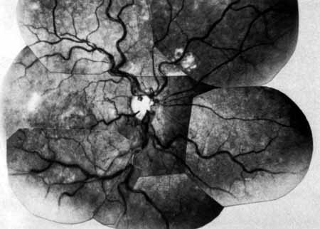
| Fig. 6 Fundus picture 4 years after central retinal vein occlusion. Moderate venous engorgement and small dot hemorrhages remain; microaneurysms, dot hemorrhages, and residual edema are present in the macula. Some sheathing of vessels is seen along with patchy edema residues. Note collateral channels on the retinal surface temporal to the disc and tortuous vessels on the surface of the disc representing collateral cilioretinal communications. |