|
|
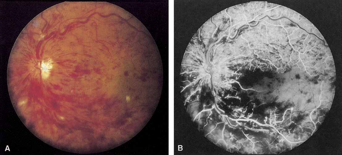
| Fig. 1 A. “Blood and thunder” appearance of a central retinal vein occlusion. B. Intravenous fluorescein angiogram shows this occlusion is primarily ischemic or nonperfused. The fact that there is more nonperfusion in the inferior half of the fundus compared with the superior half is unusual. |